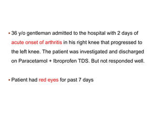
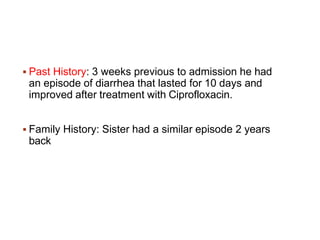
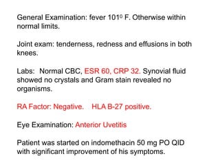
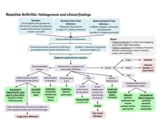
Presented By: Dr Bushu Harna
Fellow OrthoRheumatology
 36 y/o gentleman admitted to the hospital with 2 days of
acute onset of arthritis in his right knee that progressed to
the left knee. The patient was investigated and discharged
on Paracetamol + Ibroprofen TDS. But not responded well.
 Patient had red eyes for past 7 days
 Past History: 3 weeks previous to admission he had
an episode of diarrhea that lasted for 10 days and
improved after treatment with Ciprofloxacin.
 Family History: Sister had a similar episode 2 years
back
General Examination: fever 1010 F. Otherwise within
normal limits.
Joint exam: tenderness, redness and effusions in both
knees.
Labs: Normal CBC, ESR 60, CRP 32. Synovial fluid
showed no crystals and Gram stain revealed no
organisms.
RA Factor: Negative. HLA B-27 positive.
Eye Examination: Anterior Uvetitis
Patient was started on indomethacin 50 mg PO QID
with significant improvement of his symptoms.
 “Reactive Arthritis (ReA) is an infectious induced systemic
illness characterized by an ASEPTIC inflammatory joint
involvement occurring in a genetically predisposed patient
with a bacterial infection localized in a distant
organ/system”.
What’s So Reactive??
to infection
 First described by Hans Reiter in 1916. Also known as
Reiter Syndrome
 Triad: Arthritis, Urethritis, Conjunctivitis
 Name changed to Reactive Arthritis
Postinfectious
arthritis
nongonococca
l
 Aberrant autoimmune resp
uo
rn
et
sh
eritis
conjunctivitis
EPIDEMIOLOGY
 Occurs in 20-40 years.
 Begins with an enteric, urogenital or upper respiratory
tract infections.
 M:F::3:1
 Arthritis occurs few days to 6 weeks after infection
 30-70% positive for HLA-B27.
 Incidence more in urethritis, cervicitis and infectious
diarrhea.
 Incidence varies widely (1% to 20%).
 Frequency varies after infection with Salmonella,
Shigella, Campylobacter or Yersinia.
 1. Microbes-host interaction.
Components of triggering bacteria includes protein and
nucleic acid. These can be found in the synovium and
circulatory monocytes.
 2. Role of immune system
In patients with ReA, they have an elevated production of
Th2 cytokines, such us IL-10 and a possible decrease
production in Th1 cytokines.
Macrophages, CD4+ and CD8+ lymphocytes are
activated in the joints of the patients.
Some bacterial antigens like heat shock protein 60
present in Chlamydia and Yersinia.
Molecular cross reactive has been also associated
 All these factors cause a decrease in the effective
clearance of bacteria.
3. Immunogenetics
HLA-B27 probably works as an antigen presenting
molecule.
Some arthritogenic peptide from chlamydia and yersinia
can be presented by HLA-B27 leading to stimulation of
CD8+ T cells.
 IFN-gamma: low level
 IL-10: High level
 Causative organisms
 Chlamydial trachomatis
 Ureaplasma urealyticum
 Salmonella enteritidis
 Salmonella typhimurium
 Shigella flexneri
 Shigella dysenteriae
 Campylobacter jejuni
 Yersinia enterocolitica
 Streptococcus SP
Urethritis
Enteritis
 Less common association:
 Chlamydia pneumoniae
 Neisseria meningitidis serogroup B
 Bacillus cereus
 Pseudomonas
 Clostridium difficile
 Borrelia burgdorferi
 Escherichia coli
 Helicobacter pillory
 Lactobacillus
 Brucella abortus
 Hafnia alvei
Clinical Manifestations:
 Infection: History: diarrhea, urethritis, cervicitis,
STDs
 Postenteric ReA is described equally in men an women.
 The episode of diarrhea is usually prolonged.
 Postchlamydial is most common in men.
 In females episodes of cervicitis, vaginitis can precede.
 In patients with postchlamydial disease, urethritis is
usually mild, painless and nonpurulent.
 Conjunctivitis is usually observed very early, before
the onset of arthritis. Uveitis is less common but
occurs in 15% of patients with chronic persistent
disease.
 Skin manifestations include: Keratoderma
blenorrhagica, Circinate balanitis and oral ulcers.
 Less common patients can develop valvulitis, rhythm
disturbances.
Arthritis pattern
 Asymmetric mono or oligo arthritis often of lower
limbs including knees, ankles and feet.
 Musculoskeletal: peripheral arthritis, inflammatory
pain in cervical, thoracic, and lumbar spine
 Sacroilitis
 Enthesitis: plantar facisitis, heel pain, patellar tendon
insertion pain, GT hip pain, base 5th MT, MT head pain
 Sausage digits: IP joints with digital tendonitis and
multiple entheseal lesion
 Predictors of more severe disease: hip arthritis,
ESR>30, poor response to NSAIDS, Lumbar spine
stiffness, dactylitis, oligoarthritis, <16 years.
 In HLA-B27 positive patients: more severity and
increased markers like ESR and CRP.
 Eye: conjunctivitis, anterior uveitis, episcleritis and
keratitis
 Genitourinary: urethritis, cervicitis, prostatitis,
cystitis, circinate balanitis, salpingo-oophoritis
 Mucosal and skin: mucosal ulcers, keratoderma
blenorrhagica, erythema nodosum
 Cardiac
 Nail changes: onycholysis, subungual keratosis, nail
pits
 Mono or oligo arthropathy: RA, AS, Ps arthritis
 Gonococcal arthritis
 Gouty arthritis
 Septic arthritis
 Rheumatic fever
 Tubercular arthritis
 Secondary syphilis
 Rare: IBD, celiac, whipple disease and behcet
disease, immunotherapy related arthropathy
Bases on observational data from Germany and Scandinavia
*
 Mono or oligo arthritis
 Exclusion of other diagnosis
Both criteria present: probability is 40%.
 Evidence of previous infection
Then chances are 60%.
 If the bacteria can be culture, then 70%
 If there is history of symptomatic preceding infection with
Chlamdydia trachomatis, then 90%
*Sieper J, Rudwaleit M, Braun J, et al. Diagnosing reactive arthritis: Role of clinical
Definitive: Both Major criteria + 1 Minor Criteria
Probable: Both Major criteria
 Lab: raised ESR and CRP
 Neutrophilic leukocytosis
 HLA-B27
 Rule out: RF, ANA, Gram staining and culture of the joint
fluid, HIV
 Radiographs: Not required to diagnose. A large bulky
paravertebral area of ossification "floating osteophyte" is often seen.
Early juxta-articular osteoporosis, uniform joint space loss and
fusiform soft tissue swelling.
 Ultrasound
 Scintigraphy
 MRI
 Others: ECG, ophthalmological examination, urine
and stool examination, Nucleic acid amplification
tests
 Histopathological dermal features similar to
psoriasis.
 Synovial fluid reveals large macrophages, reiter cell
that have phagocytosed neutrophils, lymphocytes and
plasma cells. Extensive pannus formation is rare.
 Goal
1. Decrease pain and inflammation
2. Minimize disability
3. Prevent relapse or progression to chronic disease
 Team effort
 Patient education
 Early diagnosis and treatment
 Acute: RICE, NSAIDs, orthotics
 Intra-articular glucocorticoid injection
 Systemic corticosteroids and Topical Steroids
 Antibiotics
 DMARDs: Methotrexate, SSZ
 Anti-TNF agents: Etanercept, Infliximab
 It can be self limiting, recurrent or chronic
 Depends on triggering pathogen and genetic
background of the host.
 Chronic arthritis (>6months) occurs in 4-19% patients.
(arthritis caused by salmonella or shigella).
 Chronic relapsing arthritis is seen in 6-8% patients
(induced by salmonella, shigella or chlamydia).
 Good history taking: Past history and family
history
 Good General Physical Examination: EYE,
NAIL, SKIN
 Exclude other Spondyloarthropathy
 Involve Physician & Rheumatologist
 NSAIDs, Steroids and DMARDs form the main
basis of treatment.
 Kim PS, Klausmeier TL, Orr DP. Reactive arthritis: a review. Journal of Adolescent Health. 2009
Apr 1;44(4):309-15.
 Cheeti A, Chakraborty RK, Ramphul K. Reactive Arthritis (Reiter Syndrome). InStatPearls
[Internet] 2020 Mar 13. StatPearls Publishing.
 Stavropoulos PG, Soura E, Kanelleas A, Katsambas A, Antoniou C. Reactive arthritis. Journal of
the European Academy of Dermatology and Venereology. 2015 Mar;29(3):415-24.
 García-Kutzbach A, Chacón-Súchite J, García-Ferrer H, Iraheta I. Reactive arthritis: update
2018. Clinical Rheumatology. 2018 Apr 1;37(4):869-74.
 Colmegna I, Cuchacovich R, Espinoza LR. HLA-B27-associated reactive arthritis: pathogenetic
and clinical considerations. Clinical microbiology reviews. 2004 Apr 1;17(2):348-69.
 Carter JD. Treating reactive arthritis: insights for the clinician. Therapeutic advances in
musculoskeletal disease. 2010 Feb;2(1):45-54.
 Mukherjee S, Kar M. Reactive arthritis: current perspectives. J Ind Acad Clin Med. 2000
Oct;1:233-8.
 Selmi C, Gershwin ME. Diagnosis and classification of reactive arthritis. Autoimmunity reviews.
2014 Apr 1;13(4-5):546-9.
 HARRISON B, SILMAN A, SYMMONS D. Diagnostic evaluation of classification criteria for RA
and reactive arthritis. Annals of the Rheumatic Diseases. 2000 May 1;59(5):397-8.
reactivearthritis-2bbbbbbb01029161901.pptx

reactivearthritis-2bbbbbbb01029161901.pptx

  • 1.
    Presented By: DrBushu Harna Fellow OrthoRheumatology
  • 2.
     36 y/ogentleman admitted to the hospital with 2 days of acute onset of arthritis in his right knee that progressed to the left knee. The patient was investigated and discharged on Paracetamol + Ibroprofen TDS. But not responded well.  Patient had red eyes for past 7 days
  • 3.
     Past History:3 weeks previous to admission he had an episode of diarrhea that lasted for 10 days and improved after treatment with Ciprofloxacin.  Family History: Sister had a similar episode 2 years back
  • 4.
    General Examination: fever1010 F. Otherwise within normal limits. Joint exam: tenderness, redness and effusions in both knees. Labs: Normal CBC, ESR 60, CRP 32. Synovial fluid showed no crystals and Gram stain revealed no organisms. RA Factor: Negative. HLA B-27 positive. Eye Examination: Anterior Uvetitis Patient was started on indomethacin 50 mg PO QID with significant improvement of his symptoms.
  • 5.
     “Reactive Arthritis(ReA) is an infectious induced systemic illness characterized by an ASEPTIC inflammatory joint involvement occurring in a genetically predisposed patient with a bacterial infection localized in a distant organ/system”.
  • 6.
    What’s So Reactive?? toinfection  First described by Hans Reiter in 1916. Also known as Reiter Syndrome  Triad: Arthritis, Urethritis, Conjunctivitis  Name changed to Reactive Arthritis Postinfectious arthritis nongonococca l  Aberrant autoimmune resp uo rn et sh eritis conjunctivitis
  • 7.
    EPIDEMIOLOGY  Occurs in20-40 years.  Begins with an enteric, urogenital or upper respiratory tract infections.  M:F::3:1  Arthritis occurs few days to 6 weeks after infection  30-70% positive for HLA-B27.  Incidence more in urethritis, cervicitis and infectious diarrhea.  Incidence varies widely (1% to 20%).  Frequency varies after infection with Salmonella, Shigella, Campylobacter or Yersinia.
  • 8.
     1. Microbes-hostinteraction. Components of triggering bacteria includes protein and nucleic acid. These can be found in the synovium and circulatory monocytes.
  • 9.
     2. Roleof immune system In patients with ReA, they have an elevated production of Th2 cytokines, such us IL-10 and a possible decrease production in Th1 cytokines. Macrophages, CD4+ and CD8+ lymphocytes are activated in the joints of the patients. Some bacterial antigens like heat shock protein 60 present in Chlamydia and Yersinia. Molecular cross reactive has been also associated  All these factors cause a decrease in the effective clearance of bacteria.
  • 10.
    3. Immunogenetics HLA-B27 probablyworks as an antigen presenting molecule. Some arthritogenic peptide from chlamydia and yersinia can be presented by HLA-B27 leading to stimulation of CD8+ T cells.  IFN-gamma: low level  IL-10: High level
  • 11.
     Causative organisms Chlamydial trachomatis  Ureaplasma urealyticum  Salmonella enteritidis  Salmonella typhimurium  Shigella flexneri  Shigella dysenteriae  Campylobacter jejuni  Yersinia enterocolitica  Streptococcus SP Urethritis Enteritis
  • 12.
     Less commonassociation:  Chlamydia pneumoniae  Neisseria meningitidis serogroup B  Bacillus cereus  Pseudomonas  Clostridium difficile  Borrelia burgdorferi  Escherichia coli  Helicobacter pillory  Lactobacillus  Brucella abortus  Hafnia alvei
  • 13.
    Clinical Manifestations:  Infection:History: diarrhea, urethritis, cervicitis, STDs  Postenteric ReA is described equally in men an women.  The episode of diarrhea is usually prolonged.  Postchlamydial is most common in men.  In females episodes of cervicitis, vaginitis can precede.
  • 14.
     In patientswith postchlamydial disease, urethritis is usually mild, painless and nonpurulent.  Conjunctivitis is usually observed very early, before the onset of arthritis. Uveitis is less common but occurs in 15% of patients with chronic persistent disease.  Skin manifestations include: Keratoderma blenorrhagica, Circinate balanitis and oral ulcers.  Less common patients can develop valvulitis, rhythm disturbances.
  • 15.
    Arthritis pattern  Asymmetricmono or oligo arthritis often of lower limbs including knees, ankles and feet.  Musculoskeletal: peripheral arthritis, inflammatory pain in cervical, thoracic, and lumbar spine  Sacroilitis  Enthesitis: plantar facisitis, heel pain, patellar tendon insertion pain, GT hip pain, base 5th MT, MT head pain  Sausage digits: IP joints with digital tendonitis and multiple entheseal lesion
  • 16.
     Predictors ofmore severe disease: hip arthritis, ESR>30, poor response to NSAIDS, Lumbar spine stiffness, dactylitis, oligoarthritis, <16 years.  In HLA-B27 positive patients: more severity and increased markers like ESR and CRP.
  • 17.
     Eye: conjunctivitis,anterior uveitis, episcleritis and keratitis  Genitourinary: urethritis, cervicitis, prostatitis, cystitis, circinate balanitis, salpingo-oophoritis  Mucosal and skin: mucosal ulcers, keratoderma blenorrhagica, erythema nodosum  Cardiac  Nail changes: onycholysis, subungual keratosis, nail pits
  • 18.
     Mono oroligo arthropathy: RA, AS, Ps arthritis  Gonococcal arthritis  Gouty arthritis  Septic arthritis  Rheumatic fever  Tubercular arthritis  Secondary syphilis  Rare: IBD, celiac, whipple disease and behcet disease, immunotherapy related arthropathy
  • 19.
    Bases on observationaldata from Germany and Scandinavia *  Mono or oligo arthritis  Exclusion of other diagnosis Both criteria present: probability is 40%.  Evidence of previous infection Then chances are 60%.  If the bacteria can be culture, then 70%  If there is history of symptomatic preceding infection with Chlamdydia trachomatis, then 90% *Sieper J, Rudwaleit M, Braun J, et al. Diagnosing reactive arthritis: Role of clinical
  • 20.
    Definitive: Both Majorcriteria + 1 Minor Criteria Probable: Both Major criteria
  • 21.
     Lab: raisedESR and CRP  Neutrophilic leukocytosis  HLA-B27  Rule out: RF, ANA, Gram staining and culture of the joint fluid, HIV  Radiographs: Not required to diagnose. A large bulky paravertebral area of ossification "floating osteophyte" is often seen. Early juxta-articular osteoporosis, uniform joint space loss and fusiform soft tissue swelling.  Ultrasound  Scintigraphy  MRI
  • 22.
     Others: ECG,ophthalmological examination, urine and stool examination, Nucleic acid amplification tests  Histopathological dermal features similar to psoriasis.  Synovial fluid reveals large macrophages, reiter cell that have phagocytosed neutrophils, lymphocytes and plasma cells. Extensive pannus formation is rare.
  • 24.
     Goal 1. Decreasepain and inflammation 2. Minimize disability 3. Prevent relapse or progression to chronic disease  Team effort  Patient education  Early diagnosis and treatment
  • 25.
     Acute: RICE,NSAIDs, orthotics  Intra-articular glucocorticoid injection  Systemic corticosteroids and Topical Steroids  Antibiotics  DMARDs: Methotrexate, SSZ  Anti-TNF agents: Etanercept, Infliximab
  • 26.
     It canbe self limiting, recurrent or chronic  Depends on triggering pathogen and genetic background of the host.  Chronic arthritis (>6months) occurs in 4-19% patients. (arthritis caused by salmonella or shigella).  Chronic relapsing arthritis is seen in 6-8% patients (induced by salmonella, shigella or chlamydia).
  • 28.
     Good historytaking: Past history and family history  Good General Physical Examination: EYE, NAIL, SKIN  Exclude other Spondyloarthropathy  Involve Physician & Rheumatologist  NSAIDs, Steroids and DMARDs form the main basis of treatment.
  • 29.
     Kim PS,Klausmeier TL, Orr DP. Reactive arthritis: a review. Journal of Adolescent Health. 2009 Apr 1;44(4):309-15.  Cheeti A, Chakraborty RK, Ramphul K. Reactive Arthritis (Reiter Syndrome). InStatPearls [Internet] 2020 Mar 13. StatPearls Publishing.  Stavropoulos PG, Soura E, Kanelleas A, Katsambas A, Antoniou C. Reactive arthritis. Journal of the European Academy of Dermatology and Venereology. 2015 Mar;29(3):415-24.  García-Kutzbach A, Chacón-Súchite J, García-Ferrer H, Iraheta I. Reactive arthritis: update 2018. Clinical Rheumatology. 2018 Apr 1;37(4):869-74.  Colmegna I, Cuchacovich R, Espinoza LR. HLA-B27-associated reactive arthritis: pathogenetic and clinical considerations. Clinical microbiology reviews. 2004 Apr 1;17(2):348-69.  Carter JD. Treating reactive arthritis: insights for the clinician. Therapeutic advances in musculoskeletal disease. 2010 Feb;2(1):45-54.  Mukherjee S, Kar M. Reactive arthritis: current perspectives. J Ind Acad Clin Med. 2000 Oct;1:233-8.  Selmi C, Gershwin ME. Diagnosis and classification of reactive arthritis. Autoimmunity reviews. 2014 Apr 1;13(4-5):546-9.  HARRISON B, SILMAN A, SYMMONS D. Diagnostic evaluation of classification criteria for RA and reactive arthritis. Annals of the Rheumatic Diseases. 2000 May 1;59(5):397-8.