The document discusses the purpose, rationale, and importance of obturation after root canal treatment. Obturation functions to prevent reinfection by acting as a barrier and sealing any surviving bacterial cells. Failure to eliminate etiological factors and prevent further contamination can lead to treatment failure. At least 5mm of gutta percha retained in the root provides an adequate apical seal and reduces leakage. Coronal leakage through the restoration is also a significant cause of treatment failure if the access cavity is left unfilled and exposed to fluids over time.
The document discusses obturation of the root canal system. It states that the objectives of obturation are to eliminate all avenues of leakage and seal any remaining irritants in the root canal. It discusses factors related to the appropriate length, timing, and preparation for obturation. Components of root canal filling include core materials like gutta-percha and root canal sealers. Various types of sealers are discussed, including zinc oxide eugenol, calcium hydroxide, and glass ionomer sealers. Removal of the smear layer before obturation is also addressed.
This document discusses the objectives and process of obturation in root canal treatment. It states that obturation aims to seal the root canal system to prevent reinfection by eliminating pathways for leakage. It also discusses the importance of removing the smear layer before obturation using chemicals like MTAD or a combination of EDTA and sodium hypochlorite. The document examines the factors that influence the timing of obturation and reviews materials commonly used, including gutta-percha and various sealers. It provides guidelines for an ideal obturation material and compares methods like lateral condensation versus thermoplasticized techniques.
This document outlines the basics of root canal treatment through a lecture given by Dr. Syed Mukhtar-un- Nisar Andrabi. It begins with definitions of endodontics and discusses root canal anatomy, microbiology, and the step-by-step root canal treatment procedure. The treatment procedure involves access preparation, shaping and cleaning, irrigation, obturation, and post endodontic restoration. Case examples are also provided to demonstrate successful root canal treatments. The goal of root canal treatment is to eliminate infection and prevent reinfection to allow healing of periapical tissues. Proper diagnosis, instrumentation, obturation and restoration are essential for optimal treatment outcomes.
The document discusses the preparation of the root canal system through cleaning and shaping. It defines cleaning as the removal of contents from the root canal to eliminate bacteria, and shaping as the mechanical process of establishing a continuous taper to the canal to allow for better instrumentation, irrigation, and obturation. The objectives of preparation are to remove all irritants from the canal biologically and to develop a tapered conical form that maintains the original canal anatomy mechanically. Principles of preparation include outlining the canal shape, removing debris, and developing retention and resistance forms through appropriate tapers and diameters.
Obturation Of Root Canal Obturation Of Root Canalvasanthatpuram
The document discusses obturation, which is the filling of the root canal system after chemomechanical preparation. Obturation aims to provide an impermeable seal within the root canal system to prevent reinfection from oral or apical microleakage. Ideal obturation extends to the cementodentinal junction and has a tapered funnel-like shape reflecting the root's morphology. Materials used for obturation include gutta-percha, silver cones, and various sealers. Gutta-percha is the most common material due to its biocompatibility and plasticity which allows it to fill canal irregularities.
This chapter discusses principles of cleaning and shaping root canals. It addresses debates around the extent of apical preparation and debates termination of cleaning and shaping. Larger apical preparations allow for better irrigation and debris removal but risk over-instrumentation. Maintaining apical patency is questioned as it does not improve bacteria reduction and risks extruding debris. Various instrumentation techniques are described. The goal of cleaning is to reduce rather than eliminate irritants from the canal due to anatomical complexities. Shaping aims to facilitate cleaning and provide space for obturation.
Obturation and the Importance of Coronal SealAlwaleed Fahad
The coronal seal of an endodontically treated tooth is essential for the long-term success of the treatment. Microorganisms from the oral cavity can reinfect the root canal system through gaps in the temporary or permanent restoration. Several studies have shown that within a few weeks of leaving the access cavity open, bacteria from the saliva can travel down the entire length of the root canal. Placement of an adequate permanent restoration is critical to prevent reinfection of the root canal system and promote healing of the periapical tissues. The technical quality of the coronal restoration has a greater impact on apical periodontal health than the quality of the root canal filling.
A detailed presentation on Endodontic failures starting from the basics in case selection to final prosthesis. Good for Post Graduates and Under Graduates.
The document discusses missed canals during root canal treatment. It states that missed canals are one of the main causes of endodontic treatment failure, with an overall prevalence of 18%. Missed canals were most common in maxillary first molars at 40.6%. The document provides strategies for locating canals such as knowledge of anatomy, radiographs, CBCT, magnification, access cavity modification, ultrasonics, staining techniques, and presents common sites where canals are missed such as maxillary premolars, mandibular incisors, and mandibular molars. It emphasizes the importance of locating all canals to prevent complications and improve treatment outcomes.
This document discusses root canal morphology and its relationship to endodontic procedures. It begins by explaining that the root canal system takes on numerous complex configurations that are not always evident on radiographs. A thorough understanding of root canal anatomy is important for successful endodontic treatment. The document then describes various anatomical structures like accessory canals, lateral canals, furcation canals and apical deltas. It discusses how tools like operating microscopes and multiple angled radiographs can help visualize complex root canal anatomies. Finally, it summarizes research on the varying root canal configurations that have been observed in different tooth types.
Biomechanical preparation1/ rotary endodontic courses by indian dental academyIndian dental academy
Indian Dental Academy: will be one of the most relevant and exciting training center with best faculty and flexible training programs for dental professionals who wish to advance in their dental practice,Offers certified courses in Dental implants,Orthodontics,Endodontics,Cosmetic Dentistry, Prosthetic Dentistry, Periodontics and General Dentistry.
Nonvital pulp therapy in pediatric dentistryPriyank Pareek
The document discusses techniques for pulpectomy procedures in primary teeth. It describes removing necrotic pulp tissue from the root canals and coronal portion of teeth to maintain the tooth. Key steps include isolating the tooth, removing caries, accessing and cleaning canals, drying canals, and filling canals with zinc oxide eugenol. The goals are to resolve infection, show healing on x-rays, and allow normal resorption and eruption of permanent teeth.
Principles of intra coronal and radicular preparationIAU Dent
This document discusses the principles of endodontic cavity preparation. It is divided into three phases: cleaning and shaping, disinfection, and obturation. The objectives of cleaning and shaping are to remove all contents from the root canal that could harbor microorganisms or lead to periapical issues. The root canal must be prepared to a continuously tapering shape that maintains the original anatomy and position of the foramen. Cavity preparation has coronal and radicular divisions, following Black's principles modified for endodontics. These include outline form to follow canal anatomy, convenience form for instrument access, and resistance form to prevent overfilling. Thorough cleaning is achieved through instrumentation and irrigation to eliminate debris and bacteria.
Devitalized extirpation, amputation and combined methodLinda Jenhani
The document discusses root canal preparation techniques. It describes cleaning and shaping objectives to remove debris and bacteria while creating a tapered canal for filling. Irrigation solutions like sodium hypochlorite and EDTA are discussed for cleaning. Instrumentation with nickel titanium or stainless steel files of varying tapers, tips, lengths, and sizes is described. The crown-down technique is outlined, starting with larger files coronally and progressing apically with smaller files. Estimating canal size and determining working length is also summarized.
smear layer in endodontics/ rotary endodontic courses by indian dental academyIndian dental academy
Indian Dental Academy: will be one of the most relevant and exciting training center with best faculty and flexible training programs for dental professionals who wish to advance in their dental practice,Offers certified courses in Dental implants,Orthodontics,Endodontics,Cosmetic Dentistry, Prosthetic Dentistry, Periodontics and General Dentistry.
The double cord technique involves placing a small diameter cord in the gingival sulcus first, leaving it in place, and then packing a larger diameter cord over the first cord to provide additional retraction and hemostasis for making impressions of multiple prepared teeth or when the gingival tissues are compromised. The small inner cord provides retraction while the outer cord provides additional hemostasis and tissue displacement needed for accurate impressions.
Instrument seperation and its managementNivedha Tina
This document discusses factors related to endodontic instrument separation, including prevalence, incidence, contributing factors, and management techniques. It covers topics such as tooth, instrument, operator, and patient factors that influence separation as well as techniques to prevent separation. The document provides an overview of considerations for removing separated instruments and discusses how canal morphology, curvature, and location within the canal impact separation and removal success rates.
This document discusses various procedural complications that can occur during endodontic treatment. It identifies the three main factors for endodontic treatment success as diagnosis and treatment planning, cleaning and shaping of the root canal, and obturation. It then describes multiple mishaps that can occur related to diagnosis, anesthesia, isolation, access opening, irrigation, instrumentation, obturation, and surgical procedures. Specific radiographic issues and complications involving perforations, ledges, transportation, over-instrumentation, and under-preparation of the canal are examined. Prevention and management strategies are provided for many of the potential procedural complications.
Procedural complications in endodontics can occur during various stages including diagnosis, instrumentation, obturation, and post-treatment. Common issues involve missed canals, ledges, perforations, over-instrumentation, underfilling, and overfilling. Successful treatment depends on accurate diagnosis and treatment planning, thorough cleaning and shaping of the root canal system, and proper filling of the entire root canal space. Preventing complications requires following principles such as using pre-curved instruments, limiting pressure, and taking radiographs to confirm working length and detect any procedural errors.
Cleaning and shaping the root canal systemParth Thakkar
The document discusses various techniques for cleaning and shaping the root canal during endodontic treatment. The objectives are to remove infected tissue, provide access for irrigants and medicaments, create space for obturation, and maintain tooth integrity. Key steps discussed include determining working length, instrumentation using techniques like step-back preparation, and irrigation. Factors that can affect working length determination and techniques to enhance cleaning and shaping are also outlined.
This document discusses endodontic mishaps and procedural accidents that can occur during root canal treatment. It begins by defining endodontic mishaps and classifying them into categories such as access related, instrumentation related, and obturation related mishaps. Specific mishaps like treating the wrong tooth, missed canals, ledge formation, and perforations are described in detail. The document emphasizes the importance of recognizing mishaps, correcting them properly, and preventing future errors through careful diagnosis, instrumentation techniques, and quality control measures.
This case report summarizes several cases where additional root canals were found during endodontic treatment that had initially been missed. It describes the complex root canal anatomy that can vary between different types of teeth and emphasizes that clinicians must be aware of potential extra canals. The report presents clinical cases where a second mesiobuccal canal, distobuccal canal, palatal canal, and second distal canal were successfully located and treated. It discusses techniques for identifying missed canals, including modifications to access cavities, use of magnification, explorers, stains, and transillumination. Locating all canals is important to achieve successful root canal treatment outcomes.
126finalseminar-190228072850.ppt fod gingival retractiondryogeshkumar1212
This document discusses various methods for gingival retraction to displace gingival tissues away from tooth margins prior to dental impressions. It describes both mechanical methods using materials like copper bands, rubber dams, and cotton twills as well as chemico-mechanical methods using retraction cords impregnated with chemicals. Retraction cords are classified based on material, diameter, and chemical treatment. Common chemicals used include vasoconstrictors like epinephrine to control bleeding as well as coagulants. The document outlines techniques for placing retraction cords and selecting the appropriate cord based on the clinical situation.
The document discusses the importance of root canal obturation and provides details on the process. It begins by defining obturation as filling the cleaned and shaped root canal space. A key goal is to hermetically seal the root canal system to prevent reinfection. The challenges include filling an irregular space that varies between teeth. Historically, materials like gold, gutta-percha and various pastes have been used. Proper preparation, including smear layer removal, affects the quality of sealing. Debate remains around how far to instrument and fill within the canal but terminating at the apical constriction or 1mm short of the radiographic apex is often recommended.
Endodontic surgery is performed to address issues like failed root canal treatments, procedural errors, anatomical variations, and biopsies. It involves raising a surgical flap, resecting the root tip, preparing and filling the root end cavity. Potential complications include swelling, pain, nerve damage and infection. A variety of materials can be used for the root end filling including zinc oxide eugenol cements, MTA, composites and glass ionomer cements. The goal is to provide an apical seal to prevent reinfection from microbes remaining in the root canal system.
This document discusses various biological considerations related to dental restorations and periodontal health. It covers topics such as biologic width, margin placement, overhangs, and correcting biologic width violations. Key points include that supragingival margins have the least impact on the periodontium, biologic width violations can lead to inflammation and bone loss, and overhangs increase plaque and pathogens if placed subgingivally. The document provides guidelines for proper margin placement based on sulcular depth to avoid future recession. It also discusses procedures for tissue retraction and electrosurgery when placing subgingival margins.
Indian Dental Academy: will be one of the most relevant and exciting training center with best faculty and flexible training programs for dental professionals who wish to advance in their dental practice,Offers certified courses in Dental implants,Orthodontics,Endodontics,Cosmetic Dentistry, Prosthetic Dentistry, Periodontics and General Dentistry.
Similar to Purpose, rationale, and importance of obturation.pdf (20)
TEST BANK For Community Health Nursing A Canadian Perspective, 5th Edition by...Donc Test
TEST BANK For Community Health Nursing A Canadian Perspective, 5th Edition by Stamler, Verified Chapters 1 - 33, Complete Newest Version Community Health Nursing A Canadian Perspective, 5th Edition by Stamler, Verified Chapters 1 - 33, Complete Newest Version Community Health Nursing A Canadian Perspective, 5th Edition by Stamler Community Health Nursing A Canadian Perspective, 5th Edition TEST BANK by Stamler Test Bank For Community Health Nursing A Canadian Perspective, 5th Edition Pdf Chapters Download Test Bank For Community Health Nursing A Canadian Perspective, 5th Edition Pdf Download Stuvia Test Bank For Community Health Nursing A Canadian Perspective, 5th Edition Study Guide Test Bank For Community Health Nursing A Canadian Perspective, 5th Edition Ebook Download Stuvia Test Bank For Community Health Nursing A Canadian Perspective, 5th Edition Questions and Answers Quizlet Test Bank For Community Health Nursing A Canadian Perspective, 5th Edition Studocu Test Bank For Community Health Nursing A Canadian Perspective, 5th Edition Quizlet Test Bank For Community Health Nursing A Canadian Perspective, 5th Edition Stuvia Community Health Nursing A Canadian Perspective, 5th Edition Pdf Chapters Download Community Health Nursing A Canadian Perspective, 5th Edition Pdf Download Course Hero Community Health Nursing A Canadian Perspective, 5th Edition Answers Quizlet Community Health Nursing A Canadian Perspective, 5th Edition Ebook Download Course hero Community Health Nursing A Canadian Perspective, 5th Edition Questions and Answers Community Health Nursing A Canadian Perspective, 5th Edition Studocu Community Health Nursing A Canadian Perspective, 5th Edition Quizlet Community Health Nursing A Canadian Perspective, 5th Edition Stuvia Community Health Nursing A Canadian Perspective, 5th Edition Test Bank Pdf Chapters Download Community Health Nursing A Canadian Perspective, 5th Edition Test Bank Pdf Download Stuvia Community Health Nursing A Canadian Perspective, 5th Edition Test Bank Study Guide Questions and Answers Community Health Nursing A Canadian Perspective, 5th Edition Test Bank Ebook Download Stuvia Community Health Nursing A Canadian Perspective, 5th Edition Test Bank Questions Quizlet Community Health Nursing A Canadian Perspective, 5th Edition Test Bank Studocu Community Health Nursing A Canadian Perspective, 5th Edition Test Bank Quizlet Community Health Nursing A Canadian Perspective, 5th Edition Test Bank Stuvia
Promoting Wellbeing - Applied Social Psychology - Psychology SuperNotesPsychoTech Services
A proprietary approach developed by bringing together the best of learning theories from Psychology, design principles from the world of visualization, and pedagogical methods from over a decade of training experience, that enables you to: Learn better, faster!
Here is the updated list of Top Best Ayurvedic medicine for Gas and Indigestion and those are Gas-O-Go Syp for Dyspepsia | Lavizyme Syrup for Acidity | Yumzyme Hepatoprotective Capsules etc
8 Surprising Reasons To Meditate 40 Minutes A Day That Can Change Your Life.pptxHolistified Wellness
We’re talking about Vedic Meditation, a form of meditation that has been around for at least 5,000 years. Back then, the people who lived in the Indus Valley, now known as India and Pakistan, practised meditation as a fundamental part of daily life. This knowledge that has given us yoga and Ayurveda, was known as Veda, hence the name Vedic. And though there are some written records, the practice has been passed down verbally from generation to generation.
Integrating Ayurveda into Parkinson’s Management: A Holistic ApproachAyurveda ForAll
Explore the benefits of combining Ayurveda with conventional Parkinson's treatments. Learn how a holistic approach can manage symptoms, enhance well-being, and balance body energies. Discover the steps to safely integrate Ayurvedic practices into your Parkinson’s care plan, including expert guidance on diet, herbal remedies, and lifestyle modifications.
share - Lions, tigers, AI and health misinformation, oh my!.pptxTina Purnat
• Pitfalls and pivots needed to use AI effectively in public health
• Evidence-based strategies to address health misinformation effectively
• Building trust with communities online and offline
• Equipping health professionals to address questions, concerns and health misinformation
• Assessing risk and mitigating harm from adverse health narratives in communities, health workforce and health system
Adhd Medication Shortage Uk - trinexpharmacy.comreignlana06
The UK is currently facing a Adhd Medication Shortage Uk, which has left many patients and their families grappling with uncertainty and frustration. ADHD, or Attention Deficit Hyperactivity Disorder, is a chronic condition that requires consistent medication to manage effectively. This shortage has highlighted the critical role these medications play in the daily lives of those affected by ADHD. Contact : +1 (747) 209 – 3649 E-mail : sales@trinexpharmacy.com
Purpose, rationale, and importance of obturation.pdf
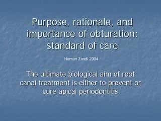
1. Purpose,
Purpose, rationale
rationale, and
, and
importance
importance of
of obturation:
obturation:
standard
standard of
of care
care
Homan Zandi 2004
The
The ultimate
ultimate biological
biological aim
aim of
of root
root
canal
canal treatment
treatment is
is either
either to
to prevent
prevent or
or
cure
cure apical
apical periodontitis
periodontitis
2. Functions
Functions of
of the
the root
root filling
filling
„
„ Preventing
Preventing the
the
reinfection
reinfection by
by acting
acting
as a
as a barrier
barrier
„
„ Sealing
Sealing any
any surviving
surviving
bacterial
bacterial cells
cells and
and
their
their irritants
irritants
„
„ Stopping
Stopping influx
influx of
of
periapical
periapical tissue
tissue
fluids
fluids
3. Failure to eliminate these
Failure to eliminate these etiological factors
etiological factors
and to prevent further irritation via continued
contamination of the root canal system are
the
the prime causes of failure
prime causes of failure of
of nonsurgical
nonsurgical and
and
surgical root canal treatment
4. „
„ Three
Three-
-dimensional (3
dimensional (3-
-D) obturation
D) obturation
„
„ Radiographic evaluation
Radiographic evaluation
„
„ Poor correlation between the quality of the root
Poor correlation between the quality of the root
canal obturation and what is viewed on a
canal obturation and what is viewed on a buccal
buccal
radiograph
radiograph
„
„ When the root filling is
When the root filling is radiograpically
radiograpically
acceptable, the likelihood of leakage is still
rather high
rather high
32. „
„ The
The success
success of
of endodontic
endodontic therapy
therapy is
is
commonly
commonly thought
thought of
of in terms
in terms of
of an
an
adequate
adequate apical
apical seal
seal
„
„ However
However,
, the
the coronal
coronal seal
seal achieved
achieved by
by the
the
restoration
restoration may
may be
be considered
considered as
as important
important
for
for the
the ultimate
ultimate success
success of
of endodontic
endodontic
treatment
treatment (Marshall et al,
(Marshall et al, Swanson
Swanson et al,
et al,
Torabinejad
Torabinejad et al,
et al, Magura
Magura et al.
et al. Khayat
Khayat et
et
al,
al, Ray
Ray et al,
et al, Tronstad
Tronstad et al)
et al)
33. Strindberg, in 1956,
Strindberg, in 1956,
considered
considered that
that the
the
most
most common
common cause
cause of
of
failure
failure was
was leakage
leakage of
of
tissue
tissue fluids
fluids apically
apically
around
around inadequate
inadequate root
root
fillings
fillings
Ingle
Ingle in 1965
in 1965 found
found
that
that of
of 104
104 failed
failed
cases, 66
cases, 66 were
were
associated
associated with
with a
a poor
poor
apical
apical seal
seal
34.
35. How much gutta
How much gutta-
-percha should
percha should
be retained to maintain the
apical seal?
apical seal?
36. Camp et al (1983)
Camp et al (1983)
determined
determined that
that
when
when 4 mm
4 mm of
of gutta
gutta
percha
percha was
was retained
retained
only
only 1
1 of
of 89
89
specimens
specimens showed
showed
leakage
leakage,
, whereas
whereas
32
32 of
of 89
89 specimens
specimens
leaked
leaked when
when 2 mm
2 mm
of
of gutta
gutta percha
percha was
was
retained
retained
38. Zmener
Zmener (1980)
(1980)
found
found that
that in
in root
root
canals
canals sealed
sealed with
with
lateral
lateral
condensation
condensation
technique
technique,
, leakage
leakage
was
was reduced
reduced when
when
more
more than
than 4 mm
4 mm of
of
gutta
gutta-
-percha
percha
remained
remained in
in the
the
apical
apical portion
portion
39. „
„ Portell
Portell et al (1982)
et al (1982) determined
determined that
that most
most of
of
the
the specimens
specimens with
with only
only 3 mm
3 mm of
of apical
apical
gutta
gutta percha
percha had
had some
some leakage
leakage
„
„ Mattison
Mattison et al (1984)
et al (1984) found
found significant
significant
differences
differences between
between 3, 5, and 7 mm
3, 5, and 7 mm of
of gutta
gutta
percha
percha, and
, and they
they concluded
concluded that
that at
at least
least 5
5
mm
mm of
of gutta
gutta percha
percha is
is necessary
necessary for an
for an
adequate
adequate apical
apical seal
seal
40. Post
Post space
space preparation
preparation and
and
leakage
leakage
„
„ During
During the
the mechanical
mechanical preparation
preparation of
of the
the
post
post space
space it is
it is possible
possible that
that the
the root
root filling
filling
may
may be
be twisted
twisted or
or vibrated
vibrated,
, with
with disruption
disruption
of
of the
the seal
seal
41. „
„ Provided
Provided a minimum
a minimum of
of 5 mm
5 mm of
of sound
sound
apical
apical root
root filling
filling is
is left
left in
in situ
situ,
, studies have
studies have
shown
shown that
that removal
removal of
of laterally
laterally condensed
condensed
gutta
gutta percha
percha does
does not
not affect
affect the
the apical
apical seal
seal,
,
irrespective
irrespective of
of whether
whether the
the post
post space
space is
is
prepared
prepared immediately
immediately after
after obturation or is
obturation or is
delayed
delayed (
(Zmener
Zmener 1980,
1980, Neagley
Neagley 1969,
1969,
Bourgeois et al 1981)
42. Endodontic success
Endodontic success
„
„ It is generally accepted that the success
It is generally accepted that the success
rate of the treatment is positively
correlated with the criteria for
correlated with the criteria for good
good
technical quality of the root filling
43.
44. „
„ Even in a good root filling performed
Even in a good root filling performed
under optimal condition, the
under optimal condition, the coronal
coronal
leakage
leakage will be consistent and extensive if
will be consistent and extensive if
the access cavity is
the access cavity is left unfilled
left unfilled and thus
and thus
exposed to fluids
exposed to fluids
45.
46.
47.
48.
49.
50.
51.
52.
53. Obturated
Obturated root
root canals
canals can
can be
be recontaminated
recontaminated
by
by micro
micro-
-organisms
organisms in a
in a number
number of
of ways
ways:
:
„
„ Delay
Delay in
in placing
placing a
a coronal
coronal restoration
restoration.
.
Temporary
Temporary materials
materials will
will dissolve
dissolve slowly
slowly after
after
in time in
in time in the
the presence
presence of
of saliva and
saliva and the
the seal
seal
may
may break
break down
down. A
. A temporary
temporary restoration
restoration of
of
inadequate
inadequate thickness
thickness will
will eventually
eventually leak
leak
54. „
„ Fracture
Fracture of
of the
the coronal
coronal restoration
restoration and /or
and /or
the
the tooth
tooth
„
„ Preparation
Preparation of
of post
post space
space when
when the
the
remaining
remaining apical
apical section
section of
of the
the root
root filling
filling is
is
of
of inadequate
inadequate density
density and / or
and / or length
length
55. Coronal
Coronal leakage
leakage…
…
„
„ Marshall &
Marshall & Massler
Massler, in 1961,
, in 1961, carried
carried out
out a
a
leakage
leakage study
study using
using a
a radioactive
radioactive tracer
tracer
and
and showed
showed that
that coronal
coronal leakage
leakage occurred
occurred
despite
despite the
the presence
presence of
of a
a coronal
coronal dressing
dressing
56. Leakage
Leakage of
of endodontic obturation
endodontic obturation
materials
materials are
are measured
measured by:
by:
„
„ Dyes (
Dyes (Swanson
Swanson et al, Madison et al)
et al, Madison et al)
„
„ Radioactive
Radioactive isotopes (Marshall et al)
isotopes (Marshall et al)
„
„ Bacteria
Bacteria (Mortensen et al,
(Mortensen et al, Goldman
Goldman et al,
et al,
Torabinejad
Torabinejad
et al)
et al)
„
„ Fluid
Fluid filtration
filtration method
method (
(Derksen
Derksen et al)
et al)
57. Allison
Allison et al, in
et al, in
1979
1979 made
made brief
brief
reference
reference to
to the
the
possibility
possibility that
that a
a
poor
poor coronal
coronal seal
seal
might
might contribute
contribute
to
to clinical
clinical failure
failure
58. Swanson
Swanson &
&
Madison, in 1987,
Madison, in 1987,
did
did an in
an in vitro
vitro study
study
where
where they
they showed
showed
that
that after
after only
only 3
3
days
days exposure
exposure to
to
artificial
artificial saliva
saliva there
there
was
was extensive
extensive
coronal
coronal leakage
leakage of
of a
a
tracer dye
tracer dye through
through
aparently
aparently sound
sound
root
root filling
filling
59. Madison &
Madison &
Wilcox
Wilcox, in 1988,
, in 1988,
confirmed
confirmed that
that
exposure
exposure of
of root
root
canals
canals to
to the
the
oral
oral environment
environment
allowed
allowed coronal
coronal
leakage
leakage to
to take
take
place
place, in
, in some
some
cases
cases along
along the
the
whole
whole length
length of
of
the
the root
root canals
canals
60. Torabinejad
Torabinejad et al, in
et al, in
1990,
1990, found
found that
that
50%
50% of
of single
single-
-rooted
rooted
teeth
teeth,
, root
root filled
filled
using
using lateral
lateral
condensation
condensation of
of
gutta
gutta percha
percha and a
and a
sealer
sealer cement
cement,
, were
were
contaminated
contaminated with
with
bacteria
bacteria along
along the
the
whole
whole length
length of
of the
the
root
root after
after 19
19 days
days or
or
42
42 days
days,
, depending
depending
upon
upon the
the
contaminating
contaminating
organism
organism
61. Khayat
Khayat et al, in 1993,
et al, in 1993,
have
have shown
shown that
that root
root
canals
canals obturated
obturated with
with
gutta
gutta percha
percha and
and Roth
Roth’
’s
s
sealer
sealer,
, using
using either
either
lateral
lateral condensation
condensation or
or
vertical
vertical condensation
condensation
were
were contaminated
contaminated
apically
apically with
with bacteria
bacteria
from saliva
from saliva exposed
exposed to
to
the
the coronal
coronal part
part of
of the
the
root
root canal
canal only
only. All
. All
canals
canals were
were
contaminated
contaminated within
within 30
30
days
days of
of exposure
exposure
63. What is more important?
What is more important?
„
„ A good root filling or a good coronal
A good root filling or a good coronal
restoration
restoration
64. „
„ 1010
1010 endodontically
endodontically treated teeth examined
treated teeth examined
radiographically
radiographically
„
„ Good
Good endodontic
endodontic treatment (GE)
treatment (GE)
„
„ Poor
Poor endodontic
endodontic treatment (PE)
treatment (PE)
„
„ Good restoration (GR)
Good restoration (GR)
„
„ Poor restoration (PR)
Poor restoration (PR)
„
„ Absence of
Absence of periraducular
periraducular inflammation (API)
inflammation (API)
„
„ Presence of
Presence of periradicular
periradicular inflammation (PPI)
inflammation (PPI)
65.
66.
67. Conclusion:
Conclusion:
The technical quality of the
The technical quality of the coronal
coronal
restoration
restoration was significantly more important
was significantly more important
than the technical quality of the
than the technical quality of the endodontic
endodontic
treatment
treatment for apical periodontal health
for apical periodontal health
68. „
„ Duplicate the study by Ray & Trope
Duplicate the study by Ray & Trope
93. Microleakage
Actual bacterial penetration through
obturating materials may not be
necessary to cause treatment failure.
More important may be leakage of
bacterial by-products
Bacterial metabolites, toxins and
degradation products are much
smaller than bacteria and could
penetrate faster
Hovland & Dumsha, in 1985, showed
that most leakage occurs between the
root canal sealer and the wall of the
root canal
94. Prokaryptic
cells (bacteria)
are the smallest
of the
unicellular
organisms.
They are, for
the most part,
approximately
1 to 1.5 µm
wide and 2 to 6
µm long Escherichia coli is
approximately 1 µm in
diameter
95. Bacterial
Bacterial mechanism
mechanism of
of tissue
tissue
damage
damage and
and bacterial
bacterial products
products
Bacterial factors for
colonization and
growth
Bacterial factors for
colonization and
growth
Bacterial factors for
invasion and tissue
damage
Bacterial factors for
invasion and tissue
damage
96. Bacterial
Bacterial factors
factors for
for invasion
invasion
and
and tissue
tissue damage
damage
Direct
Direct Indirect
Indirect
Cytotoxic
products
Cytotoxic
products
Enzymes
Enzymes Inflammatory
response
Inflammatory
response
Tissue damage
Tissue damage