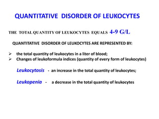
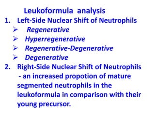
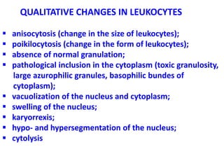
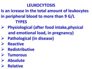
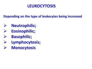
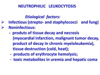
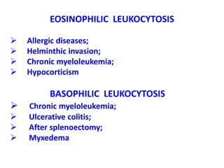
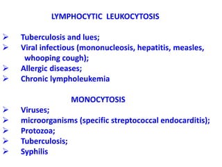
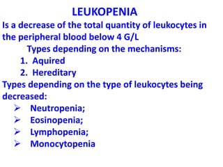
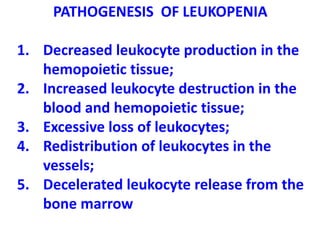
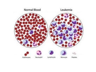
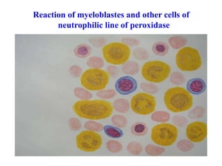
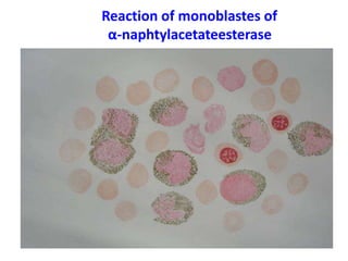
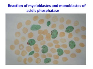
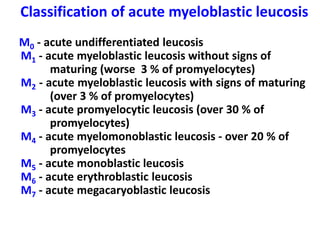
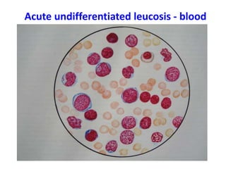
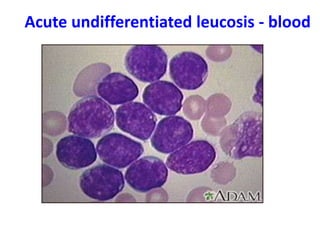
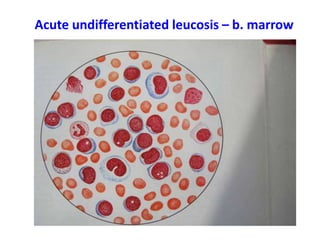
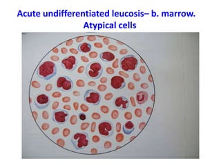
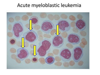
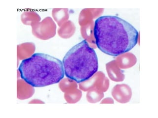
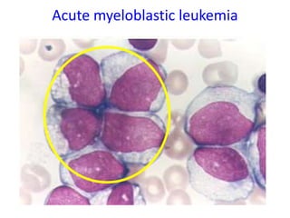
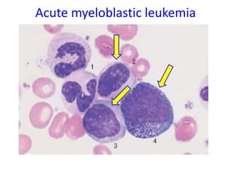
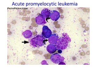
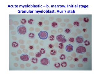
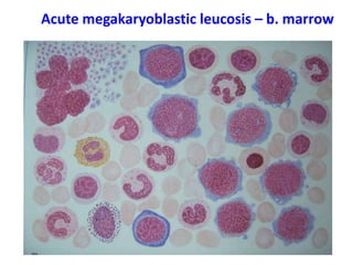
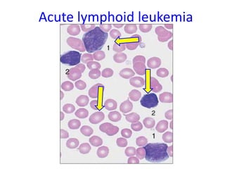
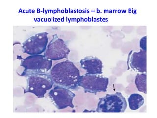
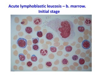
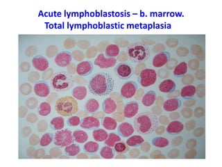
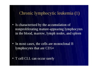
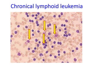
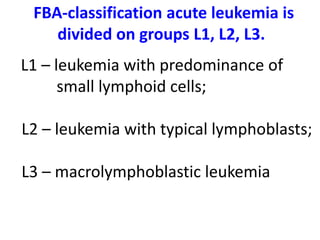
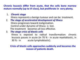
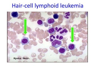
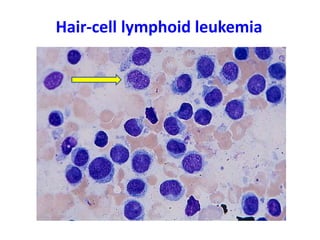
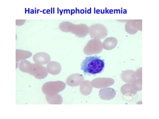
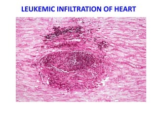
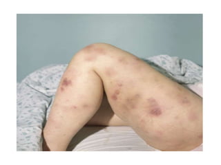
PP
PATHOLOGY OF LEUKOCYTES
LEUКЕМІА
Professor Yu.I. Bondarenko
PATHOLOGY OF LEUKOCYTES
LEUКЕМІА
Professor Yu.I. Bondarenko
SCHEME OF BLOODFORMING
QUANTITATIVE DISORDER OF LEUKOCYTES
THE TOTAL QUANTITY OF LEUKOCYTES EQUALS 4-9 G/L
QUANTITATIVE DISORDER OF LEUKOCYTES ARE REPRESENTED BY:
 the total quantity of leukocytes in a liter of blood;
 Changes of leukoformula indices (quantity of every form of leukocytes)
Leukocytosis - an increase in the total quantity of leukocytes;
Leukopenia - a decrease in the total quantity of leukocytes
Leukoformula analysis
1. Left-Side Nuclear Shift of Neutrophils
 Regenerative
 Hyperregenerative
 Regenerative-Degenerative
 Degenerative
2. Right-Side Nuclear Shift of Neutrophils
- an increased propotion of mature
segmented neutrophils in the
leukoformula in comparison with their
young precursor.
QUALITATIVE CHANGES IN LEUKOCYTES
 anisocytosis (change in the size of leukocytes);
 poikilocytosis (change in the form of leukocytes);
 absence of normal granulation;
 pathological inclusion in the cytoplasm (toxic granulosity,
large azurophilic granules, basophilic bundes of
cytoplasm);
 vacuolization of the nucleus and cytoplasm;
 swelling of the nucleus;
 karyorrexis;
 hypo- and hypersegmentation of the nucleus;
 cytolysis
LEUKOCYTOSIS
Is an icrease in the total amount of leukocytes
in peripheral blood to more than 9 G/L
TYPES
 Physiological (after food intake,physical
and emotional load, in pregnancy)
 Pathological (in disease)
 Reactive
 Redistributive
 Tumorous
 Absolute
 Relative
LEUKOCYTOSIS
Depending on the type of leukocytes being increased
 Neutrophilic;
 Eosinophilic;
 Basophilic;
 Lymphocytosis;
 Monocytosis
NEUTROPHILIC LEUKOCYTOSIS
Etiological factors:
 Infectious (strepto- and staphylococci and fung)
 Noninfectious:
- produts of tissue decay and necrosis
(myocardial infarction, malignant tumor decay,
product of decay in chronic myeloleukemia),
tissue destruction (cold, heat);
- products of erythrocyte hemolysis;
- toxic metabolites in uremia and hepatic coma
EOSINOPHILIC LEUKOCYTOSIS
 Allergic diseases;
 Helminthic invasion;
 Chronic myeloleukemia;
 Hypocorticism
BASOPHILIC LEUKOCYTOSIS
 Chronic myeloleukemia;
 Ulcerative colitis;
 After splenoectomy;
 Myxedema
LYMPHOCYTIC LEUKOCYTOSIS
 Tuberculosis and lues;
 Viral infectious (mononucleosis, hepatitis, measles,
whooping cough);
 Allergic diseases;
 Chronic lympholeukemia
MONOCYTOSIS
 Viruses;
 microorganisms (specific streptococcal endocarditis);
 Protozoa;
 Tuberculosis;
 Syphilis
LEUKOPENIA
Is a decrease of the total quantity of leukocytes in
the peripheral blood below 4 G/L
Types depending on the mechanisms:
1. Aquired
2. Hereditary
Types depending on the type of leukocytes being
decreased:
 Neutropenia;
 Eosinopenia;
 Lymphopenia;
 Monocytopenia
ETIOLOGY OF LEUKOPENIA
 Physical (ionizing radiation);
 Chemical;
- poisons;
- medicines (aspirin, amidopyrin,
cytostatics, barbiturates, glucocorticoids);
- Vitamin B12 and folic acid deficiency;
 Biological;
- infectious;
- immune;
- hormonal (stress);
- genetic (mutation)
PATHOGENESIS OF LEUKOPENIA
1. Decreased leukocyte production in the
hemopoietic tissue;
2. Increased leukocyte destruction in the
blood and hemopoietic tissue;
3. Excessive loss of leukocytes;
4. Redistribution of leukocytes in the
vessels;
5. Decelerated leukocyte release from the
bone marrow
LEUКЕМІА
Leukemia (leucosis) is a tumour, which arises
from bloodforming cells and is primary
damages bone marrow.
The most characteristic signs of leucosis is the
filling bone marrow by malignant cells of the local
origin. It can be leucocytes and their predecessors,
erythroblasts, megacaryoblasts.
Classification of leukemia
1. Acute leukemia
2. Chronic leukemia
Acute leucosis is characterized disorder of
bloodforming cells differentiation, do not go
further ІV classes.
The growth up of cells, which do not
mature, lead to accumulation blast cells ІІ, ІІІ
and ІV classes. They more and more take part
of bone marrow at the expense of volume,
which should be occupied normal hemopoietic
elements.
FAB-classification
created by French, American and British experts.
It is constructed on stable morphological and
cytochemical characteristics of leucosis cells. These
characteristics reflect features them metabolism.
According to modern conception all acute leucosis
divide on two groups – myeloblast and lymphoblast.
They are represented by many nosologic forms.
Acute myeloblastic leucosis differentiate on five
cytochemical signs – presence or absence in leucosis
cells of the following substances: peroxidase, acid
phosphatase, unspecific esterase, lipids and glycogen.
Reaction of myeloblastes and other cells of
neutrophilic line of peroxidase
Reaction of monoblastes of
α-naphtylacetateesterase
Reaction of myeloblastes and monoblastes of
acidic phosphatase
Reaction with black sudan of lipids
Acidic sulfurated mucopolysaccharides reaction
Classification of acute myeloblastic leucosis
M0 - acute undifferentiated leucosis
M1 - acute myeloblastic leucosis without signs of
maturing (worse 3 % of promyelocytes)
M2 - acute myeloblastic leucosis with signs of maturing
(over 3 % of promyelocytes)
M3 - acute promyelocytic leucosis (over 30 % of
promyelocytes)
M4 - acute myelomonoblastic leucosis - over 20 % of
promyelocytes
M5 - acute monoblastic leucosis
M6 - acute erythroblastic leucosis
M7 - acute megacaryoblastic leucosis
Acute undifferentiated leucosis - blood
Acute undifferentiated leucosis – b. marrow
Acute undifferentiated leucosis– b. marrow.
Аtypical cells
Acute myeloblastic leukemia
Acute promyelocytic leukemia
Acute myeloblastic – b. marrow. Initial stage.
Granular myeloblast. Aur’s stab
Acute megakaryoblastic leucosis – b. marrow
Complete hematological picture of chronic leucosis.
In the blood present as the maturing cells as an
abundance cells of all classes – young, transition and
mature. Hiatus leukemicus is absent. In particular, in
blood of chronic myelocytic leucosis will be the next cells
– predecessor ІІ and ІІІ classes, myeloblasts (ІV classes),
cell V classes – promyelocytes myelocytes
metamyelocytes stick nucleus neutrophils and mature
cells of the VІ class (neutrophils). The picture of
peripheral blood of chronic lymphocytic leucosis is
characterized by the following features: there are a lot of
mature lymphocytes, there are prolymphocytes and
lymphoblasts, and also desroyed lymphoid cells
(Gumprecht’s bodies or shadows of lymphocytes).
Classification of acute lymphoblastic leucosis
1. Acute leucosis of general type
( from cells-predecessors of B
lymphocytes)
2. T-lymphoblastic leucosis
3. B-lymphoblastic leucosis.
Acute lymphoid leukemia
Acute В-lymphoblastosis – b. marrow Big
vacuolized lymphoblastes
Acute lymphoblastic leucosis – b. marrow.
Initial stage
Acute lymphoblastosis – b. marrow.
Тоtal lymphoblastic metaplasia
Chronical lymphoid leukemia
FBA-classification acute leukemia is
divided on groups L1, L2, L3.
L1 – leukemia with predominance of
small lymphoid cells;
L2 – leukemia with typical lymphoblasts;
L3 – macrolymphoblastic leukemia
Chronic leucosis differ from acute, that the cells bone marrow
mature normally (up to VІ class), but proliferate in very plenty.
1. Chronic stage
Illness represents a benign tumour and can be treatment.
2. The stage of accelerated development of illness
Illness progresses toward malignisation.
Control under dynamic of illness is lost.
The treatment becomes all less effective.
3. The stage crisis of blastic cells
Illness is exposed to radical transformation: chronic
leucosis passes in acute (in 70 % - in acute myeloblastic, in
30 % - in acute lymphoblastic).
Crisis of blastic cells approaches suddenly and becomes the
reason of patients death.
Classification of chronic myeloid leukemia
1. Chronic myeloleucosis
2. Chronic monocytic leucosis
3. Chronic erythromyelosis
4. Chronic megacaryocytic leucosis
5. Eosinophilic leucosis
Acute myeloblastic leukemia – bone marrow
Chronical myeloleucosis – blood.
Stage of crisis of blastic cells
Classification of chronic lymphoid
leukemia
1. B - cell leucosis
2. T - cell leucosis
3. Haircell leucosis
Hair-cell lymphoid leukemia
Etiology and pathogenesis of the leucosis
On modern conception, leucosis arise on
genetic, mutational basis.
The question is about specific bloodforming cells
mutation, which lead to superexpression of cell
oncogenes, or protooncogenes. These genes are an
integral part of cells genome and answer for proliferation
of cells in norma. Cell oncogenes vitally are necessary. At
the some time cell oncogenes have latent blastomogenic
potentions.
To major etiological factors, which capable to transform
protooncogenes in active oncogenes are the chemical
agents, ionising rays and retroviruses. It is know a few
mechanisms of the cell oncogenes activation.
1. Chromosomal aberrations
2. Genic amplification
3. Point mutations
4. Viral transduction
5. Insertional mutagenesis
6. Transactivation of transcription
Mechanisms of protooncogene activation
Leucosogenic action of ionising rays.
Increase of frequency leucosis took place after
nuclear bombardment of Hiroshima and Nagasaki in
1945. The leucosis was fixed also in case of use ionising
radiation with the medical purpose, myelomic illness,
lymphogranulomatosis, autoimmune diseases, some
dermatosis.
The approximately 25-35 % of cells, mainly
lymphocytes, after therapeutical iradiation contain
chromosomal aberrations as ring chromosomes, dicentric
chromosomes and acentric of fragments.
It is known leucosogenic action of radioactive isotopes.
The radioactive phosphorum, which is used for treatment
erythremia, caused acute leucosis in 15-18 % of the
patients.
Chromosomal aberrations.
The precise correlation between localization of
oncogenes and specific translocations of chromosomes
is marked. It is established, that cell oncogenes
frequently placed just in those sites chromosomes,
where it is easy and naturally there are their breaks with
consequent translocations of deleted fragments. The
translocations can be original activators
protooncogenes.
To the present time in chromosomes of malignant cells
more than 80 points are registered, where the breaks
are observed. The analysis of distribution of these
malignant spesific points and localization
protooncogenes in genome of that person testify that
the majority protooncogenes placed just in zones of
specific breaks chromosomes.
Chromosomal role aberrations in activation of
protooncogenes represent chromosomal and genes of
illness, which are characterized by increased instability
of chromosomes.
1. Dawn’s illness;
2. Fankony’s anemia;
3. Blum’s syndrome;
4. Louis-Bar’s syndrome and etc.
It is established, that among patients with Down’s
illness the frequency leucosis in 20 times is higher, than
among persones without Down’s illness. Fankony’s
anemia the diverse deviations karyotype from norm are
found: chromatide breaks, acentric fragmentation,
dicentric chromosomes, chromatide exchanges. In
children with the Blum’s syndrome large percent of
breaks chromosomes, as Fankony’s anemia is observed.
Every type of leucosis are characterized
specific chromosomal aberrations.
The most better is investigated translocation 9/22,
characterized for chronic myelocytic leucosis. This
anomaly the first time was described in 1960 in
Philadelphia (USA). Changed chromosome named
philadelphian. That chromosome derivated in result
reciprocal translocation between 9-th and 22-nd
chromosome. Long shoulder of 9-th chromosome
contains protooncogene abl (Abelson’s), which in
mice causes leucosis, and long shoulder of 22-nd
chromosome contains protooncogene sis, which
causes sarcoma in haired monkeys.
Expression of Abelson’s oncogene in bone marrow to a cell
stipulate appearance in it special oncoprotein with molecular
weight 210 kD and by thyrosine activity. This oncoproteine is
coded simultaneously Abelson’s oncogene from 9-th chromosome
and site 22-nd chromosome, which adjoins to the point of break.
The data about a role of chromosome aberrations in leucosis
etiology can be generalized as follows.
The anomalies karyotype only when can cause leucosis, if they
seize chromosome locuses, where are located protooncogenes.
The activation stipulates these protooncogenes pathological
proliferation and leucosis. Each chromosome has so-called fragile
sites, which can be identified by means of differential colouring.
Just here there are deletion, inversions and translocation, which
become by the initiators of activation protooncogenes more often.
Virus transduction.
 On leucosogenic properties retroviruses
divide on two groups – fast-transformed (viruses
acute leucosis) and slow- transformed (viruses of
chronic leucosis).
Retroviruses of the acute leucosis differ by that
their gene has an additional gene. It represents cells
oncogene, which was snatched out from genome of
cell and is built in virus RNA. It is uncellular and virus
oncogene.
 This additional gene consider as the specific
factor, which causes malignant transformation of a
cell, and the process of massage cells oncogene
through a virus is named virus transduction.
After repeated introduce in a cell virus (form
cells) oncogene shows high propensity to
expression.
The first reason – it is seized by virus without
surrounding regulatoring repressors genes.
The second reason – DNA-copy retrovirus is not
absolutely exactly reading out return transcriptase.
When the again created virus particles are introduced
into the following cell, their DNA-copies with an
additional gene (virus oncogene) are built in cell by the
gene and easily expression or because mutational virus
oncogene becomes inaccessible repressoring gene to
environment, or because this environment in general is
absent.
Insertion of provirus.
Most of viruses leucosis belongs not to fast
transformational, and to slow transformational
retroviruses.
They do not contain oncogenes and induce
experimental leucosis in an animal less effectively,
than fast transformational.
Slow transformational retroviruses cause
transformation of cells because their DNA-copies
are inserted in cells by a gene near to cells
oncogene.
The presence of another's DNA sometime
activates cells oncogene up to very high level
expression.
Genes amplification.
This increase of copies of separate genes in reply to
change of the external environment.
 In leucosis cells are detected amplificated of copy
some protooncogenes.
 In itself amplification of oncogene does not
concern to initiating events in leucogenesis.
 It is connected to a progression already of initiated
cells. But in any case amplification of gene results
in increase of level expressed RNA.
Major chain of pathogenesis leucosis is oppression by
leucosis cells normal hemopoiesis.
1. Leucosis cells are capable produced in
redundant amount colonialstimulation factor –
stimulator of myelopoiesis.
2. This factor acts on leucosis cells stronger, than on
the normal predecessors hemopoiesis.
Clinical signs of leukemia
1. Metaplastic anemia
2. Thrombocytopenia and hemorrhagic
syndrome
3. Inhibition of immune
4. Decrease of resistance to infectious
agent
LEUKEMIC INFILTRATION OF HEART
Presentation on Leukocytosis Leukemia.pptx

Presentation on Leukocytosis Leukemia.pptx

  • 1.
    PP PATHOLOGY OF LEUKOCYTES LEUКЕМІА ProfessorYu.I. Bondarenko PATHOLOGY OF LEUKOCYTES LEUКЕМІА Professor Yu.I. Bondarenko
  • 2.
  • 3.
    QUANTITATIVE DISORDER OFLEUKOCYTES THE TOTAL QUANTITY OF LEUKOCYTES EQUALS 4-9 G/L QUANTITATIVE DISORDER OF LEUKOCYTES ARE REPRESENTED BY:  the total quantity of leukocytes in a liter of blood;  Changes of leukoformula indices (quantity of every form of leukocytes) Leukocytosis - an increase in the total quantity of leukocytes; Leukopenia - a decrease in the total quantity of leukocytes
  • 4.
    Leukoformula analysis 1. Left-SideNuclear Shift of Neutrophils  Regenerative  Hyperregenerative  Regenerative-Degenerative  Degenerative 2. Right-Side Nuclear Shift of Neutrophils - an increased propotion of mature segmented neutrophils in the leukoformula in comparison with their young precursor.
  • 5.
    QUALITATIVE CHANGES INLEUKOCYTES  anisocytosis (change in the size of leukocytes);  poikilocytosis (change in the form of leukocytes);  absence of normal granulation;  pathological inclusion in the cytoplasm (toxic granulosity, large azurophilic granules, basophilic bundes of cytoplasm);  vacuolization of the nucleus and cytoplasm;  swelling of the nucleus;  karyorrexis;  hypo- and hypersegmentation of the nucleus;  cytolysis
  • 6.
    LEUKOCYTOSIS Is an icreasein the total amount of leukocytes in peripheral blood to more than 9 G/L TYPES  Physiological (after food intake,physical and emotional load, in pregnancy)  Pathological (in disease)  Reactive  Redistributive  Tumorous  Absolute  Relative
  • 7.
    LEUKOCYTOSIS Depending on thetype of leukocytes being increased  Neutrophilic;  Eosinophilic;  Basophilic;  Lymphocytosis;  Monocytosis
  • 8.
    NEUTROPHILIC LEUKOCYTOSIS Etiological factors: Infectious (strepto- and staphylococci and fung)  Noninfectious: - produts of tissue decay and necrosis (myocardial infarction, malignant tumor decay, product of decay in chronic myeloleukemia), tissue destruction (cold, heat); - products of erythrocyte hemolysis; - toxic metabolites in uremia and hepatic coma
  • 9.
    EOSINOPHILIC LEUKOCYTOSIS  Allergicdiseases;  Helminthic invasion;  Chronic myeloleukemia;  Hypocorticism BASOPHILIC LEUKOCYTOSIS  Chronic myeloleukemia;  Ulcerative colitis;  After splenoectomy;  Myxedema
  • 10.
    LYMPHOCYTIC LEUKOCYTOSIS  Tuberculosisand lues;  Viral infectious (mononucleosis, hepatitis, measles, whooping cough);  Allergic diseases;  Chronic lympholeukemia MONOCYTOSIS  Viruses;  microorganisms (specific streptococcal endocarditis);  Protozoa;  Tuberculosis;  Syphilis
  • 11.
    LEUKOPENIA Is a decreaseof the total quantity of leukocytes in the peripheral blood below 4 G/L Types depending on the mechanisms: 1. Aquired 2. Hereditary Types depending on the type of leukocytes being decreased:  Neutropenia;  Eosinopenia;  Lymphopenia;  Monocytopenia
  • 12.
    ETIOLOGY OF LEUKOPENIA Physical (ionizing radiation);  Chemical; - poisons; - medicines (aspirin, amidopyrin, cytostatics, barbiturates, glucocorticoids); - Vitamin B12 and folic acid deficiency;  Biological; - infectious; - immune; - hormonal (stress); - genetic (mutation)
  • 13.
    PATHOGENESIS OF LEUKOPENIA 1.Decreased leukocyte production in the hemopoietic tissue; 2. Increased leukocyte destruction in the blood and hemopoietic tissue; 3. Excessive loss of leukocytes; 4. Redistribution of leukocytes in the vessels; 5. Decelerated leukocyte release from the bone marrow
  • 14.
    LEUКЕМІА Leukemia (leucosis) isa tumour, which arises from bloodforming cells and is primary damages bone marrow. The most characteristic signs of leucosis is the filling bone marrow by malignant cells of the local origin. It can be leucocytes and their predecessors, erythroblasts, megacaryoblasts.
  • 15.
    Classification of leukemia 1.Acute leukemia 2. Chronic leukemia Acute leucosis is characterized disorder of bloodforming cells differentiation, do not go further ІV classes. The growth up of cells, which do not mature, lead to accumulation blast cells ІІ, ІІІ and ІV classes. They more and more take part of bone marrow at the expense of volume, which should be occupied normal hemopoietic elements.
  • 16.
    FAB-classification created by French,American and British experts. It is constructed on stable morphological and cytochemical characteristics of leucosis cells. These characteristics reflect features them metabolism. According to modern conception all acute leucosis divide on two groups – myeloblast and lymphoblast. They are represented by many nosologic forms. Acute myeloblastic leucosis differentiate on five cytochemical signs – presence or absence in leucosis cells of the following substances: peroxidase, acid phosphatase, unspecific esterase, lipids and glycogen.
  • 19.
    Reaction of myeloblastesand other cells of neutrophilic line of peroxidase
  • 20.
    Reaction of monoblastesof α-naphtylacetateesterase
  • 21.
    Reaction of myeloblastesand monoblastes of acidic phosphatase
  • 22.
    Reaction with blacksudan of lipids
  • 23.
  • 24.
    Classification of acutemyeloblastic leucosis M0 - acute undifferentiated leucosis M1 - acute myeloblastic leucosis without signs of maturing (worse 3 % of promyelocytes) M2 - acute myeloblastic leucosis with signs of maturing (over 3 % of promyelocytes) M3 - acute promyelocytic leucosis (over 30 % of promyelocytes) M4 - acute myelomonoblastic leucosis - over 20 % of promyelocytes M5 - acute monoblastic leucosis M6 - acute erythroblastic leucosis M7 - acute megacaryoblastic leucosis
  • 25.
  • 26.
  • 27.
  • 28.
    Acute undifferentiated leucosis–b. marrow. Аtypical cells
  • 29.
  • 31.
  • 32.
  • 33.
  • 34.
    Acute myeloblastic –b. marrow. Initial stage. Granular myeloblast. Aur’s stab
  • 35.
  • 36.
    Complete hematological pictureof chronic leucosis. In the blood present as the maturing cells as an abundance cells of all classes – young, transition and mature. Hiatus leukemicus is absent. In particular, in blood of chronic myelocytic leucosis will be the next cells – predecessor ІІ and ІІІ classes, myeloblasts (ІV classes), cell V classes – promyelocytes myelocytes metamyelocytes stick nucleus neutrophils and mature cells of the VІ class (neutrophils). The picture of peripheral blood of chronic lymphocytic leucosis is characterized by the following features: there are a lot of mature lymphocytes, there are prolymphocytes and lymphoblasts, and also desroyed lymphoid cells (Gumprecht’s bodies or shadows of lymphocytes).
  • 37.
    Classification of acutelymphoblastic leucosis 1. Acute leucosis of general type ( from cells-predecessors of B lymphocytes) 2. T-lymphoblastic leucosis 3. B-lymphoblastic leucosis.
  • 38.
  • 39.
    Acute В-lymphoblastosis –b. marrow Big vacuolized lymphoblastes
  • 40.
    Acute lymphoblastic leucosis– b. marrow. Initial stage
  • 41.
    Acute lymphoblastosis –b. marrow. Тоtal lymphoblastic metaplasia
  • 43.
  • 44.
    FBA-classification acute leukemiais divided on groups L1, L2, L3. L1 – leukemia with predominance of small lymphoid cells; L2 – leukemia with typical lymphoblasts; L3 – macrolymphoblastic leukemia
  • 45.
    Chronic leucosis differfrom acute, that the cells bone marrow mature normally (up to VІ class), but proliferate in very plenty. 1. Chronic stage Illness represents a benign tumour and can be treatment. 2. The stage of accelerated development of illness Illness progresses toward malignisation. Control under dynamic of illness is lost. The treatment becomes all less effective. 3. The stage crisis of blastic cells Illness is exposed to radical transformation: chronic leucosis passes in acute (in 70 % - in acute myeloblastic, in 30 % - in acute lymphoblastic). Crisis of blastic cells approaches suddenly and becomes the reason of patients death.
  • 46.
    Classification of chronicmyeloid leukemia 1. Chronic myeloleucosis 2. Chronic monocytic leucosis 3. Chronic erythromyelosis 4. Chronic megacaryocytic leucosis 5. Eosinophilic leucosis
  • 48.
  • 49.
    Chronical myeloleucosis –blood. Stage of crisis of blastic cells
  • 50.
    Classification of chroniclymphoid leukemia 1. B - cell leucosis 2. T - cell leucosis 3. Haircell leucosis
  • 51.
  • 52.
  • 53.
  • 54.
    Etiology and pathogenesisof the leucosis On modern conception, leucosis arise on genetic, mutational basis. The question is about specific bloodforming cells mutation, which lead to superexpression of cell oncogenes, or protooncogenes. These genes are an integral part of cells genome and answer for proliferation of cells in norma. Cell oncogenes vitally are necessary. At the some time cell oncogenes have latent blastomogenic potentions. To major etiological factors, which capable to transform protooncogenes in active oncogenes are the chemical agents, ionising rays and retroviruses. It is know a few mechanisms of the cell oncogenes activation.
  • 55.
    1. Chromosomal aberrations 2.Genic amplification 3. Point mutations 4. Viral transduction 5. Insertional mutagenesis 6. Transactivation of transcription Mechanisms of protooncogene activation
  • 56.
    Leucosogenic action ofionising rays. Increase of frequency leucosis took place after nuclear bombardment of Hiroshima and Nagasaki in 1945. The leucosis was fixed also in case of use ionising radiation with the medical purpose, myelomic illness, lymphogranulomatosis, autoimmune diseases, some dermatosis. The approximately 25-35 % of cells, mainly lymphocytes, after therapeutical iradiation contain chromosomal aberrations as ring chromosomes, dicentric chromosomes and acentric of fragments. It is known leucosogenic action of radioactive isotopes. The radioactive phosphorum, which is used for treatment erythremia, caused acute leucosis in 15-18 % of the patients.
  • 57.
    Chromosomal aberrations. The precisecorrelation between localization of oncogenes and specific translocations of chromosomes is marked. It is established, that cell oncogenes frequently placed just in those sites chromosomes, where it is easy and naturally there are their breaks with consequent translocations of deleted fragments. The translocations can be original activators protooncogenes. To the present time in chromosomes of malignant cells more than 80 points are registered, where the breaks are observed. The analysis of distribution of these malignant spesific points and localization protooncogenes in genome of that person testify that the majority protooncogenes placed just in zones of specific breaks chromosomes.
  • 58.
    Chromosomal role aberrationsin activation of protooncogenes represent chromosomal and genes of illness, which are characterized by increased instability of chromosomes. 1. Dawn’s illness; 2. Fankony’s anemia; 3. Blum’s syndrome; 4. Louis-Bar’s syndrome and etc. It is established, that among patients with Down’s illness the frequency leucosis in 20 times is higher, than among persones without Down’s illness. Fankony’s anemia the diverse deviations karyotype from norm are found: chromatide breaks, acentric fragmentation, dicentric chromosomes, chromatide exchanges. In children with the Blum’s syndrome large percent of breaks chromosomes, as Fankony’s anemia is observed.
  • 59.
    Every type ofleucosis are characterized specific chromosomal aberrations. The most better is investigated translocation 9/22, characterized for chronic myelocytic leucosis. This anomaly the first time was described in 1960 in Philadelphia (USA). Changed chromosome named philadelphian. That chromosome derivated in result reciprocal translocation between 9-th and 22-nd chromosome. Long shoulder of 9-th chromosome contains protooncogene abl (Abelson’s), which in mice causes leucosis, and long shoulder of 22-nd chromosome contains protooncogene sis, which causes sarcoma in haired monkeys.
  • 60.
    Expression of Abelson’soncogene in bone marrow to a cell stipulate appearance in it special oncoprotein with molecular weight 210 kD and by thyrosine activity. This oncoproteine is coded simultaneously Abelson’s oncogene from 9-th chromosome and site 22-nd chromosome, which adjoins to the point of break. The data about a role of chromosome aberrations in leucosis etiology can be generalized as follows. The anomalies karyotype only when can cause leucosis, if they seize chromosome locuses, where are located protooncogenes. The activation stipulates these protooncogenes pathological proliferation and leucosis. Each chromosome has so-called fragile sites, which can be identified by means of differential colouring. Just here there are deletion, inversions and translocation, which become by the initiators of activation protooncogenes more often.
  • 61.
    Virus transduction.  Onleucosogenic properties retroviruses divide on two groups – fast-transformed (viruses acute leucosis) and slow- transformed (viruses of chronic leucosis). Retroviruses of the acute leucosis differ by that their gene has an additional gene. It represents cells oncogene, which was snatched out from genome of cell and is built in virus RNA. It is uncellular and virus oncogene.  This additional gene consider as the specific factor, which causes malignant transformation of a cell, and the process of massage cells oncogene through a virus is named virus transduction.
  • 62.
    After repeated introducein a cell virus (form cells) oncogene shows high propensity to expression. The first reason – it is seized by virus without surrounding regulatoring repressors genes. The second reason – DNA-copy retrovirus is not absolutely exactly reading out return transcriptase. When the again created virus particles are introduced into the following cell, their DNA-copies with an additional gene (virus oncogene) are built in cell by the gene and easily expression or because mutational virus oncogene becomes inaccessible repressoring gene to environment, or because this environment in general is absent.
  • 63.
    Insertion of provirus. Mostof viruses leucosis belongs not to fast transformational, and to slow transformational retroviruses. They do not contain oncogenes and induce experimental leucosis in an animal less effectively, than fast transformational. Slow transformational retroviruses cause transformation of cells because their DNA-copies are inserted in cells by a gene near to cells oncogene. The presence of another's DNA sometime activates cells oncogene up to very high level expression.
  • 64.
    Genes amplification. This increaseof copies of separate genes in reply to change of the external environment.  In leucosis cells are detected amplificated of copy some protooncogenes.  In itself amplification of oncogene does not concern to initiating events in leucogenesis.  It is connected to a progression already of initiated cells. But in any case amplification of gene results in increase of level expressed RNA.
  • 65.
    Major chain ofpathogenesis leucosis is oppression by leucosis cells normal hemopoiesis. 1. Leucosis cells are capable produced in redundant amount colonialstimulation factor – stimulator of myelopoiesis. 2. This factor acts on leucosis cells stronger, than on the normal predecessors hemopoiesis.
  • 66.
    Clinical signs ofleukemia 1. Metaplastic anemia 2. Thrombocytopenia and hemorrhagic syndrome 3. Inhibition of immune 4. Decrease of resistance to infectious agent
  • 67.