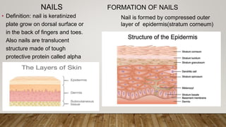
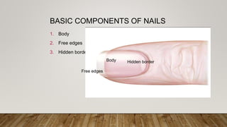
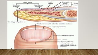
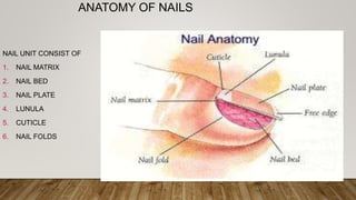
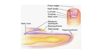
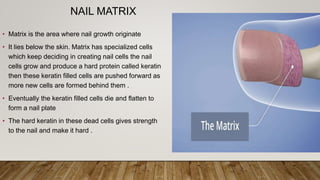
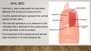
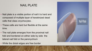
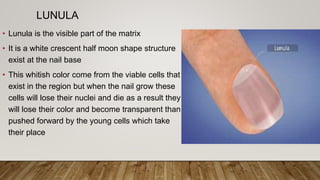
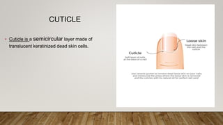
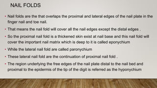
DR MUNAVVAR SAEED
PG.SCHOLAR
DEPT OF DERMATOLOGY
ANATOMY OF NAILS
NAILS
• Definition: nail is keratinized
plate grow on dorsal surface or
in the back of fingers and toes.
Also nails are translucent
structure made of tough
protective protein called alpha
keratin
Nail is formed by compressed outer
layer of epidermis(stratum corneum)
FORMATION OF NAILS
BASIC COMPONENTS OF NAILS
1. Body
2. Free edges
3. Hidden border
Body Hidden border
Free edges
ANATOMY OF NAILS
NAIL UNIT CONSIST OF
1. NAIL MATRIX
2. NAIL BED
3. NAIL PLATE
4. LUNULA
5. CUTICLE
6. NAIL FOLDS
NAIL MATRIX
• Matrix is the area where nail growth originate
• It lies below the skin. Matrix has specialized cells
which keep deciding in creating nail cells the nail
cells grow and produce a hard protein called keratin
then these keratin filled cells are pushed forward as
more new cells are formed behind them .
• Eventually the keratin filled cells die and flatten to
form a nail plate
• The hard keratin in these dead cells gives strength
to the nail and make it hard .
NAIL BED
• Nail bed a area underneath the nail plate (
Between the lunula and hyponychium)
• It is thin epidermal layer represent the ventral
portion of nail matrix
• The nail bed epithelium is so adherent to the
nail plate that is attached to the undersurface
of the nail when a nail is avulsed.
• It is composed of thin epidermal and dermal
layer but there is no subcutaneous fat.
NAIL PLATE
• Nail plate is a visible portion of nail it is hard and
composed of multiple layer of keratinized dead
cells that class onychocytes .
• These cells are hard but flexible at the same
time.
• The nail plate emerges from the proximal nail
fold and bordered on either side by side the
lateral nail fold or the paronychium
• While the distal edges are free border
LUNULA
• Lunula is the visible part of the matrix
• It is a white crescent half moon shape structure
exist at the nail base
• This whitish color come from the viable cells that
exist in the region but when the nail grow these
cells will lose their nuclei and die as a result they
will lose their color and become transparent than
pushed forward by the young cells which take
their place
CUTICLE
• Cuticle is a semicircular layer made of
translucent keratinized dead skin cells.
NAIL FOLDS
• Nail folds are the that overlaps the proximal and lateral edges of the nail plate in the
finger nail and toe nail.
• That means the nail fold will cover all the nail edges except the distal edges .
• So the proximal nail fold is a thickened skin exist at nail base and this nail fold will
cover the important nail matrix which is deep to it is called eponychium
• While the lateral nail fold are called paronychium
• These lateral nail fold are the continuation of proximal nail fold .
• The region underlying the free edges of the nail plate distal to the nail bed and
proximal to the epidermis of the tip of the digit is referred as the hyponychium
NAIL GROWTH
• Fingers nail grow about 1mm/week (3.5mm in a month)
• While toe nail grow slower than finger nails 1.6mm in
month
• Growth of nail gradually decrease with age
FUNCTIONS OF NAILS
•Help to grasp and manipulate objects
•Help in pincer grip
•Protect terminal phalanx and fingertip
•Serve an aesthetic and cosmetic purpose

Presentation nail nd hair.pptx for study

  • 1.
    DR MUNAVVAR SAEED PG.SCHOLAR DEPTOF DERMATOLOGY ANATOMY OF NAILS
  • 2.
    NAILS • Definition: nailis keratinized plate grow on dorsal surface or in the back of fingers and toes. Also nails are translucent structure made of tough protective protein called alpha keratin Nail is formed by compressed outer layer of epidermis(stratum corneum) FORMATION OF NAILS
  • 3.
    BASIC COMPONENTS OFNAILS 1. Body 2. Free edges 3. Hidden border Body Hidden border Free edges
  • 5.
    ANATOMY OF NAILS NAILUNIT CONSIST OF 1. NAIL MATRIX 2. NAIL BED 3. NAIL PLATE 4. LUNULA 5. CUTICLE 6. NAIL FOLDS
  • 7.
    NAIL MATRIX • Matrixis the area where nail growth originate • It lies below the skin. Matrix has specialized cells which keep deciding in creating nail cells the nail cells grow and produce a hard protein called keratin then these keratin filled cells are pushed forward as more new cells are formed behind them . • Eventually the keratin filled cells die and flatten to form a nail plate • The hard keratin in these dead cells gives strength to the nail and make it hard .
  • 8.
    NAIL BED • Nailbed a area underneath the nail plate ( Between the lunula and hyponychium) • It is thin epidermal layer represent the ventral portion of nail matrix • The nail bed epithelium is so adherent to the nail plate that is attached to the undersurface of the nail when a nail is avulsed. • It is composed of thin epidermal and dermal layer but there is no subcutaneous fat.
  • 9.
    NAIL PLATE • Nailplate is a visible portion of nail it is hard and composed of multiple layer of keratinized dead cells that class onychocytes . • These cells are hard but flexible at the same time. • The nail plate emerges from the proximal nail fold and bordered on either side by side the lateral nail fold or the paronychium • While the distal edges are free border
  • 10.
    LUNULA • Lunula isthe visible part of the matrix • It is a white crescent half moon shape structure exist at the nail base • This whitish color come from the viable cells that exist in the region but when the nail grow these cells will lose their nuclei and die as a result they will lose their color and become transparent than pushed forward by the young cells which take their place
  • 11.
    CUTICLE • Cuticle isa semicircular layer made of translucent keratinized dead skin cells.
  • 12.
    NAIL FOLDS • Nailfolds are the that overlaps the proximal and lateral edges of the nail plate in the finger nail and toe nail. • That means the nail fold will cover all the nail edges except the distal edges . • So the proximal nail fold is a thickened skin exist at nail base and this nail fold will cover the important nail matrix which is deep to it is called eponychium • While the lateral nail fold are called paronychium • These lateral nail fold are the continuation of proximal nail fold . • The region underlying the free edges of the nail plate distal to the nail bed and proximal to the epidermis of the tip of the digit is referred as the hyponychium
  • 13.
    NAIL GROWTH • Fingersnail grow about 1mm/week (3.5mm in a month) • While toe nail grow slower than finger nails 1.6mm in month • Growth of nail gradually decrease with age
  • 14.
    FUNCTIONS OF NAILS •Helpto grasp and manipulate objects •Help in pincer grip •Protect terminal phalanx and fingertip •Serve an aesthetic and cosmetic purpose