5. • If you has a biopsy and you saw dermal
melanocytes
• What would you think ?
6.
7.
8.
9.
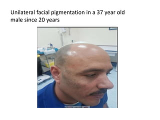
10. Nevus of Ota is a macula-dermal discoloration
occurring due
to failure of melanocytes migration from the neural
crest to derma-
epidermal junction.
More common in Asian and African population
Ophthalmic, Maxillary and Mandibular divisions of
trigeminal Nerve are involved out of which maxillary is
most common.
11. The color of the skin lesion is brown or blue, the
diameter of the area is 1–10
cm or larger.
Pathophysiology is yet to be confirmed even if it has
been postulated that Nevus of Ota and other dermal
melanocytic disorders such as nevus of lto, blue
nevus and mongolion spots may represent
melanocytes that have not migrated completely from
neural crest to the epidermis during embryonic stage
The two peak ages of onset in early infancy and in early
adolescence suggest that hormones are a factor in the
development of this condition.
12. Clinical Features
Mostly asymptomatic, can be associated with melanoma
in rare
Cases.
Ophthalmic division have ocular complications
like glaucoma, melanoma associated with cilliary
body,iris and optic nerve is also reported.
13. Histopathology Features
Skin biopsy reveals histopathologic view of dermal
melanocytes, bipolar or oval in shape, scattered in the
upper and middle portions of the dermis.
Treatment
Topical therapy is of no value in the medical
treatment of nevi of Ota. Previous treatment
modalities, including cryotherapy, dermabrasion,
and microsurgery, can be associated with scarring.
Development of the Q-Switched Nd: YAG laser
(QSYL) and the Q-switched ruby laser (QSRL) has
enabled complete, scar less
18. What it is ?
The Mongolian spot is a development condition
exclusively involving the skin. The blue color is
caused by melanocytes,which is melanin-containing
cells, that are deep under the skin.
Mostly seen on Asians, African Americans ,
Hispanics, and Native Americans.
19. Distribution
FORMS- Mongolian spots are mostly blue, bluish-
gray, bluish-green or blue-black. They flat skin
markings that appear at birth or shortly after or
during the infantile age or when born. It can have
very many shapes and sizes to these spots.
DISTRIBUTION-mostly located at the base of the
spine, on the lower back and buttocks. They can also
appear on the shoulders, upper back, arms,
wrists, legs, ankles. Palms, soles, face
and head are usually spared.
20. Percentages
90 % percent of Native Americans and children
more than 80% percent of Asians
more than 70% percent of Hispanics
They are rare in fair-skinned children — appearing in
just less than 10 % percent.
21. What you can do about it ?
There's really nothing you can do about it, you just
have to wait they eventually disappear at the age of
2.
Fewer than 5% of children with Mongolian spots still
have any by the time they're adults.
22. A normal stratum corneum and epidermis with no
increased pigmentation.
A few solitary wavy dendritic melanocytes containing
melanin granules in the mid-dermis
23.
24. Blue nevus
Common or classic blue nevus is a small (<1 cm)
gray-blue or blue-black macule, papule, or plaque usually
located on the head, neck, presacral region, or distal
extremities
It is almost in- variably acquired during the second decade
of life; most patients belong to phototypes III–IV
The cellular variant is a much larger blue- black nodular
lesion whose typical location is the gluteal region. The
scalp and the extremities are less commonly affected.
30. A normal stratum corneum and epidermis with no
increased pigmentation
A few solitary wavy dendritic melanocytes containing
melanin granules in the mid-dermis
31. A 55 year old male presented with these lesions
32. A 55 year old male presented with these lesions
33. A 55 year old male presented with these lesions
41. • They are clinically & pathologically related &
are described often as a continuum
• Tuberoeruptive :- pink-yellow
papules/nodules on the extensor surfaces ,
esp. elbows & knees
• Tuberous :- lesions are larger than
tuberoeruptive xanthomas ( size › 3 cm )
• Seen in familial hypercholesterolemia, familial
dysbetalipoproteinemias
TUBEROUS/ TUBEROERUPTIVE
XANTHOMAS
46. Plane xanthoma
Yellow- orange, non inflammatory macules, papules, plaques & patches which
are circumscribed/diffuse
Sites can give rise to clues for certain underlying diseases
53. A 51 year old female with hypopigmented lesions diagnosed as vitiligo
54. A 51 year old female with hypopigmented lesions diagnosed as vitiligo
55.
56. Various Cutaneous Manifestations of Mycosis Fungoides
Panel A shows patch-or-plaque MF affecting the lower
trunk. The patches are thin, slightly scaly,
erythematous lesions typically greater
than 4 cm in diameter and distributed in sun-shielded
areas such as those covered by a bathing suit or
intertriginous regions. Plaques are thicker than
patches.
57. Various Cutaneous Manifestations of Mycosis Fungoides
Panel B shows pagetoid reticulosis, a variant of mycosis fungoides that typically
consists of a single patch or plaque located in an acral area.
58. Various Cutaneous Manifestations of Mycosis Fungoides
Panel C shows syringotropic mycosis fungoides, which is
manifested as papules 1 to 3 mm in diameter distributed
in the eccrine ducts, indicating the propensity of
lymphoma cells to accumulate in these locations.
59. Various Cutaneous Manifestations of Mycosis Fungoides
Panel D shows follicular mycosis fungoides, in which
lesions characterized by alopecia develop. In a similar
variant, there is mucin deposition in the follicles.
60. Various Cutaneous Manifestations of Mycosis Fungoides
Panel E shows hypopigmented mycosis fungoides.
This variant is more noticeable in persons with dark
pigmentation and may be more common in childhood
and adolescence than in adulthood. Hypopigmentation to
full depigmentation occurs in patches.
61. Various Cutaneous Manifestations of Mycosis Fungoides
Panel F shows erythrodermic mycosis fungoides.
This variant may evolve from patch-or-plaque mycosis
fungoides and eventually involve more than 80 percent
of the body-surface area. It may also arise
spontaneously, as in the Sézary syndrome.
62. Various Cutaneous Manifestations of Mycosis Fungoides
Panel G shows the Sézary syndrome.
In its most florid form, the diffuse infiltration of the skin
may produce the exaggerated facial lines, resulting in
"leonine facies." The Sézary syndrome is also associated
with atypical lymphocytes on the blood smear.
63. Various Cutaneous Manifestations of Mycosis Fungoides
Panel H shows a mycosis fungoides tumor.
Such tumors define the T3 stage of disease and may
arise at the site of plaques or appear on their own,
without being preceded by a patch-or-plaque lesion.
64. MYCOSIS FUNGOIDES SHEIKHA
OTHER FEATURES OF MF
Skin Hair Follicles could be extensively infiltrated.
Mucin might be deposited Follicular MF
Pagetoid reticulosis is a verrucous variant of MF
Affecting acral sites like hands & feet.
Extreme atypical LC epidermotropism verrucae
Granulomatous slack skin pendulous folds
of slack or lax skin “macrophage-mediated
destruction of dermal elastic fibers”
Many MF have only skin problems.
15% have extracutaneous disease;
LN, Visceral sites “Lung, Oral cavity, CNS, etc”
could be affected.