Download to read offline

![Copyright © Lippincott Williams Wilkins. Unauthorized reproduction of this article is prohibited.
suffering, as well as dramatically reducing the
resulting healthcare burden. Recent human brain-
imaging studies in back pain populations suggest
the possibility of identifying individuals at risk for
pain chronification, pioneering a new field: ‘the
science of chronic pain prevention.’
CHRONIC BACK PAIN IS A MAJOR
HEALTH PROBLEM
Low back pain is a public health problem affecting
between 70 and 85% of adults at some point in their
lives [1]. The annual prevalence of chronic low back
pain ranges from 15 to 45%, with point prevalence
averaging 30% [2]. In the United States, chronic and
acute back pain are the most common causes of
activity limitations in people under 45 years of
age, and the second most frequent reason for visits
to physicians [3,4]. Data from other Western
countries are similar. An article by Deyo [5] states
that: ‘. . .. Clearly, back pain is one of society’s most
significant nonlethal medical conditions. And yet
the prevalence of back pain is perhaps matched in
degree only by the lingering mystery accompanying
it.’
CURRENT MANAGEMENT OF LOW BACK
PAIN REMAINS INADEQUATE
The majority (90%) of individuals with acute low
back pain (0–7 days of back pain) recover full func-
tion within days or weeks, with little or no lingering
pain. Yet a small number of individuals with acute
low back pain (approximately 5% or less) go on to
develop subacute back pain (SBP, 4–12 weeks
of back pain), then chronic low back pain (CBP,
3–6 months of back pain) [1]. A high percentage
of these patients fail to respond to treatment, con-
tinue to have debilitating degrees of pain, are sig-
nificantly limited in their functional capacity, and
become emotionally altered by the suffering con-
ferred by the chronic pain state.
Treatments for persistent back pain are unreli-
able and typically inadequate. Placebo-controlled
trials conducted to date support the use of NSAIDs
and antidepressants in treating back pain, but the
effectiveness is usually not sufficient to be clinically
significant (25% decrease in pain from placebo).
For example, a meta-analysis of NSAID studies found
no evidence that these drugs were effective in treat-
ing low back pain once it had become chronic [6]
(see also [7,8]). A systematic review of antidepress-
ant treatment for chronic back pain also concluded
that these compounds produce only moderate
symptom reduction [9], and a review concluded
that: ‘Many drugs used for back pain are no more,
or only slightly more, effective than placebos. . . no
drug regimen can be legitimately recommended for
back pain’ [10]. The WHO Advisory Panel likewise
concluded that ‘there is no single treatment superior
to others for relieving chronic back pain’ [11].
EXISTING BIOMARKERS OF BACK PAIN
HAVE LOW PREDICTIVE POWER
To date, previously explored psychological, physio-
logical, and genetic risk factors have failed to
provide clinically meaningful predictive value in
determining who will transition to pain chronicity.
However, recent evidence from our laboratory has
revealed that brain biomarkers may critically drive
the transition to the chronic pain state.
Anatomical and genetic risk factors
The probability that the cause of back pain can be
identified by radiography is less than 1% [12].
Nevertheless, the histological composition of her-
niated disc material seems to correlate with clinical
symptoms, such as reported pain [13]. Another
cause of pain and radicular symptoms seems due
to pressure on the nerve from ligamentum flavum
and facet joints [14]. However, the low incidence of
these biomarkers suggests that they are unlikely
targets for prevention-focused interventions.
Findings from two adult female twin studies
indicate that 50–70% of the variation in disc
degenerative processes is due to genetic factors
[15,16]. A similar Danish study also showed a
genetic influence, albeit with more modest results
[17]. Other experiments have identified a number
of candidate genes that underlie disc degeneration
and pain [18]. However, these genetic factors
remain uninformative in providing novel targets
for therapy.
Psychosocial factors
Given that peripheral physical factors have failed to
show a strong relationship with back pain, a long list
KEY POINTS
End-organ parameters are not adequate to explain the
transition to chronic pain.
Brain function and structure reorganizes with chronic
pain on local and global scales.
The brain’s emotional response to injury appears to
predict who transitions to chronicity.
Predicting transition to chronic pain Apkarian et al.
1350-7540 ß 2013 Wolters Kluwer Health | Lippincott Williams Wilkins www.co-neurology.com 361](https://image.slidesharecdn.com/predictingtransitiontochronicpain-160317223637/85/Predicting-transition-to-chronic-pain-2-320.jpg)
![Copyright © Lippincott Williams Wilkins. Unauthorized reproduction of this article is prohibited.
of psychosocial and demographic factors have been
studied. Cumulatively, however, these factors pro-
vide weak predictions regarding chronic pain.
Depression is ranked as one of the strongest predic-
tors of low back pain, and this association has been
observed by multiple studies. One of the largest
studies involved a national survey (n ¼ 91 347) and
2-year follow-up survey (n ¼ 55 690), and the find-
ings indicated that depression and low back pain are
interrelated (correlation of 0.4), with associational
odds ratios (ORs) increasing with the intensity of
back pain and the severity of depression [19] (see
also [20]). Regarding psychosocial factors, the best
predictors have been intensity and duration of pain,
expectations of recovery and perception of health
change, obesity, workplace risk factors [21,22], and
smoking [23]. Yet attempts to create models
of chronic back pain based upon psychosocial
parameters have been unproductive [24–27]. In
summary, there are no dominant physical or psy-
chosocial parameters that can substantially explain
chronic pain.
Direct examination of the brain in chronic
pain
Over the last 10 years, our research group has pio-
neered the development of brain-imaging methods
that can be specifically used to study brain function
in humans with chronic pain. A large portion of this
work targets the brain of patients with CBP. This
group produced the first study demonstrating that
cortical grey matter density decreases regionally in
CBP [28]; since this work was published, over
50 studies have described similar brain morpho-
logical changes across various chronic pain con-
ditions. It was argued that this pattern of changes
in brain morphometry may be related to the shift in
CBP pain perception from sensory (nociceptive)
to emotional (hedonic) areas of the brain. This
hypothesis was corroborated by evidence that
CBP patients exhibit impaired emotional decision-
makingindirectproportion tothemagnitudeoftheir
back pain [29], implying that the emotionally salient
nature of the back pain interferes with other
emotional tasks. This hypothesis was further sup-
ported by functional imaging, wherein we sought
to characterize the actual pain experienced by back
pain sufferers by identifying brain regions related to
fluctuations of spontaneous (unprovoked) back pain.
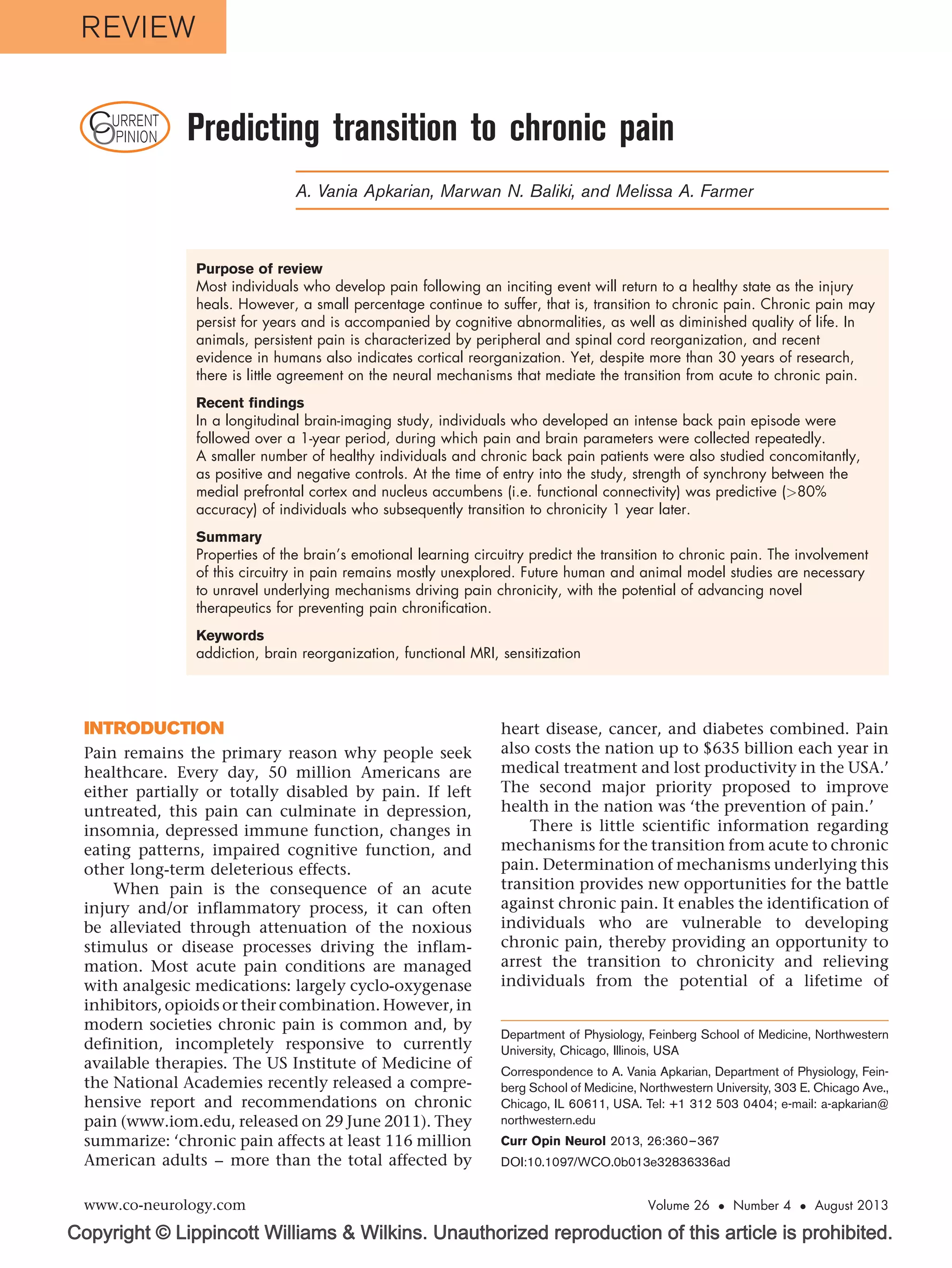
This approach yielded the novel finding that the
spontaneous pain of CBP engages the medial pre-
frontal cortex (mPFC), a brain region that modulates
emotional evaluation relative to the self (Fig. 1a)
[30]. Furthermore, this work revealed a double dis-
sociation between acute thermal pain applied to the
back and spontaneous back pain representations in
the brain, with the former encoded primarily in the
insula and the latter in the mPFC. More recently it
wasshownthatbrainactivityelicitedby thermalpain
is equivalent between healthy controls and CBP
patients in the brain areas that are thought to encode
painful stimulus information or the perception of
pain; the only brain activity that differentiated these
groups was localized in the bilateral nucleus accum-
bens (NAc). NAc activity encoded a salience signal at
the onset of painful thermal stimuli, as well as an
analgesia-related reward signal at stimulus offset.
This analgesia-related reward signal was reversed in
direction in CBP, indicating the abnormal valuation
of acute pain relief. Furthermore, the strength
of functional connectivity between the mPFC and
NAc was proportional to the magnitude of back pain
in the CPB group (Fig. 1b) [31].
PREDICTING TRANSITION TO CHRONIC
PAIN
Given that brain anatomy and functional properties
are distinct in chronic pain patients, it was necessary
to determine the temporal causal relationship
between brain reorganization and the transition
to chronic pain. Therefore, a longitudinal observa-
tional brain-imaging study was undertaken [32
],
wherein SBP patients were tracked over a year as
they transitioned to either persistent pain (SBPp;
persisting to chronicity) or into recovery (SBPr)
(Fig. 1c), thereby enabling comparisons of brain
parameters during this time window, and in con-
trast to healthy and CBP patients.
Longitudinal tracking of these participants’ pain
demonstrated that 50% of SBP patients continue to
suffer from the same magnitude of back pain 1 year
later (Fig. 1c). Thus, this is the back pain population
most vulnerable to pain chronicity, and, impor-
tantly, preventing transition to chronic pain within
this group would represent a large impact on the
future prevalence of chronic pain in society.
We observed that grey matter density decreases
only in SBPp patients, but that this is a slow process
and is preceded by functional connectivity differ-
ences that are detectable by the first brain scan
session. That is, as soon as the patients are scanned,
the functional connectivity strength between the
medial prefrontal cortex and nucleus accumbens
(mPFC–NAc) at this time distinguishes between
SBPp and SBPr (P 10À3
) and predicts the groupings
at high accuracy (81% at 1 year from the first scans)
(Fig. 1c). This is the first time that a specific brain
circuitry in humans pinpoints the transition from
acute to chronic back pain. The identified circuit is
fully consistent with earlier human brain-imaging
Neuroimaging
362 www.co-neurology.com Volume 26 Number 4 August 2013](https://image.slidesharecdn.com/predictingtransitiontochronicpain-160317223637/85/Predicting-transition-to-chronic-pain-3-320.jpg)
![Copyright © Lippincott Williams Wilkins. Unauthorized reproduction of this article is prohibited.
Z-value
Pain intensity
mPFC
[–36 44 18]
Spontaneous pain
Healthy
R = 0.86
CBP
mPFc–NAc(z-score)
Painintensity(VAS)
Visit1
Visit2
Visit3
Visit4
Time (weeks)
1 – Specificity
Time (months)
Series 4/1
Series 3/1
Series 2/1
2
−1
0
1
2
(a)
(b)
(c)
(d)
Healthy
CBP
y = 10
6.00
z = 0
−2
−2
0
0 10 20 30 40
3.0 8.0
z-score
SBPp SBPr
50 60
0.0
7
6
5
4
3
2
1
0
0 3 6 9 12
0.0
0.0
1.0
1.0
0.5
0.5
Visit 2, D = 0.68
Visit 3, D = 0.84
Visit 4, D = 0.81
SBPr
Z(r)mPFC–NAc
Oddsratio
Sensitivity
SBPp
0.2
0.6
0.4
20
*
+
*
+
*
*
*
+
40
60
80
0 20 40 60 80 100
0
2
4
6
8
0
2
4
4 6 8 10 1 3 5 7 9
R2 = 0.81
mPFC
[−18 64 16]
Pain intensity
Z-value
FIGURE 1. Brain circuitry predicting pain chronification. (a) Medial prefrontal cortex (mPFC) activity reflects intensity of back
pain in two separate groups of patients. Adapted with permission from [30]. (b) Functional connectivity of nucleus accumbens
(NAc) in healthy individuals and in chronic low back pain (CBP) patients, during a thermal pain task. In healthy individuals
connectivity is mainly to insula, whereas in CBP it preferentially connects to mPFC. Scatter plot shows that mPFC–NAc
connectivity strength in CBP reflects magnitude of back pain. Adapted with permission from [31]. (c) Left panel: Change in
back pain over 1 year in subacute back pain (SBP) patients. SBP was subdivided to recovering (SBPr, grey triangles) and
persisting (SBPp, black squares). Ordinate is mean Æ SD pain intensity assessed by visual analog scale (VAS; 0–100).
Horizontal bars represent the range of time from visit 1 for SBPp (black), SBPr (grey), and healthy controls (blue). Red lines are
the mean. Right panel: Top: whole-brain functional connectivity contrast for NAc (green) between recovering and persistent
SBP (SBPp SBPr) identifies mPFC. Bar graph: mean and SD of mPFC–NAc connectivity strength. Receiver operator curves
and probability of discrimination D are shown for predicting SBPp and SBPr groups, at three time points from initial brain
scans. Note visit 2 is 3 months, and visits 3 and 4 are 6 and 12 months from initial scan. Thus, predicting future pain
chronicity seems to improve at longer times from scan. (d) Odds ratio (OR) for predicting SBPp and SBPr based on functional
connectivity at different time intervals. Series 2/1 is OR for prediction based on visits 1 and 2, series 3/1 is based on visits
1,2, and 3, whereas series 4/1 is based on visits 1, 2, 3, 4. In all cases, the longer the time interval the better is the
prediction. Adapted with permission from [32
].
Predicting transition to chronic pain Apkarian et al.
1350-7540 ß 2013 Wolters Kluwer Health | Lippincott Williams Wilkins www.co-neurology.com 363](https://image.slidesharecdn.com/predictingtransitiontochronicpain-160317223637/85/Predicting-transition-to-chronic-pain-4-320.jpg)
![Copyright © Lippincott Williams Wilkins. Unauthorized reproduction of this article is prohibited.
studies, earlier rodent studies [33–35,36
,37], as well
as recent studies in rodents in other laboratories
[38–41]. This result identifies the more critical
elements that lead to pain chronicity.
The receiver operator curves in Fig. 1c show that
the prediction of pain chronification seems to vary
with time elapsed from the measurement of suba-
cute brain activity and the determinations of per-
sistent and recovery groups. As the brain activity
and pain measures were available at all four visits,
we calculated the time dependence of prediction
accuracy (Fig. 1d). The result illustrates that the
longer the interval between brain activity measure-
ment and the pain outcomes, the better the predic-
tion. This observation is counterintuitive to the
general principles of prediction from a pure infor-
mation theoretical viewpoint, given that one
expects prediction to be more accurate the shorter
the interval between a causal measurement and
related outcome. As the Nobel Prize physicist Niels
Bohr stated, ‘Making a prediction is hard, especially
about the future.’ This same statement is also
occasionally credited to baseball legend Yogi Berra.
Practically this fact was recently best illustrated by
Nate Silver’s FiveThirtyEight (www.fivethirtyeight.
com) forecasting the 2012 presidential elections, in
which the most salient change in time regarding
predictions was the continuous shrinkage of related
confidence intervals with the approach of day of
election. In contrast to quantum physics, batting
averages, and presidential elections, the prediction
for pain chronification shows an opposite pattern.
Yet this is exactly expected if it is in fact predicting
chronification, that is a brain reorganization that
only evolves slowly in time, but is causally depend-
ent on the initial brain response to the inciting
event.
The single brain state parameter, mPFC–NAc
connectivity, predicted pain chronification with
an OR of around 6. However, when this parameter
was combined with early versus late analgesic use
(regardless of type of analgesic), the overall OR
increased to about 12, and, importantly, the model
revealed a protective role of medication use against
pain chronification only when the early treatment
variable was included in the model with the brain
parameter. The treatment parameter by itself con-
ferred no significant predictive value for estimating
future pain chronification. The observation suggests
that treatment outcome is contingent on the brain
state. However, as the multiple regression model was
a post-hoc exploratory analysis, it remains to be
more rigorously tested. To generalize from this first
and only longitudinal brain-imaging study to other
chronic pain conditions, we presume that the pro-
perties of the brain corticolimbic circuitry play a
critical role for pain chronification, in which the
specifics of the underlying functional networks
remain to be elucidated.
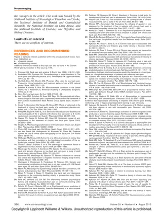
A MODEL FOR PAIN CHRONIFICATION
The above results, together with functional MRI
studies in other chronic pain conditions [42–46]
and additional brain morphometry studies [44,47],
have prompted the proposal of a new mechanistic
model for the transition to chronicity. This model
was developed in three review articles [48–50]. It
proposesthatlearningmechanismswithinthelimbic
circuitry give rise to the transition from acute to
chronic pain and render the pain more emotional
(Fig. 2). The model suggests that one might charac-
terize chronic pain as a pathologically emotional
state.
IMPLICATIONS
The brain circuitry identified as being critical for
pain chronification can be cast within the rubric of
appetitive and aversive motivational learning,
wherein aversive motivation involves behaviors
and drives to escape hedonically unpleasant con-
ditions, with the primary example being pain. As
pain provides a teaching signal that enables indi-
viduals to avoid future actions and environments
associated with harm [38,48], it is an aversive moti-
vation and its relief gives rise to negative reinforce-
ment. Motivational information provided by
nociceptive inputs should contribute to the activity
of circuitry involved in predicting the utility and
costs of competing goals, as well as to behavioral
decisions made in the presence of conflict [51–54].
Neural mechanisms of reward valuation and appe-
titive motivation engage the NAc, ventral tegmental
area, and prefrontal cortex [55]. Furthermore, both
dopaminergic projections from ventral tegmentum
to the NAc and to the cortex, as well as glutamatergic
inputs to the NAc from the amygdala, hippocampus,
and prefrontal cortex, a network collectively known
as the mesolimbic–prefrontal circuit [56,57], parti-
cipates in appetitive behaviors instructed by con-
ditioned cues. Accumulating evidence by our group
and others now shows that this system is also
intimately engaged with noxious stimuli, pain
[31,40,41,58–60], and pain relief [61
,62
]. More-
over, we show that the responses of this system to
painful stimuli are distorted in chronic pain [31] and
that its connectivity predicts the development of
pain chronification [32
].
Dysfunction of the mesolimbic–prefrontal net-
work is a hallmark of addiction, and the cortico-
striatal circuit is a subportion of this circuit.
Moreover, all substances of abuse self-administered
Neuroimaging
364 www.co-neurology.com Volume 26 Number 4 August 2013](https://image.slidesharecdn.com/predictingtransitiontochronicpain-160317223637/85/Predicting-transition-to-chronic-pain-5-320.jpg)
![Copyright © Lippincott Williams Wilkins. Unauthorized reproduction of this article is prohibited.
by humans that can result in addiction are believed
to exert their reinforcing effects by increasing dopa-
minergic tone in the NAc [63]. The persistent nature
of addiction is associated with activity-induced
plasticity of neurons within the ventral tegmentum
and NAc, with dysfunction of prefrontal activity,
with long-term downregulation of dopamine recep-
tors and dopamine production, as well as enhanced
glutamatergic transmission from prefrontal regions
to the NAc [64].
CONCLUSION
Given that we have identified the main components
of this same circuitry in pain chronification, we
assume close parallels between mechanisms leading
to addiction and those leading to pain chronifica-
tion. We therefore hypothesize that the transition
to chronic pain is dependent on activity-induced
plasticity of the corticostriatal circuit, leading to
reorganization of the network such that aversive
cues lead to aberrantly elevated and prolonged net-
work activity. Within this framework, chronic pain
and addictive behavior are viewed as distinct yet
mechanistically related manifestations of reorganiza-
tion of the brain’s motivational learning circuitry
[65,66
]. Thus, similar to addiction, chronic pain
may be viewed as a brain disease state, and we
presume that the sequelae of the transition to
chronic pain within the corticostriatal system
should exhibit strong parallels to the reorganiza-
tion observed for addiction. A corollary to this
hypothesis is the idea that chronic pain may be
prevented either by blunting the aversive drive
impinging on the corticostriatal circuit or by block-
ing the reorganization of the latter circuit, or by
combining both approaches. As recent evidence
shows that the striatum can modulate nociception
by direct or indirect descending pathways [41,67],
the corticostriatal circuit may also be involved in
spinal cord sensitization. Given this conceptualiz-
ation, clinicians and scientists face the challenge of
managing, reversing, and ideally preventing the
dysfunction of pain addiction.
Acknowledgements
The authors would like to thank Howard Fields, Thomas
J. Schnitzer, and Marco Martina for their contributions to
Pain suffering
(emotional self)
mPFC
(congnitive decision)
LPFC
Perception
Insula
ACC
Nociception
NAc
(motivation)
(arousal)
Amyg
(memory)
Hippo
FIGURE 2. A model regarding brain circuitry involved in the transition from acute to chronic pain. Nociceptive information,
perhaps distorted by peripheral and spinal cord sensitization processes, impinges on limbic circuitry. The interaction of limbic
circuitry with prefrontal processes determines the level at which a certain pain condition transitions to a more emotional state.
The limbic circuitry also provides learning/modulation signals to the rest of the cortex inducing functional and anatomical
distortions that reflect the suffering and coping strategies. Adapted from [50]. LPFC, lateral prefrontal cortex; mPFC, medial
prefrontal cortex; ACC, anterior cingulate cortex; NAc, nucleus accumbens; Amyg, amygdala; Hippo, hippocampus.
Predicting transition to chronic pain Apkarian et al.
1350-7540 ß 2013 Wolters Kluwer Health | Lippincott Williams Wilkins www.co-neurology.com 365](https://image.slidesharecdn.com/predictingtransitiontochronicpain-160317223637/85/Predicting-transition-to-chronic-pain-6-320.jpg)


Properties of the brain's emotional learning circuitry predict the transition to chronic pain. In a longitudinal study, individuals with acute back pain were scanned over a year-long period. The strength of functional connectivity between the medial prefrontal cortex and nucleus accumbens at the initial scan predicted whether the individual would transition to chronic pain with over 80% accuracy. Future studies are needed to understand the mechanisms driving pain chronicity, with the goal of developing novel therapies to prevent the transition to chronic pain.


