1
2
MISCELLANEOUS DISEASE
3
PRESENTER
DR. SHIREEN MANSOOR
Let's begin…
4
5
Learning objectives
 DIABETES MELLITUS
 DIABETES NEUROPATHY
 DIABETIC FOOT
6
DIABETIC MELLITUS
DIABETES MELLITUS
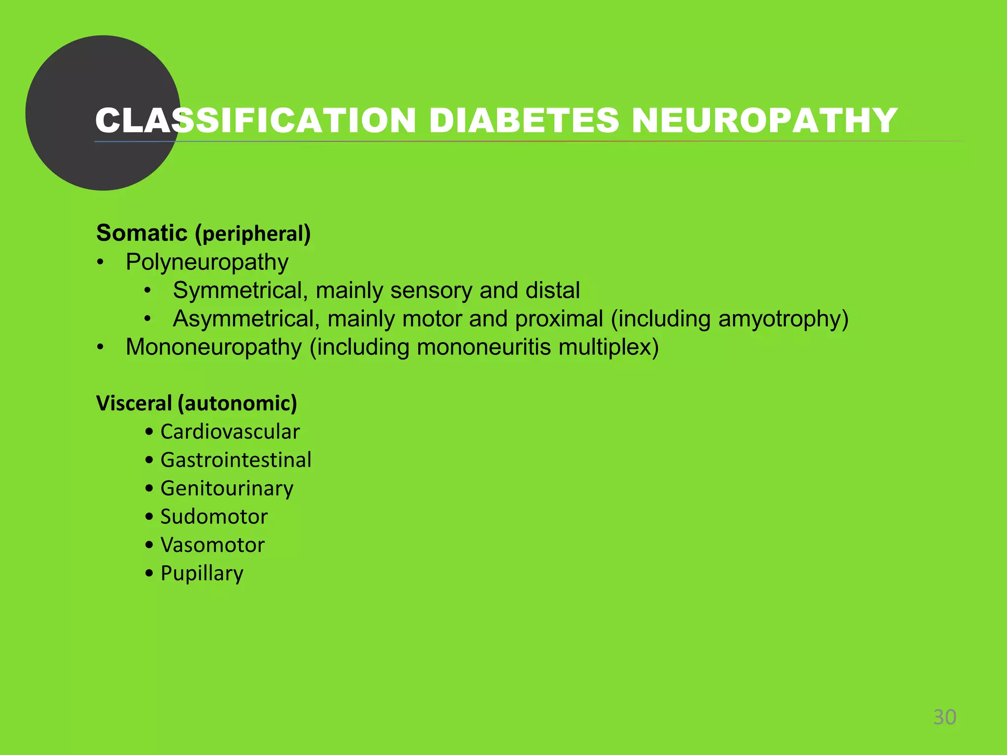
• Diabetes mellitus is a clinical syndrome characterized by an increase in plasma blood glucose
(hyperglycaemia).
• The incidence of diabetes is rising. Globally, it is estimated that 366 million people had
diabetes in 2011 (approximately 8.3% of the world population, or 3 new cases every 10
seconds), and this figure is expected to reach 552 million by 2030.
• The effects of diabetes mellitus include long–term damage, dysfunction and failure of various
organs.
4/13/2023 7
Types of Diabetes
8
• Type 1 Diabetes Mellitus
• Type 2 Diabetes Mellitus
• Gestational Diabetes
Functional Anatomy & Physiology
9
 REGULATION OF INSULIN SECRETION:
Insulin is secreted from pancreatic β cells into the portal circulation
in response to glucose and other nutrient. It reduces blood glucose
level.
Only antidiabetic hormone secreted in body.
REGULATION OF GLUCAGON SECRETION:
Pancreatic α-cells of islets of Langerhans that secrete glucagon.
Glucagon has opposite effects of insulin, stimulate Glycogenolysis.
BLOOD GLUCOSE HOMEOSTASIS:
Blood glucose concentration is maintained within narrow range.
Glucose homeostasis reflects a balance between the entry of
glucose into the circulation from the liver and intestine and the
uptake of glucose by peripheral tissues, particularly muscle and
brain.
Type 1 diabetes
10
Caused by autoimmune destruction of insulin-
producing cells (β cells) in the pancreas, resulting
in absolute insulin deficiency.
 Type 1 diabetes develops when the body’s
immune system destroys pancreatic beta
cells, the only cells in the body that make the
hormone insulin that regulates blood
glucose.
 This form of diabetes usually strikes children
and young adults, although disease onset can
occur at any age.
Type 2 diabetes
11
Characterised by resistance to the action of insulin
and an inability to produce sufficient insulin to
overcome this ‘insulin resistance’
Was previously called non-insulin-dependent
diabetes mellitus (NIDDM) or adult-onset
diabetes
It usually begins as insulin resistance, a
disorder in which the cells do not use insulin
properly. As the need for insulin rises, the
pancreas gradually loses its ability to produce
insulin.
12
Gestational Diabetes
13
 A form of glucose intolerance that is diagnosed in some women
during pregnancy.
 During pregnancy, gestational diabetes requires treatment
to normalize maternal blood glucose levels to avoid
complications in the infant.
 After pregnancy, 5% to 10% of women with gestational
diabetes are found to have type 2 diabetes.
ETIOLOGICAL CLASSIFICATION
14
 Type 1
Immune Mediated, Idiopathic
Type 1 diabetes is a T cell-mediated autoimmune disease involving destruction of
the insulin-secreting β cells in the pancreatic islets.
 Type 2
• Genetic defects of β-cell function
• Genetic defects of insulin action
• Pancreatic disease (e.g. pancreatitis)
• Excess endogenous production of hormonal antagonists to insulin
• Drug-induced e.g. corticosteroids, thiazide diuretics, phenytoin)
• Uncommon forms of immune-mediated diabetes
• Wolfram’s syndrome – diabetes insipidus, diabetes mellitus, optic atrophy, nerve
deafness; Friedreich’s ataxia; myotonic dystrophy
Presenting Complain Of DM
Hyperglycaemia
15
• High blood glucose.
• Symptoms: Thirst, Fatigue, Blurred Vision, Polyuria, Headache,
Blurred vision.
• Type 2 DM patient can be asymptomatic or present with chronic
fatigue or malaise.
• Typical type 2 diabetes occurs increasingly in obese young
people.
• Older patient may have evidence of autoimmune activity
against B cells, a slowly evolving variant of type 1
diabetes(LADA).
16
Diabetic Emergencies
17
Diabetic Ketoacidosis:
Medical emergency that principally occurs in people with type 1 diabetes. It occurs when
the body starts breaking down fat at a rate that is much too fast. The liver processes the fat
into a fuel called ketones, which causes the blood to become acidic.
The cardinal biochemical features of DKA are:
Hyperglycemia
Hyperketonaemia
 Metabolic acidosis
Treatment:
• Insulin
• Fluid Replacement
• Potassium
• Bicarbonate.
Hyperglycaemic Hyperosmolar state
18
• Hyperglycaemic hyperosmolar state (HHS) is characterised by severe
hyperglycaemia, hyperosmolality, and dehydration in the absence of
significant hyperketonaemia or acidosis.
• HHS, hyperglycaemia usually develops over a longer period (a few days to
weeks), causing more profound hyperglycaemia and dehydration.
• It typically affects elderly patients, but is seen increasingly in younger adults.
• Common precipitating factors include infection, myocardial infarction,
cerebrovascular events or drug therapy (e.g. corticosteroids).
Hypoglycaemia
19
Blood glucose level less than 70mg/dL, occur as a result of treatment with insulin.
Symptoms Of ANS: Hunger, anxiety, sweating, palpitation, trembling.
Symptoms of neuroglycopenia:speech difficulty, drowsiness and poor cordination.
Treatment depends on its severity and on whether the patient is concious.
Oral fast acting carbohydrate, carbohydrate snacks is sufficient.
TESTING
20
• URINE TESTING
o Testing the urine for glucose with dipsticks is a common screening procedure for
detecting diabetes.
o Ketone bodies can be identified by the nitroprusside reaction, which measures
acetoacetate, using either tablets or dipsticks.
o Ketonuria may be found in normal people who have been fasting or exercising
strenuously for long periods,
o Standard dipstick testing for albumin detects urinary albumin at concentrations
above 300 mg/L, but smaller amounts
• BLOOD TESTING
o Glucose
o Protein
Management of Diabetes
21
Type 1: Urgent insulin therapy and prompt referral to a specailist.
Type 2: Advice about Dietary and lifestyle modification, followed be oral antidiabetic
drugs/insulin if needed.
Drugs to reduce Hyperglycemia:
Biguanides
sulphynylureas
TZDs(Thiazolidinediones)
Alpha-glucosidase inhibitors
Insulin Therapy
22
In most patients, insulin is injected subcutaneously
several times a day into the anterior abdominal wall,
upper arms, outer thighs and buttocks.
s. It is removed mainly by the liver
and also the kidneys, so plasma insulin concentrations
are elevated in patients with liver disease or renal failure
23
Life Style Modification
24
• Undertaking regular physical activity
• Achieve adequate glycemic control
• Maintain adequate nutritional intake
• Reducing alcohol consumption
• Many people with type 2 diabetes require dietary advice for achieving weight loss, to
include caloric restriction and, in particular, reduced fat intake.
COMPLICATIONS
25
Microvascular / Neuropathic level Macrovascular
Impaired vision Myocardial ischemia
Renal failure Myocardial infarction
Sensory loss Transient ischemic attack
Pain stroke
Motor weakness claudication
Ulceration ischemia
Arthropathy
Postural hypotension
Gastrointestinal problems
26
DIABETIC
NEUROPATHY
 Nerve damage that can occur if you have diabetes.
 High blood sugar (glucose) can injure nerves throughout your body
 About 30% of patients with type 1 diabetes have developed diabetic nephropathy 20 years
after diagnosis
 Most common causes of end-stage renal failure in developed countries
27
Diabetic Neuropathy
• Diabetic neuropathy causes substantial morbidity and increases mortality.
• Diagnosed on the basis of symptoms and signs, and after the exclusion of other causes of
neuropathy
• Affects between 50 and 90% of patients with diabetes, and of these, 15–30% will have
painful diabetic neuropathy (PDN).
• Like retinopathy, neuropathy occurs secondary to metabolic disturbance
• Prevalence is related to the duration of diabetes and the degree of metabolic control.
28
Diabetic
Neuropathy(cont.)
29
SIGNS & SYMPTOMS
• Pain
• Poor circulation
• Numbness or tingling in your fingers, toes, hands, and
feet
• Sharp pains or cramps.
• Increased sensitivity to touch
30
CLASSIFICATION DIABETES NEUROPATHY
Somatic (peripheral)
• Polyneuropathy
• Symmetrical, mainly sensory and distal
• Asymmetrical, mainly motor and proximal (including amyotrophy)
• Mononeuropathy (including mononeuritis multiplex)
Visceral (autonomic)
• Cardiovascular
• Gastrointestinal
• Genitourinary
• Sudomotor
• Vasomotor
• Pupillary
31
Symmetrical sensory polyneuropathy
• frequently asymptomatic.
• most common clinical signs are diminished perception of
vibration sensation distally
• loss of tendon reflexes in the lower limbs.
• In symptomatic patients, sensory abnormalities are
predominant.
32
Symmetrical sensory
polyneuropathy(cont.)
Symptoms include
• paraesthesiae in the feet (and, rarely, in the hands)
• pain in the lower limbs (dull, aching and/or lancinating
• worse at night, and mainly felt on the anterior aspect of the
legs)
• burning sensations in the soles of the feet
• cutaneous hyperaesthesia
• an abnormal gait (commonly wide-based), often associated
with a sense of numbness in the feet.
33
Asymmetrical motor diabetic neuropathy
• Also known as ( Diabetic amyotrophy )
• presents as severe and progressive weakness and
wasting of the proximal muscles of the lower
(and occasionally the upper) limbs.
• accompanied by severe pain, mainly felt on the
anterior aspect of the leg, and hyperaesthesia
and paraesthesiae.
• Tendon reflexes may be absent on the affected
side(s).
34
Autonomic neuropathy
• not associated with peripheral somatic neuropathy.
• Parasympathetic or sympathetic nerves may be predominantly
affected in one or more visceral systems.
• Within 10 years of developing overt symptoms of autonomic
neuropathy, 30–50% of patients are dead, many from sudden
cardiorespiratory arrest.
• Patients with postural hypotension (a drop in systolic pressure
of 30 mmHg or more on standing from the supine position)
have the highest subsequent mortality.
Clinical features of autonomic neuropathy
35
Cardiovascular
• Postural hypotension
• Resting tachycardia
• Fixed heart rate
Gastrointestinal
• Dysphagia, due to oesophageal atony
• Abdominal fullness, nausea and vomiting, unstable
glycaemia, due to delayed gastric emptying (‘gastroparesis’)
• Nocturnal diarrhoea
• Fecal incontinence
Clinical features of autonomic neuropathy
36
Genitourinary
• Difficulty in micturition, urinary incontinence, recurrent
infection, due to atonic bladder
• Erectile dysfunction and retrograde ejaculation
Sudomotor
• Nocturnal sweats without hypoglycaemia
• Gustatory sweating
• Anhidrosis; fissures in the
feet
Clinical features of autonomic neuropathy
37
Vasomotor
• Feet feel cold, due to loss of skin vasomotor responses
• Dependent oedema, due to loss of vasomotor tone and
increased vascular permeability
Bullous formation
Pupillary
• Decreased pupil size
• Resistance to mydriatics
• Delayed or absent reflexes to light
MANAGEMENT
38
Pain and paraesthesiae from peripheral somatic
neuropathies
• Intensive insulin therapy (strict glycaemic control)
• Anticonvulsants (gabapentin, pregabalin, carbamazepine,
phenytoin)
• Tricyclic antidepressants (amitriptyline, imipramine)
• Other antidepressants (duloxetine)
• Substance P depleter (capsaicin – topical)
• Opiates (tramadol, oxycodone)
• Membrane stabilisers (mexiletine, IV lidocaine)
• Antioxidant (α-lipoic acid)
MANAGEMENT
39
Postural hypotension
• Support stockings
• Fludrocortisone
• NSAIDs
• α-adrenoceptor agonist (midodrine)
Diarrhoea
• Loperamide
• Broad-spectrum antibiotics
• Clonidine
• Octreotide
Atonic bladder
• Intermittent self-catheterisation
MANAGEMENT
40
Excessive sweating
• Anticholinergic drugs (propantheline, poldine, oxybutinin)
• Clonidine
• Topical antimuscarinic agent (glycopyrrolate cream)
Gastroparesis
• Dopamine antagonists (metoclopramide, domperidone)
• Erythromycin
• Gastric pacemaker; percutaneous enteral (jejunal) feeding
Constipation
• Stimulant laxatives (senna)
MANAGEMENT
41
Erectile dysfunction
• Phosphodiesterase type 5 inhibitors (sildenafil, vardenafil,
tadalafil) – oral
• Dopamine agonist (apomorphine) – sublingual
• Prostaglandin E1 (alprostadil) – injected into corpus
cavernosum or intra-urethral administration of pellets
• Vacuum tumescence devices
• Implanted penile prosthesis
• Psychological counselling; psychosexual therapy
42
DIABETIC FOOT
43
DIABETIC FOOT
• Foot is the frequent site of complications in
patients with diabetes.
• Tissue necrosis in the feet is a common reason
for hospital admission in diabetic patient.
44
ETIOLOGY
Aetiology of diabetic foot is :
• Due to trauma
• Peripheral neuropathy
• Ischemia
CLINCAL FEATURES OF DIABETIC FOOT
4/13/2023 45
NEUROPATHY ISCHEMIA
SYMPTOMS NONE
PARAESTHESIA
PAIN
NUMBNESS
NONE
CLAUDICATION
REST PAIN
STRUCTURAL DAMAGES ULCER
SEPSIS
ABSCESS
OSTEOMYELITIS
DIGITAL GANGRENE
CHARCOT JOINT
ULCER
SEPSIS
GANGRENE
Stages of diebetic
ulcer
• Grade 0 : No ulcer a high risk foot.
• Grade 1 : Superficial ulcer involving the full skin thickness but not underlying tissues.
• Grade 2 : Deep ulcers, penetrating down to ligament and muscles, but no bone
involvement or abscess formations.
• Grade 3 : Deep ulcers with cellulitis or abscess formation, often with osteomyelitis
• Grade 4 : Localized gangrene
• Grade 5 : Extensive gangrene involving the whole foot
4/13/2023 46
47
48
MANAGEMENT
• Antibiotics
• If gangrene is localized or extensive Surgical amputation will be done
49
Care of the Diabetic Foot
Preventive advice to all diabetic patients includes:
• Inspect feet every day
• Wash feet every day
• Moisturize skin if dry
• Cut or file toenails regularly
• Change socks or stockings every day
• Avoid walking barefoot
• Check footwear for foreign bodies
• Wear suitable, well-fitting shoes
• Cover minor cuts with sterile dressings
• Do not burst blisters
• Avoid over-the-counter corn/callus remedies
50

PPD on Diabetes Mellitus (1).pptx

  • 1.
  • 2.
  • 3.
  • 4.
  • 5.
    5 Learning objectives  DIABETESMELLITUS  DIABETES NEUROPATHY  DIABETIC FOOT
  • 6.
  • 7.
    DIABETES MELLITUS • Diabetesmellitus is a clinical syndrome characterized by an increase in plasma blood glucose (hyperglycaemia). • The incidence of diabetes is rising. Globally, it is estimated that 366 million people had diabetes in 2011 (approximately 8.3% of the world population, or 3 new cases every 10 seconds), and this figure is expected to reach 552 million by 2030. • The effects of diabetes mellitus include long–term damage, dysfunction and failure of various organs. 4/13/2023 7
  • 8.
    Types of Diabetes 8 •Type 1 Diabetes Mellitus • Type 2 Diabetes Mellitus • Gestational Diabetes
  • 9.
    Functional Anatomy &Physiology 9  REGULATION OF INSULIN SECRETION: Insulin is secreted from pancreatic β cells into the portal circulation in response to glucose and other nutrient. It reduces blood glucose level. Only antidiabetic hormone secreted in body. REGULATION OF GLUCAGON SECRETION: Pancreatic α-cells of islets of Langerhans that secrete glucagon. Glucagon has opposite effects of insulin, stimulate Glycogenolysis. BLOOD GLUCOSE HOMEOSTASIS: Blood glucose concentration is maintained within narrow range. Glucose homeostasis reflects a balance between the entry of glucose into the circulation from the liver and intestine and the uptake of glucose by peripheral tissues, particularly muscle and brain.
  • 10.
    Type 1 diabetes 10 Causedby autoimmune destruction of insulin- producing cells (β cells) in the pancreas, resulting in absolute insulin deficiency.  Type 1 diabetes develops when the body’s immune system destroys pancreatic beta cells, the only cells in the body that make the hormone insulin that regulates blood glucose.  This form of diabetes usually strikes children and young adults, although disease onset can occur at any age.
  • 11.
    Type 2 diabetes 11 Characterisedby resistance to the action of insulin and an inability to produce sufficient insulin to overcome this ‘insulin resistance’ Was previously called non-insulin-dependent diabetes mellitus (NIDDM) or adult-onset diabetes It usually begins as insulin resistance, a disorder in which the cells do not use insulin properly. As the need for insulin rises, the pancreas gradually loses its ability to produce insulin.
  • 12.
  • 13.
    Gestational Diabetes 13  Aform of glucose intolerance that is diagnosed in some women during pregnancy.  During pregnancy, gestational diabetes requires treatment to normalize maternal blood glucose levels to avoid complications in the infant.  After pregnancy, 5% to 10% of women with gestational diabetes are found to have type 2 diabetes.
  • 14.
    ETIOLOGICAL CLASSIFICATION 14  Type1 Immune Mediated, Idiopathic Type 1 diabetes is a T cell-mediated autoimmune disease involving destruction of the insulin-secreting β cells in the pancreatic islets.  Type 2 • Genetic defects of β-cell function • Genetic defects of insulin action • Pancreatic disease (e.g. pancreatitis) • Excess endogenous production of hormonal antagonists to insulin • Drug-induced e.g. corticosteroids, thiazide diuretics, phenytoin) • Uncommon forms of immune-mediated diabetes • Wolfram’s syndrome – diabetes insipidus, diabetes mellitus, optic atrophy, nerve deafness; Friedreich’s ataxia; myotonic dystrophy
  • 15.
    Presenting Complain OfDM Hyperglycaemia 15 • High blood glucose. • Symptoms: Thirst, Fatigue, Blurred Vision, Polyuria, Headache, Blurred vision. • Type 2 DM patient can be asymptomatic or present with chronic fatigue or malaise. • Typical type 2 diabetes occurs increasingly in obese young people. • Older patient may have evidence of autoimmune activity against B cells, a slowly evolving variant of type 1 diabetes(LADA).
  • 16.
  • 17.
    Diabetic Emergencies 17 Diabetic Ketoacidosis: Medicalemergency that principally occurs in people with type 1 diabetes. It occurs when the body starts breaking down fat at a rate that is much too fast. The liver processes the fat into a fuel called ketones, which causes the blood to become acidic. The cardinal biochemical features of DKA are: Hyperglycemia Hyperketonaemia  Metabolic acidosis Treatment: • Insulin • Fluid Replacement • Potassium • Bicarbonate.
  • 18.
    Hyperglycaemic Hyperosmolar state 18 •Hyperglycaemic hyperosmolar state (HHS) is characterised by severe hyperglycaemia, hyperosmolality, and dehydration in the absence of significant hyperketonaemia or acidosis. • HHS, hyperglycaemia usually develops over a longer period (a few days to weeks), causing more profound hyperglycaemia and dehydration. • It typically affects elderly patients, but is seen increasingly in younger adults. • Common precipitating factors include infection, myocardial infarction, cerebrovascular events or drug therapy (e.g. corticosteroids).
  • 19.
    Hypoglycaemia 19 Blood glucose levelless than 70mg/dL, occur as a result of treatment with insulin. Symptoms Of ANS: Hunger, anxiety, sweating, palpitation, trembling. Symptoms of neuroglycopenia:speech difficulty, drowsiness and poor cordination. Treatment depends on its severity and on whether the patient is concious. Oral fast acting carbohydrate, carbohydrate snacks is sufficient.
  • 20.
    TESTING 20 • URINE TESTING oTesting the urine for glucose with dipsticks is a common screening procedure for detecting diabetes. o Ketone bodies can be identified by the nitroprusside reaction, which measures acetoacetate, using either tablets or dipsticks. o Ketonuria may be found in normal people who have been fasting or exercising strenuously for long periods, o Standard dipstick testing for albumin detects urinary albumin at concentrations above 300 mg/L, but smaller amounts • BLOOD TESTING o Glucose o Protein
  • 21.
    Management of Diabetes 21 Type1: Urgent insulin therapy and prompt referral to a specailist. Type 2: Advice about Dietary and lifestyle modification, followed be oral antidiabetic drugs/insulin if needed. Drugs to reduce Hyperglycemia: Biguanides sulphynylureas TZDs(Thiazolidinediones) Alpha-glucosidase inhibitors
  • 22.
    Insulin Therapy 22 In mostpatients, insulin is injected subcutaneously several times a day into the anterior abdominal wall, upper arms, outer thighs and buttocks. s. It is removed mainly by the liver and also the kidneys, so plasma insulin concentrations are elevated in patients with liver disease or renal failure
  • 23.
  • 24.
    Life Style Modification 24 •Undertaking regular physical activity • Achieve adequate glycemic control • Maintain adequate nutritional intake • Reducing alcohol consumption • Many people with type 2 diabetes require dietary advice for achieving weight loss, to include caloric restriction and, in particular, reduced fat intake.
  • 25.
    COMPLICATIONS 25 Microvascular / Neuropathiclevel Macrovascular Impaired vision Myocardial ischemia Renal failure Myocardial infarction Sensory loss Transient ischemic attack Pain stroke Motor weakness claudication Ulceration ischemia Arthropathy Postural hypotension Gastrointestinal problems
  • 26.
  • 27.
     Nerve damagethat can occur if you have diabetes.  High blood sugar (glucose) can injure nerves throughout your body  About 30% of patients with type 1 diabetes have developed diabetic nephropathy 20 years after diagnosis  Most common causes of end-stage renal failure in developed countries 27 Diabetic Neuropathy
  • 28.
    • Diabetic neuropathycauses substantial morbidity and increases mortality. • Diagnosed on the basis of symptoms and signs, and after the exclusion of other causes of neuropathy • Affects between 50 and 90% of patients with diabetes, and of these, 15–30% will have painful diabetic neuropathy (PDN). • Like retinopathy, neuropathy occurs secondary to metabolic disturbance • Prevalence is related to the duration of diabetes and the degree of metabolic control. 28 Diabetic Neuropathy(cont.)
  • 29.
    29 SIGNS & SYMPTOMS •Pain • Poor circulation • Numbness or tingling in your fingers, toes, hands, and feet • Sharp pains or cramps. • Increased sensitivity to touch
  • 30.
    30 CLASSIFICATION DIABETES NEUROPATHY Somatic(peripheral) • Polyneuropathy • Symmetrical, mainly sensory and distal • Asymmetrical, mainly motor and proximal (including amyotrophy) • Mononeuropathy (including mononeuritis multiplex) Visceral (autonomic) • Cardiovascular • Gastrointestinal • Genitourinary • Sudomotor • Vasomotor • Pupillary
  • 31.
    31 Symmetrical sensory polyneuropathy •frequently asymptomatic. • most common clinical signs are diminished perception of vibration sensation distally • loss of tendon reflexes in the lower limbs. • In symptomatic patients, sensory abnormalities are predominant.
  • 32.
    32 Symmetrical sensory polyneuropathy(cont.) Symptoms include •paraesthesiae in the feet (and, rarely, in the hands) • pain in the lower limbs (dull, aching and/or lancinating • worse at night, and mainly felt on the anterior aspect of the legs) • burning sensations in the soles of the feet • cutaneous hyperaesthesia • an abnormal gait (commonly wide-based), often associated with a sense of numbness in the feet.
  • 33.
    33 Asymmetrical motor diabeticneuropathy • Also known as ( Diabetic amyotrophy ) • presents as severe and progressive weakness and wasting of the proximal muscles of the lower (and occasionally the upper) limbs. • accompanied by severe pain, mainly felt on the anterior aspect of the leg, and hyperaesthesia and paraesthesiae. • Tendon reflexes may be absent on the affected side(s).
  • 34.
    34 Autonomic neuropathy • notassociated with peripheral somatic neuropathy. • Parasympathetic or sympathetic nerves may be predominantly affected in one or more visceral systems. • Within 10 years of developing overt symptoms of autonomic neuropathy, 30–50% of patients are dead, many from sudden cardiorespiratory arrest. • Patients with postural hypotension (a drop in systolic pressure of 30 mmHg or more on standing from the supine position) have the highest subsequent mortality.
  • 35.
    Clinical features ofautonomic neuropathy 35 Cardiovascular • Postural hypotension • Resting tachycardia • Fixed heart rate Gastrointestinal • Dysphagia, due to oesophageal atony • Abdominal fullness, nausea and vomiting, unstable glycaemia, due to delayed gastric emptying (‘gastroparesis’) • Nocturnal diarrhoea • Fecal incontinence
  • 36.
    Clinical features ofautonomic neuropathy 36 Genitourinary • Difficulty in micturition, urinary incontinence, recurrent infection, due to atonic bladder • Erectile dysfunction and retrograde ejaculation Sudomotor • Nocturnal sweats without hypoglycaemia • Gustatory sweating • Anhidrosis; fissures in the feet
  • 37.
    Clinical features ofautonomic neuropathy 37 Vasomotor • Feet feel cold, due to loss of skin vasomotor responses • Dependent oedema, due to loss of vasomotor tone and increased vascular permeability Bullous formation Pupillary • Decreased pupil size • Resistance to mydriatics • Delayed or absent reflexes to light
  • 38.
    MANAGEMENT 38 Pain and paraesthesiaefrom peripheral somatic neuropathies • Intensive insulin therapy (strict glycaemic control) • Anticonvulsants (gabapentin, pregabalin, carbamazepine, phenytoin) • Tricyclic antidepressants (amitriptyline, imipramine) • Other antidepressants (duloxetine) • Substance P depleter (capsaicin – topical) • Opiates (tramadol, oxycodone) • Membrane stabilisers (mexiletine, IV lidocaine) • Antioxidant (α-lipoic acid)
  • 39.
    MANAGEMENT 39 Postural hypotension • Supportstockings • Fludrocortisone • NSAIDs • α-adrenoceptor agonist (midodrine) Diarrhoea • Loperamide • Broad-spectrum antibiotics • Clonidine • Octreotide Atonic bladder • Intermittent self-catheterisation
  • 40.
    MANAGEMENT 40 Excessive sweating • Anticholinergicdrugs (propantheline, poldine, oxybutinin) • Clonidine • Topical antimuscarinic agent (glycopyrrolate cream) Gastroparesis • Dopamine antagonists (metoclopramide, domperidone) • Erythromycin • Gastric pacemaker; percutaneous enteral (jejunal) feeding Constipation • Stimulant laxatives (senna)
  • 41.
    MANAGEMENT 41 Erectile dysfunction • Phosphodiesterasetype 5 inhibitors (sildenafil, vardenafil, tadalafil) – oral • Dopamine agonist (apomorphine) – sublingual • Prostaglandin E1 (alprostadil) – injected into corpus cavernosum or intra-urethral administration of pellets • Vacuum tumescence devices • Implanted penile prosthesis • Psychological counselling; psychosexual therapy
  • 42.
  • 43.
    43 DIABETIC FOOT • Footis the frequent site of complications in patients with diabetes. • Tissue necrosis in the feet is a common reason for hospital admission in diabetic patient.
  • 44.
    44 ETIOLOGY Aetiology of diabeticfoot is : • Due to trauma • Peripheral neuropathy • Ischemia
  • 45.
    CLINCAL FEATURES OFDIABETIC FOOT 4/13/2023 45 NEUROPATHY ISCHEMIA SYMPTOMS NONE PARAESTHESIA PAIN NUMBNESS NONE CLAUDICATION REST PAIN STRUCTURAL DAMAGES ULCER SEPSIS ABSCESS OSTEOMYELITIS DIGITAL GANGRENE CHARCOT JOINT ULCER SEPSIS GANGRENE
  • 46.
    Stages of diebetic ulcer •Grade 0 : No ulcer a high risk foot. • Grade 1 : Superficial ulcer involving the full skin thickness but not underlying tissues. • Grade 2 : Deep ulcers, penetrating down to ligament and muscles, but no bone involvement or abscess formations. • Grade 3 : Deep ulcers with cellulitis or abscess formation, often with osteomyelitis • Grade 4 : Localized gangrene • Grade 5 : Extensive gangrene involving the whole foot 4/13/2023 46
  • 47.
  • 48.
    48 MANAGEMENT • Antibiotics • Ifgangrene is localized or extensive Surgical amputation will be done
  • 49.
    49 Care of theDiabetic Foot Preventive advice to all diabetic patients includes: • Inspect feet every day • Wash feet every day • Moisturize skin if dry • Cut or file toenails regularly • Change socks or stockings every day • Avoid walking barefoot • Check footwear for foreign bodies • Wear suitable, well-fitting shoes • Cover minor cuts with sterile dressings • Do not burst blisters • Avoid over-the-counter corn/callus remedies
  • 50.