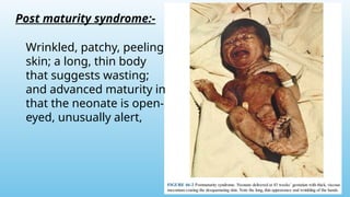
Post term
pregnanc
y
PRESENTOR :- DR
SUNNY BHALADHARE
Outline:-
 Definition
 Incidence
 Etiology
 Estimation og gestational
age
 Diagnosis
 Pathophysiological
changes
 Complications
 Management
Definition:-
The definition of post term pregnancy is
one that exceeds 420/7
weeks or is 294
≥
days from the first day of the last
menstrual period (LMP).
Pregnancies between 411/7
and 416/7
weeks
is considered as late term.
• Post-term pregnancy affects
approximately 4-15% of all pregnancies
(Arias practical guide to high risk
pregnancy and delivery)
• This rate has declining because of
improved pregnancy dating accuracy and
earlier intervention.
Incidence
Etiology:-
• Wrong dates:- Inaccurate Last menstrual period (LMP)
• Maternal factors :-
1. Nulliparous with Long mid-pregnancy cervical length.
2. Advance maternal age.
3. Biological :- due to chr 2q13 gene (associated with
gestational age).
4. Obesity .
5. Previous post term pregnancy (Alfirevic and Walkinshaw, 1994;
Mogren et al., 1999; Olesen et al., 1999)
6. Male fetus (Divon et al., 2002)
• Feto-placental factors:-
1. Anencephaly
2. Adrenal hypoplasia
3. X linked placental sulfatase deficiency – reduced
placental oestrogen synthesis
Estimation of gestational age:-
Gestational age is based on
two things
1.First day of LMP i.e. last
menstrual period.
• Fertilization is
presumed to
occur 2 weeks
after a confident
LMP
2.Measurement of embryo
or fetus at initial Ultrasound.
Diagnosis:-
1. By accurate dating of gestation.
2. Ultrasound up to 136/7
weeks’ gestation as the most
accurate method to establish gestation age(ACOG
Committee Opinion of May 2017)
3. Calculation of estimated due date (EDD) should be done
with help of LMP as well as early ultrasound.
4. Other less-reliable methods include uterine size,
fundal height measurement, Quickening and
detection of fetal heart tones on Doppler.
Pathophysiological changes seen in prolonged
pregnancy:-
1.Amniotic fluid changes:- The amniotic
fluid volume reaches its peak at around
38 weeks’ gestation after which there is a
gentle decline every week.
Fluid becomes thick due to an increased
amount of vernix caseosa.
After 38 weeks, fluid volume declines by
approximately 125 mL/week,
to an average volume of 800 mL at 40
weeks.
AFI 5 cm or SDP (single deepest
≤
pocket)<2cm is considered as
oligohydramnios – marker for intervention.
2.Placental changes:-38 weeks and beyond, the placenta starts showing
infarcts and calcifications.
Along with this, there is development of fibrinoid necrosis and
atherosclerosis in the decidual and chorionic blood vessels.
Complication
s:-
Post maturity syndrome:-
Wrinkled, patchy, peeling
skin; a long, thin body
that suggests wasting;
and advanced maturity in
that the neonate is open-
eyed, unusually alert,
Perinatal mortality and morbidity:-
• Rates of Stillbirth, Neonatal
death, and Infant Morbidity
all rise after post term
pregnancy
• Major cause for the same is
1.Gestational hypertension
2.Prolonged labor
3.Cephalopelvic disproportion
4.Meconium aspiration
syndrome
5. Birth injuries
6.Hypoxic-ischemic
encephalopathy
Management:-
Antepartum management:-
1.Accurate dating of pregnancy.
2. Fetal surveillance
3.The option of induction of labor or
expectant management, which should be
discussed with the patient with prolonged
pregnancy.
Fetal surveillance:-
1. If patient declines the induction of labour .
2.No test can completely eliminate the risk of Still birth.
3.Perinatal asphyxia can occur in the range of 40–42
weeks’ gestation
4.RCOG recommends fetal surveillance from 42 weeks .
5.ACOG recommends fetal surveillance between 41
and 42 weeks
• Nonstress Tests (NST)
• Sonography for the AFI and SDP assessment.
• Biophysical profile though sensitive has its
restrictions.
• Umbilical Doppler studies -To identify fetal
compromise due to altered fetal circulation.
Twice
weekly
Timing of
delivery:-
• The recently published
ARRIVE clinical trial on
induction of labour at 39
weeks.
• Majority of the trials
adopted a policy of
induction at 41
completed weeks (287
days) or more.
• Certain factors recommend delivery of
the baby and not wait for spontaneous
labour like
1.Maternal complications like
Hypertension and Diabetes.
2.Oligohydromnios.
3.IUGR with Oligohydramnios with
Meconium stained liquor.
4. Advance maternal age.
5. Fetal compromise as per fetal
surveillance.
Method of induction:-
1. Decision of induction should be made on the basis of Modified
Bishop score.
2. Score of >8 – suggests ripe cervix :- goes into spontaneous
labour.
3. Score<6 :-Modes of ripening of the cervix are used to increase
the chances of a vaginal delivery.
CERVIX SCORE BISHOP SCORE
MODIFIERS
0 1 2 3 Add 1point
for:
-Preeclampsia
-Each previous
vaginal delivery
Subtract 1
point for :
- Postdate
pregnancy
- Nulliparity
Cervical dilation
(cm)
0 1-2 3-4 5+
Cervical length(cm) >4 2-4 1-2 <1
Station of
presenting part
(cm in relation to
ischial spine)
-3 or
above
-2 -1,0 +1, +2
Consistency Firm Medium Soft
Position Posterior Mid Anterior
MODIFIED BISHOP SCORE
Oxytocin :-
• Ripening of cervix can be done by:-
• Membrane stripping
• Intracervical Foley catheter
• Oxytocin and
• Most effectively by
• Prostaglandins.
REGIMEN STARTING
DOSE(mU/min)
INTERVAL(min) INCREMENT(mU/min)
LOW DOSE
REGIME
1-2 30 1-2
HIGH DOSE
REGIME
4-6 15-30 4-6
Prostaglandins :-
The National Institute for Health and Care
Excellence (NICE) guidelines recommend the
vaginal PGE2 preparation as the preferred
method for induction of labour.
Misoprostol- the PGE1 is safe, effective
and therefore may be seen as the first-
line option for cervical ripening and
induction of labour.
ACOG recommends 25 mcg as every 3-6
hourly.
Combination of Foley and misoprostol
were twice as likely to deliver before a
patient induced by a single agent.
Intrapartum management:-
1. Abnormal Cardiotocography
2. Meconium aspiration
3. Fetal trauma:- prolong pregnancy are often associated
with Macrosomia ,shoulder dystocia which can cause
Brachial plexus injury, clavicle or humerus fracture.
Instrumental deliveries can cause cephalic hematomas
and skull fractures.
4.Shoulder dystocia
5.Maternal perineal injuries:- Third- and fourth-degree
perineal tears, deep vaginal lacerations and cervical tears
are seen with difficult instrumental deliveries or while
deliveries of large babies.
Prolonged fetal heart rate deceleration
before emergency cesarean delivery in a
post term pregnancy with
oligohydramnios.
Variable decelerations in a
post term pregnancy with
oligohydramnios.
Saltatory baseline fetal heart rate
showing oscillations exceeding 20
bpm and associated with
oligohydramnios in a post term
pregnancy.
Indian Perspective:-
1.Perinatal morbidity and mortality increases with an
increase in gestational age, more so after 40 weeks.
2.Asians and African Americans having a shorter
duration of pregnancy.
3.The study had found that between 40 and 41 weeks
and then beyond 41 weeks, meconium aspiration was
two and two and half times, respectively, compared to
39 completed weeks.
THANK YOU.

Postterm pregnancy (after 42weeks) .pptx

  • 1.
  • 2.
    Outline:-  Definition  Incidence Etiology  Estimation og gestational age  Diagnosis  Pathophysiological changes  Complications  Management
  • 3.
    Definition:- The definition ofpost term pregnancy is one that exceeds 420/7 weeks or is 294 ≥ days from the first day of the last menstrual period (LMP). Pregnancies between 411/7 and 416/7 weeks is considered as late term.
  • 4.
    • Post-term pregnancyaffects approximately 4-15% of all pregnancies (Arias practical guide to high risk pregnancy and delivery) • This rate has declining because of improved pregnancy dating accuracy and earlier intervention. Incidence
  • 5.
    Etiology:- • Wrong dates:-Inaccurate Last menstrual period (LMP) • Maternal factors :- 1. Nulliparous with Long mid-pregnancy cervical length. 2. Advance maternal age. 3. Biological :- due to chr 2q13 gene (associated with gestational age). 4. Obesity . 5. Previous post term pregnancy (Alfirevic and Walkinshaw, 1994; Mogren et al., 1999; Olesen et al., 1999) 6. Male fetus (Divon et al., 2002) • Feto-placental factors:- 1. Anencephaly 2. Adrenal hypoplasia 3. X linked placental sulfatase deficiency – reduced placental oestrogen synthesis
  • 6.
    Estimation of gestationalage:- Gestational age is based on two things 1.First day of LMP i.e. last menstrual period. • Fertilization is presumed to occur 2 weeks after a confident LMP
  • 7.
    2.Measurement of embryo orfetus at initial Ultrasound.
  • 8.
    Diagnosis:- 1. By accuratedating of gestation. 2. Ultrasound up to 136/7 weeks’ gestation as the most accurate method to establish gestation age(ACOG Committee Opinion of May 2017) 3. Calculation of estimated due date (EDD) should be done with help of LMP as well as early ultrasound. 4. Other less-reliable methods include uterine size, fundal height measurement, Quickening and detection of fetal heart tones on Doppler.
  • 9.
    Pathophysiological changes seenin prolonged pregnancy:- 1.Amniotic fluid changes:- The amniotic fluid volume reaches its peak at around 38 weeks’ gestation after which there is a gentle decline every week. Fluid becomes thick due to an increased amount of vernix caseosa. After 38 weeks, fluid volume declines by approximately 125 mL/week, to an average volume of 800 mL at 40 weeks. AFI 5 cm or SDP (single deepest ≤ pocket)<2cm is considered as oligohydramnios – marker for intervention.
  • 10.
    2.Placental changes:-38 weeksand beyond, the placenta starts showing infarcts and calcifications. Along with this, there is development of fibrinoid necrosis and atherosclerosis in the decidual and chorionic blood vessels.
  • 12.
  • 13.
    Post maturity syndrome:- Wrinkled,patchy, peeling skin; a long, thin body that suggests wasting; and advanced maturity in that the neonate is open- eyed, unusually alert,
  • 14.
    Perinatal mortality andmorbidity:- • Rates of Stillbirth, Neonatal death, and Infant Morbidity all rise after post term pregnancy • Major cause for the same is 1.Gestational hypertension 2.Prolonged labor 3.Cephalopelvic disproportion 4.Meconium aspiration syndrome 5. Birth injuries 6.Hypoxic-ischemic encephalopathy
  • 16.
    Management:- Antepartum management:- 1.Accurate datingof pregnancy. 2. Fetal surveillance 3.The option of induction of labor or expectant management, which should be discussed with the patient with prolonged pregnancy.
  • 17.
    Fetal surveillance:- 1. Ifpatient declines the induction of labour . 2.No test can completely eliminate the risk of Still birth. 3.Perinatal asphyxia can occur in the range of 40–42 weeks’ gestation 4.RCOG recommends fetal surveillance from 42 weeks . 5.ACOG recommends fetal surveillance between 41 and 42 weeks • Nonstress Tests (NST) • Sonography for the AFI and SDP assessment. • Biophysical profile though sensitive has its restrictions. • Umbilical Doppler studies -To identify fetal compromise due to altered fetal circulation. Twice weekly
  • 18.
    Timing of delivery:- • Therecently published ARRIVE clinical trial on induction of labour at 39 weeks. • Majority of the trials adopted a policy of induction at 41 completed weeks (287 days) or more.
  • 19.
    • Certain factorsrecommend delivery of the baby and not wait for spontaneous labour like 1.Maternal complications like Hypertension and Diabetes. 2.Oligohydromnios. 3.IUGR with Oligohydramnios with Meconium stained liquor. 4. Advance maternal age. 5. Fetal compromise as per fetal surveillance.
  • 22.
    Method of induction:- 1.Decision of induction should be made on the basis of Modified Bishop score. 2. Score of >8 – suggests ripe cervix :- goes into spontaneous labour. 3. Score<6 :-Modes of ripening of the cervix are used to increase the chances of a vaginal delivery. CERVIX SCORE BISHOP SCORE MODIFIERS 0 1 2 3 Add 1point for: -Preeclampsia -Each previous vaginal delivery Subtract 1 point for : - Postdate pregnancy - Nulliparity Cervical dilation (cm) 0 1-2 3-4 5+ Cervical length(cm) >4 2-4 1-2 <1 Station of presenting part (cm in relation to ischial spine) -3 or above -2 -1,0 +1, +2 Consistency Firm Medium Soft Position Posterior Mid Anterior MODIFIED BISHOP SCORE
  • 23.
    Oxytocin :- • Ripeningof cervix can be done by:- • Membrane stripping • Intracervical Foley catheter • Oxytocin and • Most effectively by • Prostaglandins. REGIMEN STARTING DOSE(mU/min) INTERVAL(min) INCREMENT(mU/min) LOW DOSE REGIME 1-2 30 1-2 HIGH DOSE REGIME 4-6 15-30 4-6
  • 24.
    Prostaglandins :- The NationalInstitute for Health and Care Excellence (NICE) guidelines recommend the vaginal PGE2 preparation as the preferred method for induction of labour. Misoprostol- the PGE1 is safe, effective and therefore may be seen as the first- line option for cervical ripening and induction of labour. ACOG recommends 25 mcg as every 3-6 hourly. Combination of Foley and misoprostol were twice as likely to deliver before a patient induced by a single agent.
  • 25.
    Intrapartum management:- 1. AbnormalCardiotocography 2. Meconium aspiration 3. Fetal trauma:- prolong pregnancy are often associated with Macrosomia ,shoulder dystocia which can cause Brachial plexus injury, clavicle or humerus fracture. Instrumental deliveries can cause cephalic hematomas and skull fractures. 4.Shoulder dystocia 5.Maternal perineal injuries:- Third- and fourth-degree perineal tears, deep vaginal lacerations and cervical tears are seen with difficult instrumental deliveries or while deliveries of large babies.
  • 26.
    Prolonged fetal heartrate deceleration before emergency cesarean delivery in a post term pregnancy with oligohydramnios.
  • 27.
    Variable decelerations ina post term pregnancy with oligohydramnios.
  • 28.
    Saltatory baseline fetalheart rate showing oscillations exceeding 20 bpm and associated with oligohydramnios in a post term pregnancy.
  • 29.
    Indian Perspective:- 1.Perinatal morbidityand mortality increases with an increase in gestational age, more so after 40 weeks. 2.Asians and African Americans having a shorter duration of pregnancy. 3.The study had found that between 40 and 41 weeks and then beyond 41 weeks, meconium aspiration was two and two and half times, respectively, compared to 39 completed weeks.
  • 30.