This study validated the peroxisome targeting of two pathogen defense proteins in Arabidopsis thaliana, disease resistance protein 1 (DRP1) and nudix hydrolase homologue 15 (NUDT15), which were predicted to have peroxisome targeting signals (PTS). Experiments showed that an alternative splice variant of DRP1 (DRP1.2) localized to peroxisomes due to a functional PTS1 domain, while another variant (DRP1.1) remained cytosolic. Similarly, a splice variant of NUDT15 (NUDT15.2) was targeted to peroxisomes by its PTS1, but another variant (NUDT15.1) was
Comparing Residual Integration Levels of Some IntegrationDeficient Lentiviral...inventionjournals
Lentiviral vectors (LVs) have many advantageous characteristics making them a good choice in the field of gene therapy. Nevertheless, their integration may lead to detrimental effects. To overcome this problem, lentiviral integration can be targeted through using integration-deficient lentiviral vectors (IDLVs). In this study, an integration-proficient lentiviral vector (IPLV) and a battery of IDLVs with single or multiple mutations affecting integration were produced and their integration levels were compared. eGFP time-course experiment and clonogenic assay were used to make these comparisons. It was found that there was not any significant difference between the residual integration of any of the IDLVs used in this study and that of the standard IDLV; D64V-IDLV. It can be concluded that most IDLV integration is mediated by integraseindependent mechanisms.
ANTIMICROBIAL PEPTIDES & THEIR POTENTIAL TO COMBAT ANTIMICROBIAL RESISTANCE O...Kazimierz Murzyn
Presentation given during Cost AMiCI meeting in Tallinn Nov 2017
by Maria Olivia Pereira, Assistant Professor University of Minho Department of Biological Engineering Braga, Portugal, Professor in Biomedical Engineering Principal Investigator in the Biofilm Group
Prime-ome: "A molecular approach towards defense priming"Dhanya AJ
Prime-ome is the entire set of messenger RNA (mRNA) molécules or transcripts, proteins and metabolites produced or modified by an organism or system during the different stages of priming in plants and prime-omics is the study of prime-ome.
Cloning, Expression and Purification of Vibrio parahaemolyticus L-type Lectin from White Leg Shrimp Litopenaeus vannamei for Bacterial Agglutinating
http://dx.doi.org/10.21276/SSR-IIJLS.2020.6.3.3
Objective(s):
This study aimed to find the effects of silver nanoparticles (Ag-NPs) (40 nm) on skin wound healing in mice Mus musculus when innate immune system has been suppressed.
Materials and Methods:
A group of 50 BALB/c mice of about 8 weeks (weighting 24.2±3.0 g) were randomly divided into two groups: Ag-NPs and control group, each with 25 mice. Once a day at the same time, a volume of 50 microliters from the nanosilver solution (10ppm) was applied to the wound bed in the Ag-NPs group while in the untreated (control) group no nanosilver solution was used but the wound area was washed by a physiological solution. The experiment lasted for 14. Transforming growth factor beta (TGF-β), complement component C3, and two other immune system factors involving in inflammation, namely C-reactive protein (CRP) and rheumatoid factor (RF) in sera of both groups were assessed and then confirmed by complement CH50 level of the blood.
Results:
The results show that wound healing is a complex process involving coordinated interactions between diverse immunological and biological systems and that Ag-NPs significantly accelerated wound healing and reduce scar appearance through suppression of immune system as indicated by decreasing levels of all inflammatory factors measured in this study.
Conclusion:
Exposure of mice to Ag-NPs can result in significant changes in innate immune function at the molecular levels. The study improves our understanding of nanoparticle interaction with components of the immune system and suggests that Ag-NPs have strong anti-inflammatory effects on skin wound healing and reduce scarring.
Evaluation of infection course in mice induced by L. major in presence of pos...Nanomedicine Journal (NMJ)
Abstract:
An inoculation of virulent Leishmania major is known as leishmanization (LZ) which is proven to be the most effective control measure against Cutaneous Leishmaniasis (CL). However, using LZ is restricted due to various side effects such as uncontrolled lesion development. In the present research, the efficacy of cationic nanoliposomes containing CpG oligodeoxynucleotides (CpG ODN) as an improved adjuvant delivery system was studied to diminish the lesion development and infection course of L. major after inoculation into the mice. BALB/c mice were inoculated subcutaneously (SC) with L. major plus empty DSPC, DSPC (CpG ODN), DSPC (Non CpG ODN), empty DMPC, DMPC (CpG ODN), DMPC (Non CpG ODN) or HEPES buffer. The results showed that group of mice received DMPC (CpG ODN) nanoliposomes developed a significantly smaller lesion and showed minimum number of L. major in the spleen and draining lymph nodes. In addition, using DMPC (CpG ODN) liposomes resulted in a Th1 type of immune response with a preponderance of IgG2a isotype which is concurrent with the production of DMPC (CpG) induced IFN-γ in the spleen of the mice. Taken together, the results suggested that immune modulation using DMPC (CpG ODN) nanoliposomes might be a practical approach to improve the safety of LZ
Keywords:
DMPC (CpG ODN) nanoliposomes; CpG ODN; L. major; Leishmanization; Immune response
1. University
of Stavanger
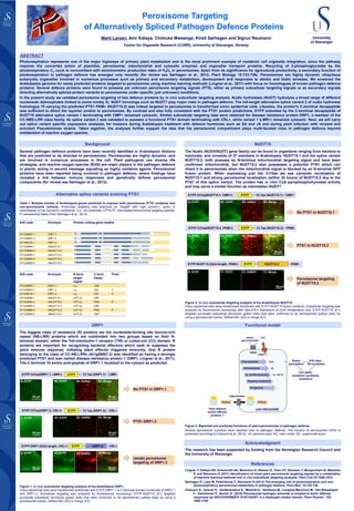
Peroxisome Targeting
of Alternatively Spliced Pathogen Defence Proteins
Center for Organelle Research (CORE), University of Stavanger, Norway
Marit Larsen, Amr Kataya, Chimuka Mwaanga, Kirsti Sørhagen and Sigrun Reumann
ABSTRACT
Photorespiration represents one of the major highways of primary plant metabolism and is the most prominent example of metabolic cell organelle integration, since the pathway
requires the concerted action of plastidial, peroxisomal, mitochondrial and cytosolic enzymes and organellar transport proteins. Recycling of 2-phosphoglycolate by the
photorespiratory C2 cycle is concomitant with stoichiometric production rates of H2O2 in peroxisomes. Apart from its significance for agricultural productivity, a secondary function of
photorespiration in pathogen defence has emerged only recently (for review see Sørhagen et al., 2013, Plant Biology 15:723-736). Peroxisomes are highly dynamic ubiquitous
eukaryotic organelles involved in numerous processes such as primary and secondary metabolism, development and responses to abiotic and biotic stresses. We screened the
Arabidopsis genome for newly predicted proteins targeted to peroxisomes using machine learning methods (Lingner et al., 2011) with focus on homologues of known pathogen defence
proteins. Several defence proteins were found to possess yet unknown peroxisome targeting signals (PTS), either as primary subcellular targeting signals or as secondary signals
directing alternatively spliced protein variants to peroxisomes under specific (yet unknown) conditions.
In the present study we validated peroxisome targeting of two defence proteins by in vivo subcellular targeting analysis. Nudix hydrolases (NUDT) hydrolyse a broad range of different
nucleoside diphosphates (linked to some moiety X). NUDT homologs such as NUDT7 play major roles in pathogen defence. The full-length alternative splice variant 2 of nudix hydrolase
homologue 15 carrying the predicted PTS1 PKM> (NUDT15.2) was indeed targeted to peroxisomes in transformed onion epidermal cells. Likewise, the protein’s C-terminal decapeptide
was sufficient to direct the reporter protein to peroxisomes. By contrast and also fully consistent with the PTS1 protein predictions, EYFP extended by the C-terminal decapeptide of
NUDT15 alternative splice variant 1 terminating with CMP> remained cytosolic. Similar subcellular targeting data were obtained for disease resistance protein DRP1, a member of the
CC-NBS-LRR class family. Its splice variant 2 was validated to possess a functional PTS1 domain terminating with CRL>, while variant 1 (LMR>) remained cytosolic. Next, we will carry
out splice variant specific expression analyses, for instance during Arabidopsis treatment with defence hormones such as SA and JA and during plant infection with virulent and
avirulent Pseudomonas strains. Taken together, the analyses further support the idea that the peroxisomal compartment plays multi-faceted roles in pathogen defence beyond
metabolism of reactive oxygen species.
Background
References
Acknowledgment
Lingner T, Kataya AR, Antonicelli GE, Benichou A, Nilssen K, Chen XY, Siemsen T, Morgenstern B, Meinicke
P, and Reumann S (2011) Identification of novel plant peroxisomal targeting signals by a combination
of machine learning methods and in vivo subcellular targeting analyses. Plant Cell 23:1556-1572.
Sørhagen K, Laxa M, Peterhänsel C, Reumann S (2013) The emerging role of photorespiration and non-
photorespiratory peroxisomal metabolism in pathogen defence. Plant Biol. 15:723-736.
Chaouch S., Queval G., Vanderauwera S., Mhamdi A., Vandorpe M., Langlois-Meurinne M., Van Breusegem
F., Saindrenan P., Noctor G. (2010) Peroxisomal hydrogen peroxide is coupled to biotic defense
responses by ISOCHORISMATE SYNTHASE1 in a daylength related manner. Plant Physiol., 153,
1692-1705.
Several pathogen defence proteins have been recently identified in Arabidopsis thaliana
that are predicted to be directed to peroxisomes. Peroxisomes are highly dynamic, and
are involved in numerous processes in the cell. Plant pathogens use diverse life
strategies, and reactive oxygen species (ROS) are major players in the pathogen defence
in plants, acting in second message signalling as highly oxidative agents. Peroxisomal
proteins have been reported being involved in pathogen defence, where findings have
revealed a link between immune responses and genetically defines peroxisomal
components (for review see Sørhagen et al., 2013).
CPK1
PEN2
Photorespiration H2O2
Indole
glucosinolates
Ca2+
Host defense
and/or effector
proteins ?
Redox
pertubation
JAJA biosynthesis
O2
-, NO (?)O2
- and NO production
Polyamine metabolism
ICS1-dep-.
SA synthesis
Cell death,
camalexin synthesis,
resistance
LEAF PEROXISOME
SA signaling
Importomer
AGI code Acronym N-term.
target.
signal
C-term.
tripep.
Pred.
AT1G58807.1 DRP1.1 n.p. LMR
AT1G58807.2 DRP1.2 n.p. CRL P
AT1G59124.1 DRP1.3 n.p. CRL P
AT1G28960.1 AtNUDT15.1 mTP (2) CMP
AT1G28960.2 AtNUDT15.2 mTP (2) PKM P
AT1G28960.3 AtNUDT15.3 mTP (2) CMP
AT1G28960.4 AtNUDT15.4 mTP (2) PKM P
AT1G28960.5 AtNUDT15.5 mTP (2) CMP
Table I: Multiple models of Arabidopsis genes predicted to express both peroxisomal (PTS1 proteins) and
non-peroxisomal variants. N-terminal targeting was predicted by TargetP with high (score=1, given in
parenthesis) or low (score=5) confidence. n.p.: not predicted; cTP/mTP: chloroplast/mitochondrial targeting peptide;
P: peroxisomal (taken from Sørhagen et al., 2013)
EYFP-DRP1.2(full-length, CRL>) CRL>EYFP DRP1.2
LMR>EYFP Ct 7aa (DRP1.1)EYFP-Ct7aa(DRP1.1, LMR>)
EYFP-Ct7aa(DRP1.2, CRL>) CRL>EYFP Ct 7aa (DRP1.2)
EYFP-NUDT15.2(full-length, PKM>) PKM>EYFP NUDT15.2
CMP>EYFP Ct 7aa (NUDT15.1)EYFP-Ct7aa(NUDT15.1, CMP>):
C. EYFP D1. EYFP
PKM>EYFP Ct 7aa (NUDT15.2)EYFP-Ct7aa(NUDT15.2, PKM>):
Figure 2: In vivo subcellular targeting analysis of the Arabidopsis NUDT15.
Onion epidermal cells were transformed biolistically with EYFP-NUDT15 fusion construct. Subcellular targeting was
analyzed by fluorescence microscopy after about18 h expression at room temperature only. EYFP-NUDT15 (E1)
targeted punctuate subcellular structures (green dots) that were confirmed to be peroxisomes (yellow dots) by
using a peroxisomal marker, DsRed-SKL (E2) in merge (E3).
Alternative splice variants evolving PTS1
DRP1
The biggest class of resistance (R) proteins are the nucleotide-binding site leucine-rich
repeat (NB-LRR) proteins which are subdivided into two groups based on their N-
terminal domain; either the Toll-interleukin-1 receptor (TIR) or coiled-coil (CC) domain. R
proteins are important for recognizing bacterial effectors which seek to suppress the
plant immune response, initiating plant effector triggered immunity. One R protein
belonging to the class of CC-NB-LRRs (At1g58807.2) was identified as having a strongly
predicted PTS1 and was named disease resistance protein 1 (DRP1, Lingner et al., 2011).
The C-terminal 10 amino acid-peptide of DRP1.1 localized to the cytosol as predicted.
NUDT15
Functional model
The Nudix (NUDX/NUDT) gene family can be found in organisms ranging from bacteria to
mammals, and consists of 27 members in Arabidopsis. NUDT15.1 and the splice variant
NUDT15.2, both possess an N-terminal mitochondrial targeting signal and have been
confirmed mitochondrial, but NUDT15.2 also possesses a potential PTS1 which can
direct it to peroxisomes when the mitochondrial signal is blocked by an N-terminal GFP
fusion protein. When expressing just the Ct10aa we see cytosolic localization of
NUDT15.1 and strong peroxisomal localization (within 24 hours) of NUDT15.2 due to the
PTS1 of this splice variant. The protein has in vitro CoA pyrophosphohyrolase activity
and may serve a similar function as mammalian NUDT7.
DRP1.1-LMR>
20 µm
20 µm
20 µm
A. EYFP B1. EYFP B2. DsRed B3. Merge
D2. DsRed D3. Merge
A. EYFP B1. EYFP B2. DsRed B3. Merge
D2. DsREDD1. EYFP D3. MergeC. EYFP
20 µm20 µm
F1. EYFP EF. MergeE. EYFP
20 µm 20 µm
E. EYFP F2. DsREDF1. EYFP F3. Merge
60 µm
F2. DsRED
The research has been supported by funding from the Norwegian Research Council and
the University of Stavanger.
PTS1/2PTS1/2
Figure 3: Reported and predicted functions of plant peroxisomes in pathogen defense.
Several peroxisome functions have reported links to pathogen defense. The function of peroxisomal H2O2 is
presented according to Chaouch et al. (2010). JA, jasmonic acid; NO, nitric oxide; O2-, superoxide anion.
No PTS1 in NUDT15.1
Peroxisome targeting
of NUDT15.2
PTS1 in NUDT15.2
No PTS1 in DRP1.1
(weak) peroxisome
targeting of DRP1.2
PTS1 DRP1.2
AGI code Acronym Protein coding gene models
AT1G58807.1 DRP1.1
AT1G58807.2 DRP1.2
AT1G59124.1 DRP1.3
AT1G28960.1 AtNUDT15.1
AT1G28960.2 AtNUDT15.2
AT1G28960.3 AtNUDT15.3
AT1G28960.4 AtNUDT15.4
AT1G28960.5 AtNUDT15.5
Figure 1: In vivo subcellular targeting analysis of the Arabidopsis DRP1.
Onion epidermal cells were transformed biolistically with EYFP-DRP1.1 or C-terminal domain constructs of DRP1.1
and DRP1.2. Subcellular targeting was analyzed by fluorescence microscopy. EYFP-NUDT15 (E1) targeted
punctuate subcellular structures (green dots) that were confirmed to be peroxisomes (yellow dots) by using a
peroxisomal marker, DsRed-SKL (E2) in merge (E3).
20 µm