Portal vein diameter and its relationship to anthropometric parameters
•
1 like•112 views

This study measured the portal vein diameter in 108 healthy individuals in Northeast India using ultrasound. The mean portal vein diameter was 8.83±2.12 mm overall, 9.17±2.33 mm in males and 8.55±1.90 mm in females. Diameter increased with weight and BMI in all adults and females but did not correlate with any body parameters in males. Diameter also increased from ages 15-60 years but decreased after 60. The study provides reference values for portal vein diameter in this population that can help identify abnormal diameters indicating disease.

Recommended
Recommended
More Related Content
What's hot
What's hot (20)
Viewers also liked
Viewers also liked (10)
Similar to Portal vein diameter and its relationship to anthropometric parameters
Similar to Portal vein diameter and its relationship to anthropometric parameters (20)
More from iosrjce
More from iosrjce (20)
Recently uploaded
Recently uploaded (20)
Portal vein diameter and its relationship to anthropometric parameters
- 1. IOSR Journal of Dental and Medical Sciences (IOSR-JDMS) e-ISSN: 2279-0853, p-ISSN: 2279-0861.Volume 14, Issue 12 Ver. I (Dec. 2015), PP 110-113 www.iosrjournals.org DOI: 10.9790/0853-14121110113 www.iosrjournals.org 110 | Page Portal vein diameter in a tertiary care centre in North-East India Nirmalya Saha1 , Ruma Sarkar2 , Moirangthem Matum Singh3 1. Assistant Professor, Department of Anatomy, Tripura Medical College & Dr. B. R. A. M. Teaching Hospital, Agartala, Tripura, India; 2. Assistant Professor, Department of Radiodiagnosis, Jawharlal Nehru Institure of Medical Sciences, Imphal, Manipur, India; 3. Professor, Department of Anatomy, Regional Institute of Medical Sciences, Imphal, Manipur. Abstract: The diameter of portal vein is variable according to gender, age, weight, height, body mass index (BMI) and with factors like, respiration, postural changes and absorptive state. Therefore, this study was conducted to determine the normal portal vein diameter and to evaluate its relationship with parameters like age, gender distribution, weight, height and body mass index (BMI). A total of 108 healthy individuals age ranged from 15 to 85 years attending Radiodiagnosis department were chosen for the study. Variables were: sex, age, weight, height, basal metabolic rate (BMI), portal vein diameter respectively. Transabdominal ultrasound was done to measure the diameter of the main portal vein with average of three times measurements. In present study, the portal vein diameter in males and females were 9.17±2.33 mm and 8.55±1.90 mm respectively. The diameter correlated with weight and BMI in total adult population and females but in males none of the body parameters were correlated significantly. KeyWords: Portal vein, Portal vein diameter, Ultrasound I. Introduction The portal vein arises from the confluence of superior mesenteric and splenic veins posterior to the neck of pancreas.1 It carries 80% of venous blood from intestine and spleen to the liver to supply up to the one half of the oxygen requirements of the hepatocyte.2 Various studies revealed that, the portal vein diameter (PVD) changes with the age and gender distribution.3, 4 The upper limit of portal vein diameter was found to be 13 mm.5 Portal hypertension is the most common complication and also one of the important cause of death in chronic liver diseases.6 Variations in the anthropometric features of various populations, races, regions are an established fact. The diameter of portal vein varies according to gender, age, weight, height, body mass index (BMI). There was a correlation between portal vein diameter and various physical parameters like age, sex and height.7 Factors like, respiration, postural changes, absorptive state influences the calibre of portal vein.5 So, a comprehensive anthropometric study on normal portal vein diameter is essential for identifying the normal from abnormal diameter to diagnose the disease state. Hence, this study was conducted to determine the normal portal vein diameter and to evaluate its relationship with different ages, gender distribution, weight, height and body mass index (BMI). II. Materials & Methods This cross-sectional study was conducted in the Department of Anatomy and Radiodiagnosis, Regional Institute Medical Sciences, Imphal, among 108 healthy individuals. Patients (49 males and 59 females), age ranged from 15 to 85 years who attended Radiodiagnosis department for conditions other than portal hypertension or any other diseases related to portal vein directly or indirectly, liver, spleen or cardiac, malaria, any abdominal surgery and pregnant lady, gastrointestinal and hematologic diseases. For this conceptualisation opinion from Medicine professional also was taken. Formal permissions from the Institutional Ethics Committee, RIMS, other concerned authorities and written informed consent from the concerned individuals were taken. Transabdominal ultrasound was done by Medison SONOACE X8 with 3.5 MHz sector curvilinear transducer probe. Study variables were: sex, age, weight, height, basal metabolic rate (BMI), portal vein diameter. The individuals were divided among five groups according to the age. Group I – 15 to 30 years, Group II – 31-45 years, Group III – 46 to 60 years, Group IV – 61 to 75 years and Group V – more than 75 years. The individuals were kept in fasting after dinner of the previous night. With an elaborate history of present & previous history of illness, history of alcohol consumption, menstrual history in females mainly to exclude pregnancy, a physical examination was done. The weight, height were measured and BMI was calculated with the formula BMI = weight (kg) / height (meter) 2 . The abdomen was scanned routinely to check sonographic exclusion criterias, lying in supine position in relaxed conditions and in quite respiration. The portal vein at porta hepatis was evaluated. The diameter of the main portal vein was measured at the crossing point anterior to the inferior vena cava (IVC) at the hilum of liver just before bifurcation into right and left. The central portion of the two cursors was fixed in the inner wall of the portal vein. The wall of the portal vein was
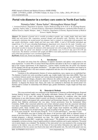
- 2. Portal vein diameter in a tertiary care centre in North-East India DOI: 10.9790/0853-14121110113 www.iosrjournals.org 111 | Page excluded from measurements. The diameter was taken in milimeter. The measurements were obtained by calculating the average of three times measurements. Fig 1: Showing portal vein diameter (D2) just before the bifurcation of the portal vein anterior to inferior vena cava. A single radiologist took all the measurements to avoid inter-observer variation. Sonographic measurements were done by the same examiner and were repeated for three times. All the individual records were entered in the in the master chart with full confidentiality. Statistical calculation was done with SPSS version 20. III. Results The present study was conducted among 49 (45.37%) males and 59 (54.63%) females. The participants were divided among five groups according to their age as mentioned. Table 1: Showing the group wise gender distribution of participants. Parameters Group Total I II III IV V Male 10 12 15 09 03 49 Female 24 22 07 04 02 59 Total 34 34 22 13 05 108 Percentage (%) 31.48 31.48 20.37 12.04 4.63 100 Table 2: Showing mean and standard deviation of body parameters and portal vein diameter. Parameters Parameters (Mean & Standard Deviation) Age (years) Weight (Kg) Height (meter) BMI= weight/height 2 Portal vein diameter (mm) Adults total (n=108) 42.30±18.01 54.22±1.07 157±8.67 21.75±2.95 8.83±2.12 Males (n=49) 48.24±18.32 58.87±1.06 163.14±7.19 22.04±1.33 9.17±2.33 Females (n=59) 37.36±16.30 50.36±9.25 153±6.72 21.51±3.15 8.55±1.90 Table 3: Showing mean and standard deviation of body parameters and portal vein diameter in Groups I to V. Parameters Parameters (Mean & Standard Deviation) Age (years) Weight (Kg) Height (meter) BMI= Weight/height 2 Portal vein diameter (mm) Group I to V Group I 23.26±5.46 53.38±8.94 156.87±7.70 21.60±2.80 8.50±1.94 Group II 38.53±4.27 55.85±1.20 157.94±9.96 22.22±3.21 9.17±2.43 Group III 52.64±4.74 56.11±8.96 158.82±7.93 22.13±2.32 9.69±2.00 Group IV 69.69±5.41 51.38±1.42 155.69±9.09 20.89±3.43 7.88±1.23 Group V 80.60±3.29 47.87±8.87 155.80±1.20 20.12±3.26 7.48±1.92
- 3. Portal vein diameter in a tertiary care centre in North-East India DOI: 10.9790/0853-14121110113 www.iosrjournals.org 112 | Page Table 4: Mean and standard deviation in males of Groups I to V. Parameters (males) Parameters (Mean & Standard Deviation) Age (years) Weight (Kg) Height (meter) BMI = weight/height 2 Portal vein diameter (mm) Group I to V Group I 23.90±4.36 57.30±6.32 163.50±5.36 21.41±1.90 8.39±2.01 Group II 38.17±4.26 65.79±1.16 167.25±8.55 23.39±2.52 10.03±2.77 Group III 52.53±4.36 57.00±8.68 161.13±7.12 21.85±2.14 10.35±1.85 Group IV 70.44±5.48 56.78±1.38 160.22±6.49 21.91±3.54 7.68±1.13 Group V 81.67±2.89 52.00±8.89 164.33±3.51 20.03±4.46 6.93±2.40 Table 5: Mean and standard deviation in females of Groups I to V. Parameters (females) Parameters (Mean & Standard Deviation) Age (years) Weight (Kg) Height (meter) BMI= weight/height 2 Portal vein diameter (mm) Group I to V Group I 23.00±5.93 51.75±9.46 154.1±6.84 21.69±3.13 8.54±1.96 Group II 38.73±4.35 50.43±8.25 152.86±5.65 21.58±3.41 8.70±2.15 Group III 52.85±5.84 54.21±9.96 153.86±7.78 22.73±2.75 8.30±1.64 Group IV 68.00±5.60 39.25±4.19 145.50±4.20 18.59±1.86 8.35±1.49 Group V 79.00±4.24 41.50±4.95 143.00±2.83 20.25±1.62 8.30±0.99 Table 6: Correlation of portal vein diameters and body parameters. Body parameters Portal vein diameter Adults total r (p value) Males r (p value) Females r (p value) Age (years) 0.015 (0.880) -0.118 (0.421) 0.067 (0.615) Weight (Kg) 0.288 (0.003) 0.204 (0.160) 0.311 (0.017) Height (meter) 0.130 (0.179) 0.083 (0.570) 0.020 (0.879) BMI (Wt/Ht 2 ) 0.322 (0.001) 0.249 (0.085) 0.379 (0.003) p value < 0.05 is significant. IV. Discussion A cross sectional gray scale ultrasound assessment of portal vein in Ethiopian population was done, age of the subjects varied from 5 to 85 years. The mean diameter of portal vein was calculated as 10.0±1.8 mm (range 8.2 to 11.8). The results concluded that gender did not have any effect on the diameter of portal vein but with increasing age the diameter also increased.4,7 Subsequently, the same results also reported in a Nigerian population establishing the mean portal vein diameter as 11.45±1.45 mm and also concluding that the diameter varies with age but not with gender.3 A Doppler ultrasonic study which is a more advanced and accurate imaging technique was conducted in Iran on 37 healthy subjects. The age varied from 20-40 years and the mean portal vein diameter was calculated as 9.36 ± 1.65 mm.8 In the present study, mean portal vein diameter was 8.83±2.12 mm (range 4.10 to 16.80 mm). The portal vein diameters in males and females were 10.5 mm (9.8- 11.2 mm) and 8.3 mm (7.7-8.9 mm) respectively. There was a statistical significance in between the values of males and females (p =0.0001).8 In current study, the PVD diameters in males and females were 9.17±2.33 mm and 8.55±1.90 mm respectively. They ranged from 4.50 to 16.80 mm and 4.10 to 13.10 mm in males and females respectively. The mean diameter was more in males than females, though there were no statistical significant changes found in gender distribution. The mean diameter increased from 15 to 60 years but gradually
- 4. Portal vein diameter in a tertiary care centre in North-East India DOI: 10.9790/0853-14121110113 www.iosrjournals.org 113 | Page decreased after 60 years in males. But in females the diameter increased up to 60 years, after that remained almost similar. No statistical significant changes were found among the groups both in males as well as females. In the previous study, in males there was no correlation between portal vein diameter and age, indicating that PVD does not vary with the age. But, height had a correlation with portal vein diameter. With increase in height PVD proportionately increases. In females there is no correlation between portal vein diameter and age as well as height, indicating that portal vein diameter had not varied with the age and height. But sex has a correlation with the portal vein diameter.7 In this present study, portal vein diameter had changed significantly with increasing weight and BMI. In males, none of the body parameters were significantly correlated with portal vein diameter. But in females, though height was not correlated with the diameter but it was significantly correlated with increasing weight and BMI. V. Conclusion In this study, portal vein diameter in adult males and females were found within the normal range. The diameter correlated with weight and BMI in total adult population and females but in males none of the body parameters were correlated significantly. Therefore, portal vein diameter in this local population may provide a reference value for assessing variation of size for clinical practice in this set up. References [1]. Namasivayam S, Kalra MK, Small WC, Saini S. Liver: normal anatomy and examination techniques. In. Gore RM, Levine MS. Text book of gastrointestinal radiology. 3rd ed. Vol. 2. China: Saunders Elsevier; 2008. P. 1527-51. [2]. Wilson SR, Withers CE. The liver. In: Rumack CM, Wilson SR, Charboneau JW, editors. Diagnostic ultrasound. 3rd ed. Vol 1(Part II). St. Louis: Elsevier Mosby; 2005. p. 75-145. [3]. Anakwue AC, Anakwue RC, Ugwu AC, Nwogu UB, Idigo FU, Agwu KK. Sonographic evaluation of normal portal vein diameter in Nigerians. Euro J Sci Res 2009;36(1):114-7. [4]. Hawaz Y, Admassie D, Kebede T. Ultrasound assessment of normal portal vein diameter in Ethiopians done at Tikur Anbessa Specialized Hospital. East Cent Afr J Surg 2012 Mar/Apr;17(1):90-3. [5]. Cosgrove DO. Liver anatomy. In: Cosgrove D, Meire H, Dewbury K, Farrant P, editors. Clinical ultrasound a comprehensive text— abdominal and general ultrasound. Vol 1. Edinburgh: Churchill Livingstone; 1994. p. 227-42. [6]. Bosch J, Garcia-Pagan JC. Complication of cirrhosis. J Hepatol 2000;32(1 Suppl):141-56. [7]. Ravi Shankar.G Shailaja Shetty , Srinath.M.G , Roopa Kulkarni. Estimation of Portal Vein Diameter in co – Relation with the Age, Sex and Height of An Individual. Anatomica Karnataka 2011;5(2):13-6. [8]. Yazdi HR, Sotoudeh H. Assessment of Normal Doppler Parameters of Portal Vein and Hepatic Artery in 37 Healthy Iranian Volunteers. Iran J Radiol. 2006;4:213–216.