This article reviews 11 cases of pituitary adenomas that hemorrhaged or infarcted in association with cardiac surgery over a 13-year period. The patients were mostly middle-aged males undergoing coronary bypass surgery who presented postoperatively with neurological deficits. Diagnosis was confirmed via CT or MRI scans showing pituitary tumors with hemorrhage. Patients received steroids and hormonal therapy. Most underwent transsphenoidal hypophysectomy and survived with minimal or resolving neurological deficits, though one patient died after craniotomy and another after deferring surgery. The cause of pituitary tumors hemorrhaging during cardiac surgery remains unclear.
SPONTANEOUS CORONARY ARTERY DISSECTION IN A PRE- MENOPAUSAL WOMAN OCCURRING J...Apollo Hospitals
SCAD is a rare presentation of acute coronary syndrome(ACS) and clinically indistinguishable from
plaque rupture. It predominantly affects young women with
no traditional cardiovascular risk factors, especially during
the post-partum and pre-menopausal period [1-3]. The
aetiology of SCAD is multifactorial and complex. Optimal
treatment strategy for SCAD is not clearely defined.
Broken Heart Syndrome: A Stress Responseasclepiuspdfs
Takotsubo cardiomyopathy, also known as broken heart syndrome, stress cardiomyopathy, or apical ballooning syndrome, is described as a type of emotional or physical stress response that may mimic acute coronary syndrome (ACS) or myocarditis. It is a form of reversible left ventricular dysfunction with characteristic apical ballooning, contributing to its’ name, along with diagnostic proof on coronary catheterization or angiography of the absence of significant coronary artery stenosis classically expected in ACS. The damage seen is typically transient, appearing to completely resolve within months with very low percentage of long-term sequelae or recurrence.
Fabry Disease (FD), also known as Anderson-Fabry disease, is an inherited X-linked disorder characterized by the absence (in men) or defi ciency (in women) in α-galactosidase A, activity that causes a progressive accumulation of glycosphingolipids within lysosomes of cells in all the major organ systems and progressive organ damage that fi rst manifests in childhood or early adulthood. End Stage Renal Disease (ESRD) is a major cause of morbidity and premature mortality in FD. We present a male patient with FD who was transplanted with kidney from a living donor and had a sudden cardiac arrest on the 4th day after operation. We suggest detailed preoperative examination including coronary angiography, echocardiography for these patients and also a multidisciplinary care is required for perioperative management of FD patients.
Guillain - Barre syndrome after acute myocardial infarction: A rare presentat...Apollo Hospitals
The association of acute coronary syndrome with any immunological mediated polyradiculopathy like Guillain–Barré syndrome is very rare. We report such a rare association of acute myocardial infarction and Guillain–Barré syndrome. Our patient underwent primary angioplasty successfully, but developed respiratory failure while in hospital. While the difficulty in weaning off from ventilator a suspicion of neuromuscular disease was made. The further investigations, including nerve conduction study confirmed a diagnosis of Guillain–Barré syndrome. Despite treatment, the patient died secondary to multi-organ dysfunction. Our case is 4th reported in the literature without use of any thrombolytic agent for such association.
Possible causes of death (Multiorgan failure)
VT/VF intraoperatively
Acute on chronic Heart failur
Respiratory failure
Acute Liver failure
Acute renal failure
Sepsis with septic shock
Concern for Intestinal infarction
Guillain–Barré syndrome after acute myocardial infarction: A rare presentationApollo Hospitals
The association of acute coronary syndrome with any immunological mediated polyradiculopathy like Guillain–Barré syndrome is very rare. We report such a rare association of acute myocardial infarction and Guillain–Barré syndrome. Our patient underwent primary angioplasty successfully, but developed respiratory failure while in hospital. While the difficulty in weaning off from ventilator a suspicion of neuromuscular disease was made. The further investigations, including nerve conduction study confirmed a diagnosis of Guillain–Barré syndrome. Despite treatment, the patient died secondary to multi-organ dysfunction. Our case is 4th reported in the literature without use of any thrombolytic agent for such association.
A Mistake that has Hurt No One: Sinus Mistakusasclepiuspdfs
There are times when we, the health care providers make a diagnosis and plan to treat that condition accordingly. In the mean time, because of a second opinion or another specialist consult might change the diagnosis completely and therefore the mode of management could change drastically. Here we present a similar case scenario for work-up of chest pains changing the diagnosis and therefore the mode of treatment. However in this process the patient did not get hurt (“Sinus Mistakus”).
Global Hospitals’ Advanced Heart, Lung & Vascular Institute provides all kinds of endovascular procedures including coronary intervention and peripheral intervention, heart surgery, heart bypass surgery as well as heart transplantation surgery in Hyderabad, Chennai, and Bangalore
ASSESSMENT AND PLANNING GUIDE FOR USE IN THE HOSPITALThe followi.docxgalerussel59292
ASSESSMENT AND PLANNING GUIDE FOR USE IN THE HOSPITAL
The following information should be included daily as it applies to your patient.
Demographic DataDate of AdmissionVital Signs
39 y/o African American male
10/28/18
BP: 115/60. Pain: 2
P: 91
T: 98.2.
RR: 22
SP02: 95
Significant Past Medical HistoryAllergies/Reactions
HTN, Hyperlipemia, Diabetes
NKA
Reason for Hospitalization and Current Diagnosis
Current Diagnosis: Acute Embolic Stroke, Cerebral Edema, R Hemiparesis, Pneumonia
Reason for hospitalization: 38 y/o male with a history of HTN presented with onset Right Sided Weakness and confusion at 11pm on 10-27-18 when he went to sleep. He woke up at 3am and he was talking gibberish to his fiancé. He went back to sleep and 2 hours later his symptoms had worsened. On 10-28-18, EMS was called by his fiancé and he was taken to the ER. His fiancé said he had taken “something” possibly cocaine. Patient was diagnosed with Acute Embolic Stroke, Cerebral Edema, R Hemiparesis and recently Pnuemonia.
Describe thepathophysiologyincluding signs, symptoms and incidence; and compare with patient findings:
· Acute Embolic Stroke:
Pathophysiology: Occurs when a blood clot that forms somewhere elsewhere in the body breaks loose and then travels to the brain through the bloodstream. The clot can lodge in an artery and blocks the flow of blood.
Common symptoms:Difficulty speaking or understanding words, numbness and tingling, temporary paralysis, blurred vision or blindness, slurred speech, dizziness, feeling faint, difficulty swallowing, nausea, sleepiness. Embolic stroke doesn’t cause any unique symptoms
Muscular symptoms: Difficulty with coordination, stiff muscles, feelings of weakness on one side or all of the body.
Cognitive symptoms: Mental confusion, an altered level of consciousness, visual agnosia
Patient Findings: Patient presented with R hemiparesis, facial drooping, slurred speech, difficulty swallowing.
· Cerebral Edema
Pathophysiology: It’s a life threatening condition that causes fluid to develop in the brain.
This fluid increases the pressure inside of the skull causing intracranial pressure (ICP). Increased ICP can reduce brain blood flow and decrease the oxygen your brain receives. The brain needs an uninterrupted flow of oxygen to function properly.
Symptoms: Headache, dizziness, nausea, lack of coordination, numbness, mood changes, memory loss, difficulty speaking, incontinence, change in consciousness, seizures, weakness in extremities
Patient Findings: Patient presented with difficulty speaking, incontinence, change in consciousness, weakness in extremities
· Hemiparesis
Pathophysiology: Hemiparesis is weakness on one side of the body. One side can still move but with reduced muscular strength.
Symptoms: Difficulty walking, standing, and maintaining your balance. You may also have numbness or tingling on your weaker side.
Patient findings: Patient has right sided weakness.
· Pneumonia
.
CHAPTER 1 SEMESTER V - ROLE OF PEADIATRIC NURSE.pdfSachin Sharma
Pediatric nurses play a vital role in the health and well-being of children. Their responsibilities are wide-ranging, and their objectives can be categorized into several key areas:
1. Direct Patient Care:
Objective: Provide comprehensive and compassionate care to infants, children, and adolescents in various healthcare settings (hospitals, clinics, etc.).
This includes tasks like:
Monitoring vital signs and physical condition.
Administering medications and treatments.
Performing procedures as directed by doctors.
Assisting with daily living activities (bathing, feeding).
Providing emotional support and pain management.
2. Health Promotion and Education:
Objective: Promote healthy behaviors and educate children, families, and communities about preventive healthcare.
This includes tasks like:
Administering vaccinations.
Providing education on nutrition, hygiene, and development.
Offering breastfeeding and childbirth support.
Counseling families on safety and injury prevention.
3. Collaboration and Advocacy:
Objective: Collaborate effectively with doctors, social workers, therapists, and other healthcare professionals to ensure coordinated care for children.
Objective: Advocate for the rights and best interests of their patients, especially when children cannot speak for themselves.
This includes tasks like:
Communicating effectively with healthcare teams.
Identifying and addressing potential risks to child welfare.
Educating families about their child's condition and treatment options.
4. Professional Development and Research:
Objective: Stay up-to-date on the latest advancements in pediatric healthcare through continuing education and research.
Objective: Contribute to improving the quality of care for children by participating in research initiatives.
This includes tasks like:
Attending workshops and conferences on pediatric nursing.
Participating in clinical trials related to child health.
Implementing evidence-based practices into their daily routines.
By fulfilling these objectives, pediatric nurses play a crucial role in ensuring the optimal health and well-being of children throughout all stages of their development.
Global launch of the Healthy Ageing and Prevention Index 2nd wave – alongside...ILC- UK
The Healthy Ageing and Prevention Index is an online tool created by ILC that ranks countries on six metrics including, life span, health span, work span, income, environmental performance, and happiness. The Index helps us understand how well countries have adapted to longevity and inform decision makers on what must be done to maximise the economic benefits that comes with living well for longer.
Alongside the 77th World Health Assembly in Geneva on 28 May 2024, we launched the second version of our Index, allowing us to track progress and give new insights into what needs to be done to keep populations healthier for longer.
The speakers included:
Professor Orazio Schillaci, Minister of Health, Italy
Dr Hans Groth, Chairman of the Board, World Demographic & Ageing Forum
Professor Ilona Kickbusch, Founder and Chair, Global Health Centre, Geneva Graduate Institute and co-chair, World Health Summit Council
Dr Natasha Azzopardi Muscat, Director, Country Health Policies and Systems Division, World Health Organisation EURO
Dr Marta Lomazzi, Executive Manager, World Federation of Public Health Associations
Dr Shyam Bishen, Head, Centre for Health and Healthcare and Member of the Executive Committee, World Economic Forum
Dr Karin Tegmark Wisell, Director General, Public Health Agency of Sweden
Leading the Way in Nephrology: Dr. David Greene's Work with Stem Cells for Ki...Dr. David Greene Arizona
As we watch Dr. Greene's continued efforts and research in Arizona, it's clear that stem cell therapy holds a promising key to unlocking new doors in the treatment of kidney disease. With each study and trial, we step closer to a world where kidney disease is no longer a life sentence but a treatable condition, thanks to pioneers like Dr. David Greene.
CRISPR-Cas9, a revolutionary gene-editing tool, holds immense potential to reshape medicine, agriculture, and our understanding of life. But like any powerful tool, it comes with ethical considerations.
Unveiling CRISPR: This naturally occurring bacterial defense system (crRNA & Cas9 protein) fights viruses. Scientists repurposed it for precise gene editing (correction, deletion, insertion) by targeting specific DNA sequences.
The Promise: CRISPR offers exciting possibilities:
Gene Therapy: Correcting genetic diseases like cystic fibrosis.
Agriculture: Engineering crops resistant to pests and harsh environments.
Research: Studying gene function to unlock new knowledge.
The Peril: Ethical concerns demand attention:
Off-target Effects: Unintended DNA edits can have unforeseen consequences.
Eugenics: Misusing CRISPR for designer babies raises social and ethical questions.
Equity: High costs could limit access to this potentially life-saving technology.
The Path Forward: Responsible development is crucial:
International Collaboration: Clear guidelines are needed for research and human trials.
Public Education: Open discussions ensure informed decisions about CRISPR.
Prioritize Safety and Ethics: Safety and ethical principles must be paramount.
CRISPR offers a powerful tool for a better future, but responsible development and addressing ethical concerns are essential. By prioritizing safety, fostering open dialogue, and ensuring equitable access, we can harness CRISPR's power for the benefit of all. (2998 characters)
Deep Leg Vein Thrombosis (DVT): Meaning, Causes, Symptoms, Treatment, and Mor...The Lifesciences Magazine
Deep Leg Vein Thrombosis occurs when a blood clot forms in one or more of the deep veins in the legs. These clots can impede blood flow, leading to severe complications.
ICH Guidelines for Pharmacovigilance.pdfNEHA GUPTA
The "ICH Guidelines for Pharmacovigilance" PDF provides a comprehensive overview of the International Council for Harmonisation of Technical Requirements for Pharmaceuticals for Human Use (ICH) guidelines related to pharmacovigilance. These guidelines aim to ensure that drugs are safe and effective for patients by monitoring and assessing adverse effects, ensuring proper reporting systems, and improving risk management practices. The document is essential for professionals in the pharmaceutical industry, regulatory authorities, and healthcare providers, offering detailed procedures and standards for pharmacovigilance activities to enhance drug safety and protect public health.
R3 Stem Cells and Kidney Repair A New Horizon in Nephrology.pptxR3 Stem Cell
R3 Stem Cells and Kidney Repair: A New Horizon in Nephrology" explores groundbreaking advancements in the use of R3 stem cells for kidney disease treatment. This insightful piece delves into the potential of these cells to regenerate damaged kidney tissue, offering new hope for patients and reshaping the future of nephrology.
Pituitary Adenomas Complicating Cardiac Surgery Summary and Review of 11 Cases.pdf
1. 125
Pituitary Adenomas Complicating
Cardiac Surgery: Summary and Review
of 11 Cases
Michael B. Pliam, M.D., Ph.D., Michael Cohen, M.D.,* Leo Cheng, M.D.,**
Matthias Spaenle, M.S.,*** Merrill H. Bronstein, M.D,t and
Thomas W. Atkin,M.D.*
Department of Cardiovascular Surgery, San Francisco Heart Institute, Seton
Medical Center, *Departments of Neurology and **Neurosurgery, Seton Medical
Center, ***Department of Neuropathology, Universitaet Bonn, Bonn, Germany,
t Clinical Associate Professor of Surgery, University of California San Francisco,
and *Department of Radiology, Seton Medical Center, Daly City, California
ABSTRACTFrom the literature and our own experience, 11 cases of hemorrhage or infarction
of a pituitary adenoma associated with cardiac surgery have been identified over a 13-year
period. Males outnumbered females by 10 to 1. Symptoms observed were headache, lethargy,
confusion, obtundation, unilateral ptosis, meiosis, and opthalmoplegia involving cranial
nerves 111, IV, and VI, visual field deficits, and hemiparesis. Diagnosis in most recent cases has
been confirmed with computerized tomography or magnetic resonance imaging. All patients
received adrenocortical steroid therapy initially. Eight patients underwent transsphenoidal hy-
pophysectomy and all survived. One patient underwent decompression craniotomy and died.
lntracranial surgery was deferred in 1 patient who survived and in another who died of a mas-
sive stroke. Residual neurological deficits were noted to be either absent, minimal, or resolv-
ing in 7 of the 9 patients who survived their initial hospitalization. While numerous mecha-
nisms have been proposed to explain the hemorrhage and necrosis of a pituitary adenoma
during heart surgery, no direct cause has been clearly identified. Surgical treatment is com-
monly necessary since untreated pituitary apoplexy is often fatal. Transsphenoidal hypo-
physectomy with decompression is the preferred method of treatment with a low perioperative
mortality and fairly good long-term prognosis. (J Card Surg 7995;70:
725-732)
A number of reports of hemorrhagic necrosis
of a pituitary adenoma complicating cardiac
surgery have appeared since 1980.1-6While
this so-called “pituitary apoplexy syndrome”
has been widely described, the incidence of as-
sociation with cardiac surgery and the precise
mechanism of pituitary tumor injury remains un-
The purpose of the present article is to
provide further insight into the diagnosis and
Address for correspondence: Michael B. Pliam, M.D., San
Francisco Heart Institute, Seton Medical Center, 1900 Sul-
livan Ave., Daly City, CA 94015. Fax: (415) 992-8388.
treatment of this condition in the cardiac surgi-
cal patient, to provide guidelines for optimal
cardiac surgical management of patients with
known pituitary tumors, and to add our own ex-
perience to the literature.
MATERIALAND METHODS
A thorough review of the literature dealing
with adenomas of the pituitary gland occurring
in cardiac surgical patients was accomplished
with the aid of a computerized search of the
National Library of Medicine’s Medline data-
2. 126 PLIAM, ET AL.
PITUITARY ADENOMA
J CARD SURG
1995;10:125-132
base including backfiles to 1966. Relevant cita-
tions in all languages were considered. Nine
cases were collected which fit these criteria: (1)
there was perioperative documentation of a pi-
tuitary adenoma by computerized axial to-
mography (CT) or examination of a resected
surgical specimen; (2) the patient underwent
cardiac surgery utilizing cardiopulmonary by-
pass; and (3) the patient developed an acute
neurological deficit in the early postoperative
period, either in part or entirely attributable to
induction of a pathological process involving an
adenomatous pituitary gland. While there are
numerous reports of spontaneous pituitary
hemorrhage and infarction due to a variety of
inciting causes, only nine cases that strictly fit
the above criteria have been included in the
present study, in addition to the two cases re-
ported here.
Each case was tabulated by author, year,
age, sex, cardiac surgical procedure, symp-
toms, neurological findings, diagnostic meth-
ods, treatment, outcome, residual neurological
deficits at follow-up, and histopathology when it
was available.
Case report 1
A 77-year-old male physician with a history of
moderate hypertension, glaucoma, and several
months of chronic cough, but no previous neu-
rological problems, developed severe progres-
sive angina over several months. Cardiac
catheterization was performed at Seton Medi-
cal Center on May 2, 1986, which demon-
strated diffuse three vessel coronary artery dis-
ease, a moderate inferior segmental wall-mo-
tion abnormality with preserved left ventricular
function, an estimated ejection fraction of 70%,
and mild pulmonary artery hypertension. During
the study, he developed intractable chest pain
and ST segment changes consistent with acute
ischemia. An intra-aortic balloon pump (IABP)
was inserted and he was taken urgently to the
operating room where he underwent a triple
coronary bypass procedure. The left internal
thoracic artery (LITA) was used to ljypass the
left anterior descending (LAD) and saphenous
vein grafts (SVG) used to bypass the diagonal
and obtuse marginal (OM) coronary arteries.
During the initial postoperative period, the
patient was hypotensive and required IABP
support, dopamine and epinephrine were to
maintain a systolic pressure of 80 to 90 mmHg.
The IABP was removed on the second postop
day and he remained stable with atrial flutter
the predominant rhythm. While his hemody-
namics stabilized, he required continued venti-
latory support. The chest X-ray showed moder-
ate left-sided atelectasis.
Twenty-four hours following surgery his sen-
sorium remained depressed; he seemed un-
able to follow commands and was at times agi-
tated. Further, he was noted to have a right
ptosis, unreactive meiosis, complete ophthal-
moplegia involving the right eye, and very mild
left hemiparesis. By the third postop day, he
was moving his left side much better and was
following occasional simple commands. Com-
plete ophthalmoplegia persisted. He was
started on dexamethasone (4 mg IV). Vascular
ultrasonography showed normal carotid and
opthalmic arteries and internal jugular veins. A
CT scan of the head performed on May 6 re-
vealed a pituitary mass with sellar invasion. At
this time, TSH was less than 1.0 pU/mL with
normal free thyroxine index, T4 and T3 uptake.
BUN was 22 mg/dL, and creatinine 3.2 mg/dL.
On May 6, he remained intubated but arous-
able with good left ventricular function and mild
renal failure. He was transferred to the Univer-
sity of California San Francisco Medical Center
where neurosurgical evaluation confirmed the
impression that the patient had sustained hem-
orrhage into a pituitary tumor. He remained
minimally responsive with persistent left hemi-
paresis and some degree of cardiopulmonary
dysfunction. He was not felt to be a candidate
for pituitary surgery. Rehabilitation proceeded
slowly and he died almost 3 months after his
coronary bypass surgery, on July 29, 1986. A
postmortem examination was not performed.
Case report 2
A 59-year-old man with unstable angina pec-
toris of several weeks duration and ECG evi-
dence of an old apical myocardial infarction
was admitted for cardiac catheterization. He
had had hepatitis 6 years earlier, with mild re-
sidual liver function abnormalities, and a history
of polio involving the right lower leg. A mild
ptosis of the right eyelid had been present for at
least 3 months and perhaps from the time that
3. J CARD SURG
1995;10:125-
132
PLIAM, ET AL. 127
PITUITARYADENOMA
ocular surgery for retinal detachment had been
done 1 year earlier. The patient had experi-
enced progressive impotence over the preced-
ing 2 years. He denied any changes in hand or
foot size and he appeared somewhat hypothy-
roid; the hair quality was normal as was the bal-
ding pattern.
Catheterization performed on the day of ad-
mission revealed: total occlusion of the proxi-
mal LAD coronary artery, with distal filling of the
LAD retrograde by collaterals from the right
coronary system; a 50% narrowing of the small
posterolateral branch of the right coronary ar-
tery; and moderate hypokinesis of the entire
anterior wall, apex, and septum.
Two days after admission, the patient under-
went single coronary bypass surgery, consist-
ing of an LlTA bypass to the LAD. The immedi-
ate postoperative course was smooth, but, on
the second postoperative day, he presented
with constant, holocranial headache,which was
worse with standing and motion, and episodes
of vomiting. Treatment with Reglan did not re-
lieve the symptoms.
Neurological evaluation revealed no Kernig
or Brudzinski signs, a completely normal men-
tal status, a partial right third cranial nerve
palsy characterized by right ptosis, moderate
vertical ophthalmoparesis, and very mild medial
rectus dysfunction. Superior oblique and lateral
rectus muscles appeared intact. Pupillary reac-
tion and the remainder of the cranial nerve ex-
amination were normal. Motor and sensory ex-
amination revealed no deficits, and reflexes
were symmetrical. A head CT scan was per-
formed, which was interpreted as negative. A
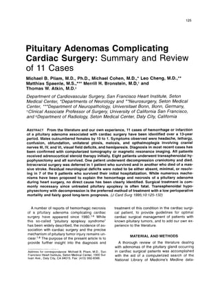
magnetic resonance imaging (MRI) scan 3
days later showed a pituitary adenoma with su-
prasellar extension and a hemorrhagic center
(Fig. 1). Transsphenoidal hypophysectomy
(TSSHX) was performed 1 week later to re-
move the necrotic papillary pituitary adenoma.
He was discharged 4 days later on medications
which included Synthyroid 100 micrograms/day
and prednisolone 10 mg/day.
RESULTS
Table 1 summarizes pertinent data obtained
from 9 cases collected from the literature and 2
cases from the present authors. Age ranged
from 55 to 77 years (mean 61.7 years). There
were 10 males and 1 female. Eight patients had
a coronary artery bypass graft (CABG) proce-
dure, 1 an aortic valve replacement (AVR), 1 a
mitral valve replacement (MVR), and 1 a dou-
ble valve replacement (AVR + MVR). Only 1 pa-
tient (case 5) was known precardiac surgery to
have a pituitary tumor diagnosed by CT and
was being treated with hormonal replacement
the rap^.^ Another (case 8) had severe head-
aches preop, and 1 patient (case 6) was hy-
pothyroid. One patient (case 11) had ptosis and
progressive impotence but the presence of a pi-
tuitary tumor was unknown preoperatively. The
remaining 9 patients were asymptomatic preop-
eratively with respect to neurological and endo-
crine abnormalities, and their pituitary tumors
were undiagnosed.
The postoperative symptoms most often ob-
served were unilateral ptosis, meiosis, and op-
thalmoplegia involving cranial nerves Ill, IV,
and VI, most often on the side corresponding to
extrasellar extension of the pituitary tumor as
could be visualized by the CT scan. Headache
was present in 4 patients, visual field deficits in
4 patients, and hemiparesis in 4 patients. Leth-
argy, confusion, or obtundation was present in
3 patients.
Diagnosis was made with the aid of CT scan
in all of the 11 patients, however, other useful
modalities included spinal tap, skull X-rays, ca-
rotid and cerebral angiography, and MRI.
Treatment included initial adrenocortical ster-
oid therapy for all patients. Hormonal therapy,
generally in the form of thyroxine, was used to
treat 4 patients, 2 of which had known endo-
crine dysfunction preoperatively. Eight patients
underwent TSSHX, and all of these survived.
One patient (case 9) underwent a decompres-
sion craniotomy with intracapsular excision of
the pituitary tumor and died shortly afterward of
cardiac failure. lntracranial surgery was de-
ferred in one patient who survived (case 1) and
in another who died of a massive stroke (case
Residual neurological deficits were noted to
be either absent, minimal, or resolving in 7 of
the 9 patients who survived their initial hospi-
talization and were seen in follow-up from 3
weeks to 2 months later. Of the remaining sur-
vivors, one had a persistent ophthalmoplegia
and panhypopituitarism and another had a per-
sistent hemiparesis (cases 8 and 7).
10).
4. 128 PLIAM. ET AL.
PITUITARYADENOMA
J CARD SURG
1995;10:125-132
Figure 1. Sagittal (top) and coronal (bottom) magnetic resonance images (700/30)
show large pituitary ade-
noma with suprasellar extension and protrusion into sphenoid sinus. Note increased signal intensity indicating
hemorrhage (arrows).
5. J CARD SURG
1995:10:125-132
PLIAM, ET AL. 129
PITUITARY ADENOMA
TABLE 1
Pituitary Adenomas Complicating Cardiac Surgery
Case Author/ Pro-
No. Year Age Sex cedure Symptoms Diagnosis Treatment Outcome Residuae Pathology
1
2
3
4
5
6
7
8
9
10
11
Peck' 68 M AVR Hdac, Leth, spinaltap,
1980 Conf, Obt X-ray, CT
Slavin' 57 M CABG Meo, Pto, CT, Angio
1984 O P k i
- 55 M MVR Meo, Pto, CT, Angio
Opleg, VFDef,
Hemip
Coope? 63 M CABG Meo, Pto, CT, Angio
1986 Opleg
- 62 M AVR + Meo, Pto, preop CT,
MVR Opleg, VFOef, pstop CT
prev Php
Opleg, prev
Hypothyr
- 55 M CABG Meo, Pto, CT
Khardori4 62 M CABG Meo, Pto, Opleg, CT x 2,
1987 VFDef, Hemip, Angio
Conf, fever
Shapiro5 60 F CABG Meo, Pto, Opleg, CT, MRI
1990 prev Hdac
Absalod 61 M CABG Hdac, nausea, C l
1993 Hemip, VFOef
Pliam* 77 M CABG Meo, Pto, Opleg, C1
1995 Conf, Hemip
- 59 M CABG Hdac, nausea, CT, MRI
Meo, Pto, Opleg,
prev Pto + Impot
steroids, survived none not available
no surg? in 3 wks
steroids, survived some eye sxs necrotic pituitary adenoma
TSSHX no major neuro
steroids, survived progressive hemorrhagic, chromophobe
TSSHX improvement adenoma
steroids, survived eye resolved necrotic hemorrhagic,
TSSHX in 2 mos chromophobe adenoma
hormones, survived resolving in sclerosing pituitary
TSSHX 3 wks adenomawith calcium
and hemorrhage
steroids, survived eye sxs part. pituitary adenoma,
hormones, resolving in no hemorrhage,
TSSHX 2 mos no necrosis
steroids, survived L hemipar., hemorrhagic infarcted,
TSSHX no vis. field pituitary adenoma
defect
steroids, survived panhypopit., hemorrhage, inflammation,
hormones, persist 111 nerve pituitary tissue
TSSHX palsy by immunostains
craniotomy died deceased, chromophobe adenoma
cardiac death with hemorrhage
steroids died deceased, not available
stroke
steroids, survived minimal eye necrotic papillary
hormones, pituitary tumor
TSSHX
Conf = confusion; Hdac = headache; Hemip = hemiparesis; lmpot = impotence; Leth = lethargy; Meo = meoisis; Obt = ob-
tunded; Opleg = ophlamoplegia; Php = panhypopituitarism; Pto = ptosis; VFDef = visual field defect; AVR = aortic valve
replacement; CABG = coronary artery bypass graft; MVR = mitral valve replacement; CT = computerized tomography;
MRI = magnetic resonance imaging; TSSHX = transsphenoidal hypophysectomy. 'Refers to present study.
Histopathology reports were available from 9
patients who underwent pituitary resection. In
DISCUSSION
each case the tumor was identified as a pitui-
tary adenoma. There were 3 chromophobe
Epidemiological perspective
adenomas, 1 papillary adenoma, and 5 tumors In a recent review, Molitch and Russellg sum-
labeled simply adenomas. Necrosis and in- marized autopsy findings in 9737 patients,
farction was noted in 5, hemorrhage in 6, but none of whom was suspected of having pitui-
neither hemorrhage nor necrosis was seen in tary disease while alive and underwent routine
the tumor of 1 patient (case 6). postmortem examination of the pituitary gland
6. 130 PLIAM, ET AL.
PITUITARY ADENOMA
J CARD SURG
1995;lO:
125-132
by sectioning. Pituitary adenomas were identi-
fied in 1065 (10.9%) of these subjects, the vast
majority of these being microadenomas, less
than 10 mm in diameter. The tumors were dis-
tributed equally throughout the age groups and
between the sexes. Only three (0.03%) of
these tumors were macroadenomas, that is,
greater than 10 mm in diameter. Thus a justifi-
able estimate of the incidence of pituitary
macroadenomas in the general population is
about 30 per 100,000 people.
Over the past decade, there have been on
average about 250,000 cardiac surgical proce-
dures performed annually in the United States.
Based on the above autopsy incidence figures,
it is reasonable to assume that approximately
75 patients per year or 750 patients over the
past decade have undergone heart surgery
who have harbored an occult pituitary
macroadenoma. Since we have only identified
ten cases over the same decade, we might
conclude that the majority (99%) of the esti-
mated 750 patients either: (1) survived cardiac
surgery without pituitary-related problems; (2)
survived with subclinical or minimally sympto-
matic pituitary injury; (3) survived significant
perioperative pituitary injury with unrecognized
neurological and/or endocrine dysfunction; or
(4) died without the discovery or reporting of pi-
tuitary injury.
Hemorrhage, necrosis, and apoplexy
The syndrome described by Sheehan and
Summers consists of infarction in a nontumor-
ous pituitary gland following obstetric shock in
which hypopituitarism is the result. This never
results in visual dysfunction or ophthalmople-
gia.lOvll While hemorrhage and infarction have
been lumped together as “hemorrhagic in-
farction” to describe many of the cases of re-
ported pituitary injury, it seems importantto dis-
criminate between these two processes which
may occur independently through separate
mechanisms. Kovacs and YaoI2 examined the
pituitary glands of 33 patients who had died
within 10 days of major cardiac surgery. Five pi-
tuitary glands (15.2%) showed ischemic ne-
crosis resembling that seen after obstetric
shock, whereas similar pituitary abnormalities
were found in about 1% to 6% of unselected
autopsy material. These authors observed that
coagulative infarction was the basic histologic
abnormality that develops in the adenohypo-
physis of heart surgery patients. While the
mechanism of decreased blood flow to the an-
terior lobe was unclear, they suggested that
various factors such as microembolism, throm-
bosis, disseminated intravascular coagulation,
vasospasm, vascular compression, platelet ag-
gregation, release of vasoactive substances
from disintegrating leukocytes, and shock re-
mained as plausible explanations. Our present
study reveals that 2 of 9 patients reviewed had
only pituitary adenoma necrosis without hemor-
rhage, lending credence to the concept that the
primary injury is that of necrosis and that hem-
orrhage occurs subsequently.
The sudden catastrophic bland or hemor-
rhagic infarction of a normal or neoplastic pitui-
tary gland may cause compression of struc-
tures adjacent to the sella with sudden loss of
visual acuity, a chiasmal field deficit, oculomo-
tor palsies, severe headache, decreased sen-
sorium, and hypopituitarism. The phenomenon
was first recognized by Baileyd3in 1898, and
was later called “pituitary apoplexy” by
Brougham et aI.l4 in 1950. Presently over 200
cases have been rep~rted.~
Varying degrees of hemorrhage and necrosis
of the adenohypophysis have been described
in association with many conditions other than
adenoma, including diabetes mellitus, arterial
hypertension, hypoparathyroidism, tuberculo-
sis, tetanus, cardiac failure, hemolytic crisis,
meningitis, temporal arteritis, and elevated in-
tracranial pressure. Usually pituitary enlarge-
ment is limited by the boundaries of an unex-
panded sella turcica so that compression of
parasellar structures does not occur and pitui-
tary apoplexy does not result. In reportedcases
where pituitary apoplexy has occurred, the lat-
eralization of signs can frequently be correlated
with the side of suprasellar expansion of ade-
noma and the severity of the clinical presenta-
tion is usually proportional to the size of the
original t ~ m o r . ~ , ~ ~ . ’ ~
Diagnosis
The differential diagnosis of a patient who
presents in the early postcardiopulmonary by-
pass period with depressed sensorium, unreac-
tive meiosis, partial or complete opthalmople-
7. J CARD SURG
1995;10:125-132
PLIAM, ET AL. 131
PITUITARY ADENOMA
gia, and some degree of hemiparesis includes:
(1) interference with the carotid circulation in
the cavernous sinus region; (2) ischemic lesion
of the oculomotor nerves in their cavernous
portion by occlusion of their nutrient arterial
supply; (3) cavernous sinus thrombosis; or (4)
a tumor of the parasellar region.
The occurrence of a relatively sudden and
profound alteration of consciousness associ-
ated with headache, cerebrospinal fluid find-
ings of a chemical meningitis, and secondary
adrenal insufficiency in a patient with skull films
and CT scan consistent with an intrasellar pitui-
tary tumor, suggested pituitary apoplexy. The
absence of bleeding on both CT scan and lum-
bar puncture suggested that the etiology of the
apoplexy was infarction rather than hemor-
rhage.
lntratumoral hemorrhage may occur without
clinical evidence of pituitary apoplexy. Areas of
hemorrhage can appear as low attentuation on
CT within the first 24 to 48 hours. MRI is more
sensitive than CT in assessing hemorrhage in
the subacute stage, and may help to differenti-
ate patients with cystic necrosis and bland in-
farction.’
Some of the less common clinical features
associated with pituitary apoplexy are the syn-
drome of inappropriate secretion of antidiuretic
hormone, hypotension, hypothermia, acute hy-
popituitarism, hemiplegia, and aphasia. Severe
hypopituitarism is uncommon, endocrine dys-
function variably present and demonstrable
only by provocative testing.’I8
The endocrine abnormalities associated with
pituitary apoplexy are those produced by the
adenoma or those resulting from the hypo-
physeal or hypothalamic damage secondary to
the hemorrhage. The hemorrhage may effec-
tively destroy the sellar contents and thus lead
to early hypopituitarism, which may respond to
steroid administration with improvement in the
level of consciousness and reversal of the arte-
rial hypotension, which sometimes is wrongly
attributed to hypothalamic dysfunction. Diabe-
tes insipidus, either transient or permanent, is
surprisingly a rather rare sequela of pituitary
apoplexy. The review by Veldhuis and Ham-
mond18 disclosed an incidence of 4% for tran-
sient and 2% for persistent diabetes insipidus
after apoplexy. Perhaps the neurohypophysis is
spared by the apoplexy or, alternatively,
enough stalk remains to permit the secretion of
adequate amounts of antidiuretic hormone
(ADH). The neurohypophysis has a separate
blood supply and is not infarcted after interrup-
tion from the portal system. It may be partially
preserved after apoplexy has destroyed the
adenohyp~physis.~
Management
Untreated pituitary apoplexy is often fatal.
Steroid replacement has been beneficial, but
still the mortality rate has been high. In 1957,
Uihlein et aI.l9 reviewed the literature and re-
ported that about two thirds of the patients
died. On the other hand, well-documented ac-
counts of the spontaneous cure of an ade-
noma, with reversal of endocrinopathy or im-
provement of ocular signs, have been re-
p ~ r t e d . ~
Not every patient requires emergent surgical
decompression after pituitary apoplexy. While
spontaneous recovery has been reported, the
clinical course is somewhat difficult to predict.
Patients with severe visual or mental impair-
ment or those with progressive deterioration re-
quire urgent surgical decompression. Ophthal-
moplegia is not an absolute surgical indication
and may disappear spontaneously, but, recov-
ery of vision is more dependent upon timely de-
compression than upon the severity of the in-
itial visual deficit. Vision is less likely to recover
than the disturbances of ocular motility. Most
patients will require long-term hormonal re-
placement. Recurrent episodes of apoplexy
may occur, but are less likely in patients who
have undergone operative decompre~sion.~
Some questions remain regarding the opti-
mal management of a patient with a known pi-
tuitary macroadenoma who requires cardiac
surgery. To the best of our knowledge, no op-
erative decompression of a pituitary tumor in
anticipation of cardiac surgery has ever been
reported. Only one patient (case 5) in this re-
view was known to have a pituitary adenoma
prior to cardiac surgery. That patient underwent
a double valve replacement and subsequently
sustained pituitary apoplexy which was suc-
cessfully treated by transsphenoidal decom-
pression hypophysectomy. However, the
epidemiological analysis presented here
strongly suggests that the majority of patients
8. 132 PLIAM, ET AL.
PITUITARYADENOMA
J CARD SURG
1995;10:125-132
who undergo cardiac surgery in the presence of
a pituitary macroadenoma probably remain as-
ymptomatic. Certainly, patients with known pi-
tuitary tumors should be treated with steroids
and appropriate hormonal replacement therapy
and be monitored expectantly for signs of in-
creased intracranial pressure and meningeal ir-
ritation throughout their postoperative course.
Should opthalmoplegia or other serious central
nervous system symptoms develop, transsphe-
noidal decompression should probably be per-
formed since this has been demonstrated to be
a safe and effective method of treatment.
CONCLUSION
Symptoms of severe headache, lethargy,
confusion, obtundation, unilateral ptosis, meio-
sis, opthalmoplegia involving cranial nerves Ill,
IV, and VI, visual field deficits, and hemiparesis
observed in patients following cardiac surgery
may indicate hemorrhage or infarction of a pi-
tuitary adenoma. The diagnosis can generally
be confirmed through CT or MRI scan. Treat-
ment of those with severe symptoms, particu-
larly if there has been an acute visual loss, can
be best accomplished by timely TSSHX decom-
pression. Operative mortality with the
transsphenoidal approach should be minimal.
The long-term outlook is generally good with
early resolution of neurological residuae, pro-
vided intervention is promptly undertaken. The
precise mechanism that triggers hemorrhage
and infarction of pituitary adenomas during car-
diac surgery remains unclear and suggests an
area for further investigation.
Acknowledgment: The authors wish to acknowledge the as-
sistance of John Volkert in the preparation of the figures.
REFERENCES
1. Peck V, Lieberman A, Pinto R, et al: Pituitary
apoplexy following open-heart surgery (case re-
port). NY State J Med 1980;80:641-643.
2.Slavin ML, Budabin M: Pituitary apoplexy asso-
ciated with cardiac surgery. Am J Opthalmol
3.Cooper DM, Bazaral MG, Furlan AJ, et al: Pitu-
tiary apoplexy: A complication of cardiac sur-
gery. Ann Thorac Surg 1986;41:547-550.
4.Khardori R, Bussing RC, Burns GM, et al: Car-
diac bypass surgery with haemorrhagic endo-
crine sequelae. Postgrad Med J 1987;63:489-
492.
5. Shapiro LM: Pituitary apoplexy following coro-
nary artery bypass surgery. J Surg Oncol 1990;
6.Absalom M, Rogers KH, Moulton RJ, et al: Pitui-
tary apoplexy after coronary artery surgery. An-
esth Analg 1993;76:648-649.
7.Cardoso ER, Petersen EW: Pituitaryapoplexy: A
review. Neurosurgery 1984;14:363-373.
8.Rovit RL, Fein JM: Pituitary apoplexy: A review
and reappraisal. J Neurosurg 1972;37:280-288.
9. Molitch MR, Russell EJ: The pituitary “incidenta-
loma”. Ann Int Med 1990;112:925-931.
10.Sheehan HL: Post-partum necrosis of the ante-
rior pituitary. J Pathol Bacteriol 1937;45:189-
214.
11. Sheehan HL, SummersVK: The syndrome of hy-
popituitarism. Q J Med 1949;18:319-325.
12. Kovacs K, Yao J: Pituitary necrosisfollowing ma-
jor heart surgery. Z Kardiol 1975;64:52-57.
13.Bailey P: Pathological report of a case of ak-
romegaly, with special referenceto the lesions in
the hypophysis cerebri and in the the thyroid
gland: and 2 cases of hemorrhage into the pitui-
tary. Phila Med J 1898;1:789-792.
14.Brougham M, Huesner AP, Adams RD, et al:
Acute degenerative changes in adenomas of the
pituitary body: With special references to pitui-
tary apoplexy. J Neurosurg 1950;7:421-439.
15.Ostrov SG,Quencer RM, Hoffman JC, et al:
Hemorrhage within pituitary adenomas: How
often associated with pituitary apoplexy syn-
drome? Am J Roentgenol 1989;l
53:l53-160.
16.McCormick WF, Halmi NS: The hypophysis in
patients with coma depasse (“respirator brain”).
Am J Clin Pathol 1970;54:374-383.
17.Reid RK, Quigley ME, Yen SSC: Pituitary apo-
plexy: A review. Arch Neurol 1985;42:712-719.
18.Veldhuis JD, Hammond JM: Endocrine function
after spontaneous infarction of the human pitui-
tary: Report, review, and reappraisal. Endocr
Rev 198O;l:lOO-107.
19.Uihlein A, Balfour WM, Donavan PI: Acute hem-
orrhage into pituitary adenomas. J Neurosurg
1984;98:291-296.
44:66-68.
1957;14:140-151.