1. Structure
Article
Structural Investigation of Rimantadine Inhibition
of the AM2-BM2 Chimera Channel
of Influenza Viruses
Rafal M. Pielak,1 Kirill Oxenoid,1 and James J. Chou1,*
1Jack and Eileen Connors Structural Biology Laboratory, Department of Biological Chemistry and Molecular Pharmacology,
Harvard Medical School, Boston, MA 02115, USA
*Correspondence: chou@cmcd.hms.harvard.edu
DOI 10.1016/j.str.2011.09.003
SUMMARY
The M2 channel of influenza A is a target of the ada-
mantane family antiviral drugs. Two different drug-
binding sites have been reported: one inside the
pore, and the other is a lipid-facing pocket. A pre-
vious study showed that a chimera of M2 variants
from influenza A and B that contains only the pore-
binding site is sensitive to amantadine inhibition,
suggesting that the primary site of inhibition is inside
the pore. To obtain atomic details of channel-drug
interaction, we determined the structures of the chi-
meric channel with and without rimantadine. Inside
the channel and near the N-terminal end, methyl
groups of Val27 and Ala30 from four subunits form
a hydrophobic pocket around the adamantane, and
the drug amino group appears to be in polar contact
with the backbone oxygen of Ala30. The structures
also reveal differences between the drug-bound
and -unbound states of the channel that can explain
drug resistance.
INTRODUCTION
The M2 proteins of influenza A and B virus, AM2 and BM2,
respectively, are transmembrane (TM) proteins that tetramerize
in the viral membrane to form channel structures that selectively
transport protons across the membrane (Mould et al., 2003; Pa-
terson et al., 2003; Pinto et al., 1992; Sugrue and Hay, 1991). The
role of proton conduction by M2 is believed to equilibrate pH
across the viral membrane during cell entry and across the
trans-Golgi membrane of infected cells during viral maturation
(Hay et al., 1985; Helenius, 1992). Proton conductance depends
on the pH and pH difference across the membrane, and
the channel is essentially in a closed conformation at pH >7.5
(Pielak and Chou, 2010a; Wang et al., 1995). The transport
activity of AM2, but not BM2, can be blocked by the adamantane
family antiviral compounds, of which the amantadine and riman-
tadine were the first effective drugs licensed for influenza treat-
ment (Davies et al., 1964). The majority of the circulating virus
strains are now resistant to these drugs (Bright et al., 2006),
and at least six single mutations in the AM2 TM region have
been reported that confer drug resistance. Therefore, it is of
interest to obtain a precise picture of drug binding for under-
standing the mechanism of drug resistance and for developing
a next-generation antiflu compound that targets M2.
Recent structural characterizations of the channel domain of
AM2 have included solution NMR structures of the wild-type
AM2 (Schnell and Chou, 2008) and the drug-resistant mutants
S31N (Pielak et al., 2009) and V27A (Pielak and Chou, 2010b),
crystal structures of AM2 at different pH values (Khurana et al.,
2009; Stouffer et al., 2008), and backbone structures of AM2
derived from solid-state NMR measurements of proteins in lipid
bilayers (Cady et al., 2010; Sharma et al., 2010). Moreover, the
structure of the BM2 channel has also been determined by solu-
tion NMR methods (Wang et al., 2009). These structural models
show that a left-handed four-helical bundle forms the channel
pore, and that tetramerization of the four TM helices is further
stabilized by intermolecular contacts between C-terminal
amphipathic helices flanking the TM domain. The packing of
Trp41 indole rings creates a channel gate, which closes off the
C-terminal end of the pore. The imidazole rings of His37, which
are essential in transporting protons, are inside the pore.
Two different drug-binding sites have been reported, leading
to proposals for two different mechanisms of drug inhibition.
The structure of the TM domain of AM2 (residues 22–46) crystal-
lized in the presence of amantadine showed electron density in
the channel pore, near Ser31 (Stouffer et al., 2008), directly
blocking the channel passage near the N-terminal end of the
pore. However, the position and orientation of amantadine could
not be defined unambiguously by the relatively low-resolution
data (3.5 A˚ ) because the diameter of the roughly spherical ada-
mantane cage is $3.5 A˚ . The solution NMR structure of a longer
channel construct (residues 18–60) showed that rimantadine
binds near the C-terminal end of the channel to an external site
consisting of Trp41, Ile42, and Arg45 from one TM helix and
Leu40, Leu43, and Asp44 from the adjacent TM helix (Schnell
and Chou, 2008). If this were the site of inhibitory binding, the
mechanism would be allosteric: drug binding would stabilize
a closed conformation of the channel. Subsequent solid-state
NMR measurements using the TM domain reconstituted in lipids
confirmed the existence of both binding sites, and reported that
the site in the pore has greater affinity for the drug than the
external site (Cady et al., 2010).
Independent of the structural studies, a functional experiment
using an AM2-BM2 fusion protein provided probably the most
convincing resolution to the controversy. In the fusion protein
Structure 19, 1655–1663, November 9, 2011 ª2011 Elsevier Ltd All rights reserved 1655
2. the N-terminal half of the channel domain is from AM2 (drug
sensitive and contains the pore-binding site), and the C-terminal
half is from BM2 (drug insensitive and does not contain the
external binding site). It was reported that proton conduction of
this AM2-BM2 chimera could still be blocked by amantadine
and rimantadine, providing a compelling argument that the func-
tional binding pocket is located in the N-terminal half of the
channel pore (Jing et al., 2008; Ohigashi et al., 2009).
Inspired by the above functional experiment, we have carried
out a structural investigation of drug binding to the AM2-BM2
fusion protein. We find that a protein construct corresponding
to the TM region of the AM2-BM2 chimera, (AM2-BM2)TM, repro-
duces functional properties unique to the wild-type AM2 channel
in a liposomal proton flux assay. The (AM2-BM2)TM protein re-
constituted in detergent micelles can be used to record high-
quality NMR spectra, and addition of rimantadine causes large
chemical shift perturbations. The high-quality NMR data provide
an extensive set of unambiguous structural restraints, which we
have used to determine high-resolution structures of the
chimeric channel in the drug-bound and -unbound states. Our
results show that rimantadine indeed binds inside the pore, sup-
ported by specific hydrophobic and polar interactions with the
pore-lining residues of the channel, and that drug binding alters
the channel conformation.
RESULTS
The AM2-BM2 Construct for Characterizing
Rimantadine Binding
Both AM2 and BM2 have unstructured N-terminal segments, a
channel-forming TM helix, and a relatively long cytoplasmic
region (Lamb et al., 1985; Paterson et al., 2003). The residue
numbers of the TM helix are 26–46 for AM2 and 5–31 for BM2.
There is almost no sequence identity between the two proteins
except the conserved histidine (His37 in AM2 and His19 in
BM2) and tryptophan (Trp41 in AM2 and Trp23 in BM2) that
are necessary for proton selectivity and ion gating of the chan-
nels (Mould et al., 2003; Tang et al., 2002; Wang et al., 1995).
Whole-cell channel recording experiments showed that replac-
ing residues 1–19 of the full-length BM2 protein, which is insen-
sitive to amantadine, with residues 1–37 of AM2 introduced
a hybrid protein sensitive to drug inhibition (Jing et al., 2008; Ohi-
gashi et al., 2009). The result suggests that the primary site of
drug action is contained in the half of the TM domain N terminal
to the centrally located His37. To characterize drug binding to
the AM2-BM2 chimera, we constructed a fusion peptide corre-
sponding to the channel-forming region of the chimera that is
amenable to structural studies. This peptide contains residues
18–37 of AM2, followed by residues 20–34 from BM2. In this
study we refer to this peptide as (AM2-BM2)TM, and number
the residues (18–52) such that they are consistent with the
residue numbering for the native AM2 channel.
Functional Relevance of the (AM2-BM2)TM Construct
The (AM2-BM2)TM construct is not a natural sequence, and thus,
the relevance of using this peptide for understanding drug inhibi-
tion needs to be investigated. The proton conduction and drug
inhibition of (AM2-BM2)TM were tested using a liposomal proton
flux assay established earlier (Pielak et al., 2009). In this assay
a known quantity of channel protein was reconstituted into
liposomes. Proton conduction was initiated by the addition of
concentrated acid to the external solution under conditions of
rapid solution mixing and then followed as an increase in pH
of the external solution as protons move down the pH gradient
into the liposomes (Experimental Procedures). This experimental
setup mimics what occurs after endocytosis of the virus, as the
host cell acidifies the endosomal compartments. Using the lipo-
some assay, we first found that (AM2-BM2)TM has similar proton
conduction properties as the native AM2 and exhibits similar
drug sensitivity (Figures 1A and 1B). The chimera and the native
AM2 channels both showed approximately 10 H+
/s/channel
conduction rate, which is $95% inhibited with 50 mM rimanta-
dine (Figures 1A and 1B) (Pielak et al., 2009). We then used a
(AM2-BM2)TM peptide with the S31N mutation, which is a preva-
lent drug-resistance mutation that accounts for the majority of all
resistant viruses (Bright et al., 2006; Hay et al., 1985), to test
whether the mutant (AM2-BM2)TM also acquires drug resistance.
Indeed, the proton conductance of the S31N chimera was not
affected by 50 mM rimantadine (Figures 1C and 1D), similar to
what we found for the S31N mutant of AM2 in the same assay
(Pielak et al., 2009). These results indicate that channels formed
by the (AM2-BM2)TM peptide in liposomes are almost identical to
native AM2 in proton conduction, rimantadine inhibition, and
rimantadine resistance.
Functional Relevance of the (AM2-BM2)TM NMR System
For structural characterization of the chimera channel, we recon-
stituted the (AM2-BM2)TM peptide in dihexanoyl-phosphatidyl-
choline (DHPC) detergent micelles and found that the protein
runs on SDS-PAGE as a stable tetramer (see Figure S1 available
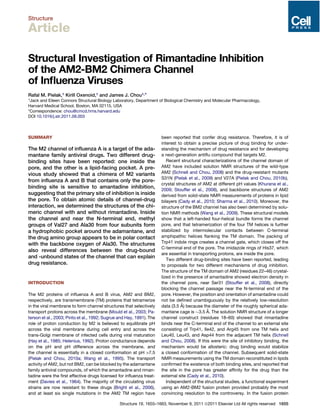
Figure 1. Functional Investigation of the (AM2-BM2)TM Channel by
the Liposomal Proton Flux Assay
(A) The conductance of the (AM2-BM2)TM channel exhibits similar conduc-
tance rate ($10 H+
/s/channel) as the wild-type AM2 channel (Pielak and Chou,
2010a).
(B) The conductance of (AM2-BM2)TM was reduced by $95% by the addition
of 50 mM rimantadine (rim).
(C) Introducing the S31N mutation to (AM2-BM2)TM did not affect its proton
conduction rate.
(D) However, the S31N mutation completely abolished inhibition by rimanta-
dine (rim).
Structure
Influenza, M2 Channel, Drug Binding, Structures
1656 Structure 19, 1655–1663, November 9, 2011 ª2011 Elsevier Ltd All rights reserved
3. online). The reconstituted tetramer generated very similar 1
H-15
N
TROSY-HSQC spectra in DHPC (see Experimental Procedures
for the full name) micelles and DMPC/DHPC bicelles (q = 0.3)
(Figure S2), suggesting that the overall conformation of the
protein is essentially the same in detergent as in a lipid environ-
ment. The NMR spectra also reflect partial structural similarities
between the chimera and the native AM2 and BM2 channels
because chemical shifts of the chimera residues that come
from the AM2 sequence (18–36) are similar to the corresponding
chemical shifts from AM2, and the same is true for those that
come from the BM2 sequence (38–52) (Figure S3).
We then tested whether the (AM2-BM2)TM tetramer exhibits
the functional properties of the native AM2 channel under the
NMR sample conditions. Upon addition of 50 mM rimantadine
to the (AM2-BM2)TM sample at pH 7.5, a new set of peaks
emerged in the TROSY-HSQC spectrum (Figures 2A and 2B).
The peaks corresponding to the drug-bound state have very
different chemical shifts for almost all residues (the largest
changes are around Ser31, $5 ppm in 15
N and $0.16 ppm in
1
H; and His37, $0.1 ppm in 15
N and $0.6 ppm in 1
H) (Figure 2B;
Figure S4). This result suggests that the drug binds specifically to
the chimeric channel in detergent micelles and that drug binding
may have induced substantial conformational change. We did
not detect spectral changes for the chimera with the S31N muta-
tion when adding the same amount of the drug to the sample
(Figures 2C and 2D), indicating that the chemical shift perturba-
tion in Figure 2B is specific to drug binding and that the NMR
system developed for the chimera is suitable for investigating
the mechanism of drug binding of the native AM2 channel.
At pH 7.5, the channel is essentially in a closed state (Pielak and
Chou, 2010a; Wang et al., 1995). We also investigated whether
the drug binds to the open state of the chimeric channel in
a more acidic buffer. At pH 6.1, the TROSY-HSQC spectrum of
(AM2-BM2)TM inDHPCmicelles(Figure2E)issignificantlydifferent
from that at pH 7.5 (Figure 2A). Addition of rimantadine also gener-
ates a new set of peaks (Figure 2F) that have similar chemical
shifts as those of the drug-bound closed state. At 50 mM rimanta-
dine and pH 6.1, the peaks corresponding to the drug-bound pop-
ulation are barely visible; even at 100 mM, they are $5-fold less
intense than those at pH 7.5. The results suggest that the drug
also binds to the open state, but with much lower affinity.
Location of the Drug-Binding Site in the Chimera
Channel
To determine the precise location of drug binding, we made
(AM2-BM2)TM peptide that is 15
N-labeled and 99.9% deuterated
at the nonlabile sites so that nuclear Overhauser enhancement
(NOE) between the protein backbone amide protons and drug
protons could be measured unambiguously. In the presence of
50 mM rimantadine, about 60% of the chimeric channels were
drug bound and 40% unbound, as judged by the relative intensi-
ties of the NMR peaks. In this case the NMR peaks of the popu-
lation not bound with drug served as an excellent internal-nega-
tive control for measuring protein-drug NOE. We recorded a 3D
15
N-edited NOESY spectrum with 250 ms NOE mixing and found
that the amide peaks of Ala30, Ser31, and Ile32 of the drug-
bound population showed intense NOE cross-peaks to the
adamantane CH2 and CH protons, whereas the peaks of the
unbound population did not (Figure 3, left panel). There was
also an NOE cross-peak between the amide of Gly17 and the
terminal methyl group (1.2 ppm) of rimantadine. Independently
of the 15
N-edited NOESY, we recorded a 3D 13
C-edited NOESY
(150 ms NOE mixing) using a uniformly 15
N- and 13
C-labeled and
nondeuterated protein for identifying NOE between the protein
methyl groups and the drug. In this spectrum we unambiguously
identified strong NOE cross-peak between the Val27 Cg1
H3
group and the adamantane CH2 group of rimantadine (Figure 3,
right panel). These NOE data indicate that the drug binds inside
the chimera channel near residue positions 27–31.
Solution Structures of the Chimera Channel
with and without Rimantadine
Using the NMR methods established previously for oligomeric,
channel-like proteins (Oxenoid and Chou, 2005; Schnell and
Figure 2. Functional Investigation of the (AM2-BM2)TM Channel by
NMR
The 1
H-15
N TROSY-HSQC spectra of the (AM2-BM2)TM (1.4 mM monomer)
reconstituted in DHPC are as follows: (A) at pH 7.5; (B) at pH 7.5 with 50 mM
rimantadine; (C) with the S31N mutation at pH 7.5; (D) same as in (C) but with
50 mM rimantadine; (E) at pH 6.1; and (F) at pH 6.1 with 100 mM rimantadine.
The red peaks correspond to resonances that have emerged upon addition of
rimantadine. Please also see Figure S1–S4.
Structure
Influenza, M2 Channel, Drug Binding, Structures
Structure 19, 1655–1663, November 9, 2011 ª2011 Elsevier Ltd All rights reserved 1657
4. Chou, 2008), we determined the high-resolution structures of
the (AM2-BM2)TM tetramer at pH 7.5 in the presence and
absence of rimantadine. Both structures were determined with
an extensive set of experimental restraints including intramolec-
ular and intermolecular distances derived from NOE, dihedral
angles from chemical shifts, and backbone bond orientations
from residual dipolar couplings (RDCs) (Table 1). For the drug-
bound structure the restraints also include 16 experimental inter-
hydrogen distances between the (AM2-BM2)TM tetramer and
rimantadine. In both cases (drug-bound and -unbound), the
structural restraints were used to generate an ensemble of 15
low-energy structures with rmsd of all heavy atoms smaller
than 1 A˚ (Figure 4A and Table 1).
In the absence of rimantadine, the (AM2-BM2)TM tetramer is a
left-handed four-helical bundle that spans about 33 A˚ in deter-
gent micelles (Figure 4B, left panel). The individual subunits
are a helical from residues 26 to 49, but the TM helices show a
bend ($17
) near His37. The AM2 and BM2 sequences fuse at
His37, and the helical bending may be necessary to satisfy the
different helix-helix packing angles in the AM2 and BM2 channel
assemblies (À21
in AM2; Schnell and Chou [2008]; and À37
in
BM2; Wang et al. [2009]). We also found that the AM2 and
BM2 regions of the chimera are structurally very similar to the
corresponding regions in the native AM2 and BM2 channels
(Figure 4B, right panel). A ring of methyl groups from Val27
constricts the N-terminal end of the channel. The imidazoles of
His37 and indoles of Trp41 are both pore lining; their packing
closes off the C-terminal end of the channel. The orientations
of the Trp41 indoles are more similar to those in the native
BM2 channel; they are roughly perpendicular to the channel
axis.
The structure of the chimera in complex with rimantadine
shows that the drug binds inside the channel, forming numerous
contacts with the side chains of Val27 and Ala30. Although the
overall structures of the drug-bound and -unbound states of
the chimera appear to be very similar, overlay of the two struc-
tures reveals substantial differences for the region of the channel
in which the drug binds. In particular in the presence of rimanta-
dine, the N-terminal regions (residues 26–34) of the four TM
helices are more closely packed than they are in the absence
of the drug (Figure 4C, left panel). Furthermore, these regions
Figure 3. Selected Regions of NOESY
Spectra for Identifying Protein-Drug NOEs
Left panel shows strips from the 3D 15
N-edited
NOESY-tr-HSQC spectrum recorded using the
15
N-, 2
H-labeled chimera in the presence of 50 mM
rimantadine.
Right panel illustrates strips from the 3D 13
C-edi-
ted NOESY-HSQC spectrum recorded using the
15
N-, 13
C-labled chimera in the presence of 50 mM
rimantadine. The resonances of the drug-bound
state are labeled in red.
of the drug-bound TM helices appeared
to have been slightly rotated around
the individual helical axes relative to the
unbound state (Figure 4C, right panel).
As a result, the pore-lining methyl groups
of Val27 and Ala30 are on average closer to the adamantane
cage of the drug.
Atomic Details of Rimantadine Binding
in the Chimera Channel
The structure of the drug-bound channel shows that eight methyl
groups of the (AM2-BM2)TM tetramer (two from each subunit:
Val27 Cg1
H3 and Ala30 Cb
H3) surround the adamantane cage
of rimantadine (Figure 5A). The four Cg1
H3 from Val27 are in
van der Waals (VDW) contact with CH2 protons on one side of
the adamantane, and the four Cb
H3 from Ala30 are at VDW
distance from both CH and CH2 protons on all sides of the ada-
mantane (Figure 5A). The eight methyl groups together form a
deep internal pocket that completely wraps around the adaman-
tane cage of the drug (Figure 5B). The nitrogen of the rimantadine
amino group is on average 2.8 A˚ from the backbone carbonyl
oxygen of Ala30 of one of the four subunits, probably forming
a hydrogen bond. The terminal methyl group of the rimantadine
is on average roughly in the middle of the pore, facing the open
space in the channel around the position of Gly34. Although
the rimantadine methyl group does not appear to have extensive
hydrophobic interaction with the protein, it is on average at $4 A˚
from the Ala30 methyl group and from the a protons of Ser31 and
Gly34 of one of the four subunits. The ensemble of structures
in Figure 4A also shows that the rimantadine is tilted relative to
the channel: its vertical axis is on average $20
from the C4
symmetry axis of the channel. This tilt angle is consistent with
the amantadine tilt in the AM2 channel observed with solid-state
NMR spectroscopy (Cady et al., 2010).
The fact that the NMR spectra are symmetric in the presence
of this asymmetric interaction indicates fast exchange between
the four different asymmetric conformations. Because the
largest chemical shift difference between bound and unbound
state is $0.5 ppm at 1
H frequency of 600 Hz, we expect the
exchange rate to be much faster than 300 sÀ1
.
DISCUSSION
We have shown that the channel formed by the (AM2-BM2)TM
peptide is a relevant and powerful experimental system for
structural investigation of the inhibition of the AM2 channel by
Structure
Influenza, M2 Channel, Drug Binding, Structures
1658 Structure 19, 1655–1663, November 9, 2011 ª2011 Elsevier Ltd All rights reserved
5. adamantane family antiviral compounds. The (AM2-BM2)TM
peptide formed channel structures that are similar to the native
AM2 in proton conduction, rimantadine inhibition, and resistance
to rimantadine. The chimera channels in DHPC micelles can also
be used to record high-quality NMR spectra for the closed and
open states or in the presence and absence of the drug.
In earlier studies we found that a region of the native AM2 that
included residues 18–60 also yielded good NMR spectra when
reconstituted in the DHPC micelles, but in that system the
channel did not bind rimantadine inside the pore (Schnell and
Chou, 2008). Instead, the drug bound at four equivalent pockets
at the helix-helix interface near the C-terminal end of the channel,
accessible from the lipid bilayer. Trp41, Ile42, and Arg45 from
one TM helix and Leu40, Leu43, and Asp44 from the adjacent
TM helix create this lipid-facing pocket. We proposed, based
on the observation, that the drug binding to the lipid-facing
pockets inhibits proton transport allosterically by stabilizing the
closed conformation of the channel. However, it was unclear
whether the pocket has high enough affinity for the drug to
play an important role in inhibition because rimantadine binding
at this site only introduced very small changes in the NMR spec-
trum (Figure S2 in Schnell and Chou, 2008). This is not the case
for drug binding inside the channel. Rimantadine binding inside
the chimera pore changes the NMR spectrum beyond recogni-
tion, which required reassigning the majority of the NMR peaks
in the present work (Figure 2B). The large chemical shift changes
are consistent with those observed for the native AM2 channels
in lipid bilayer using solid-state NMR methods (Andreas et al.,
2010; Cady and Hong, 2008). Therefore, our previous NMR
system that used residues 18–60 of AM2 reconstituted in
DHPC detergent did not support amantadine or rimantadine
binding inside the channel, for reasons that remain to be under-
stood. We can only suspect at this point that DHPC somehow
interferes with rimantadine binding, possibly by increasing the
energy barrier for the drug to enter the channel. Even for the
chimeric channel in DHPC micelles, 50 mM rimantadine only
yielded $60% channel occupancy, whereas 50 mM rimantadine
applied in the liposome assay achieved 95% inhibition. The large
discrepancy is less when considering the effective drug concen-
tration in surfactants because rimantadine partitions strongly in
detergents and lipids. The w/v of DHPC in the NMR sample is
$50-fold higher than the w/v of lipid in the liposome sample.
Thus, the effective rimantadine concentration in the NMR sample
is about 20-fold higher than that in the functional assay. We
chose DHPC because among all detergents tested, it provided
the quality of spectra that allowed us to measure NMR parame-
ters accurately.
How significant is the lipid-facing pocket? We think that
although the lipid-facing site probably does not play a significant
role in adamantane inhibition, it may still be useful in developing
new antiviral compounds because Asp44 is likely where protons
exit from the channel (Pielak and Chou, 2010b), and rimantadine
binding at the Asp44 position is known to affect the dynamics of
the tryptophan gates (Schnell and Chou, 2008).
The structure of the open state of the chimeric channel may be
substantially different from that of the closed state because
the TROSY-HSQC spectra at pH 7.5 and 6.1 are significantly
different (Figures 2A and 2E). But the spectra of the rimanta-
dine-bound state at these two pH values are similar (Figures
2B and 2F), suggesting that the closed and the open states con-
verge to the same inhibited state upon interaction with the drug.
Comparing the structures of the drug-bound and -unbound
states in Figure 4C revealed small but clear differences for the
regions wherein the rimantadine binds. We believe that the free
energy of the structural rearrangement from the uninhibited to
the inhibited state comes mostly from hydrophobic interactions
between the drug adamantane cage and Val27 and Ala30 of
the protein. It is clear from Figure 4C, right panel, that in changing
from the uninhibited to the inhibited state, Val27 and Ala30
methyl groups have reorganized to maximize VDW contacts
with the adamantane cage. This reorganization involves both
twisting and tighter packing of the TM helices.
Our NMR titration data in Figure 2 also show that although the
chemical shifts of the drug-bound state at pH 7.5 and 6.1 are
very similar, the peaks corresponding to the drug-bound popula-
tion at pH 6.1 are $5-fold weaker than those at pH 7.5 under
twice as high rimantadine concentration (Figures 2B and 2F).
Table 1. NMR and Refinement Statistics for Protein Structures
(AM2-BM2)TM
(ÀRIM)
(AM2-BM2)TM
(+RIM)
NMR Distance and Dihedral Constraints
Distance constraints
Total NOE 149 3 4 112 3 4
Intraresidue 43 3 4 23 3 4
Inter-residue 93 3 4 73 3 4
Sequential (jI À jj = 1) 67 3 4 52 3 4
Medium range (jI À jj % 4) 26 3 4 21 3 4
Long range (jI À jj R 5) 0 0
Intermolecular 13 3 4 16 3 4
Hydrogen bonds 0 0
Total dihedral angle restraints 44 3 4 44 3 4
f (TALOS) 22 3 4 22 3 4
c (TALOS) 22 3 4 22 3 4
c1 (J couplings) 0 0
Total RDCs 26 3 4 21 3 4
Backbone NH 26 3 4 21 3 4
Structure Statisticsa
Violations (mean ± SD)
Distance constraints (A˚ ) 0.064 ± 0.005 0.093 ± 0.007
Dihedral angle constraints (
) 0.838 ± 0.126 1.869 ± 0.086
RDC constraints (Hz) 1.270 ± 0.155 1.902 ± 0.084
Deviations from idealized geometry
Bond lengths (A˚ ) 0.004 ± 0.000 0.005 ± 0.000
Bond angles (
) 0.543 ± 0.016 0.704 ± 0.018
Impropers (
) 0.407 ± 0.020 0.721 ± 0.029
Average pairwise rmsd (A˚ )b
Heavy 0.98 0.83
Backbone 0.65 0.68
a
Statistics are calculated and averaged over an ensemble of the 15
lowest-energy structures.
b
The precision of the atomic coordinates is defined as the average rms
difference between the 15 final structures and their mean coordinates.
The residues 25–49 are included in this calculation.
Structure
Influenza, M2 Channel, Drug Binding, Structures
Structure 19, 1655–1663, November 9, 2011 ª2011 Elsevier Ltd All rights reserved 1659
6. This observation suggests that rimantadine binds preferentially
to the closed state of the channel. We speculate that the open
state is more dynamic, and that the increased dynamics would
require paying higher entropic cost for the drug to ‘‘lock’’ the
channel in the closed conformation. Indeed, earlier solution
NMR measurements of the native AM2 channel showed that
lowering the pH from 7.5 to 6.5 broadened most of the NMR
peaks corresponding to the TM helix, and that the resonance
broadening was due to increased conformational exchange in
the open state of the channel.
The solution structure of rimantadine binding to the chimeric
channel is overall similar to the crystal structure of amantadine
binding to the AM2 TM domain (Stouffer et al., 2008), but the
properties of the internal pockets that accommodate the ada-
mantyl cage are significantly different. In the solution structure
this pocket is completely hydrophobic, constituted by methyl
groups of valines and alanines (Figure 6A). In the crystal structure
the pocket is slightly larger, formed by methyl groups of Val27
and Ala30 as well as hydroxyl groups of Ser31 (Figure 6B).
Some of these differences may be attributed to the structural
difference between amantadine and rimantadine and/or to the
different helix-helix packing in the two structures.
Does the high-resolution structure of the drug-channel
complex explain the known drug-resistance mutations? Muta-
tions that have been reported to confer drug resistance include
L26F, V27A, A30T, S31N, G34E, and L38F, among which the
S31N and V27A account for the vast majority of resistant viruses.
The internal hydrophobic pocket specific for the adamantane
cage is composed of methyl groups of Val27 and Ala30, and
therefore, either the V27A or A30T mutation will fundamentally
change the physical or chemical properties of the pocket and,
thus, resist rimantadine binding. For example the structure of
Figure 4. Solution Structures of the (AM2-BM2)TM Channel in the Absence and Presence of Rimantadine
(A) Ensembles of 15 low-energy structures of the drug-free (left) and drug-bound (right) chimera channels, determined at pH 7.5. Rimantadine is highlighted in red.
(B) Ribbon representation of the drug-free (AM2-BM2)TM tetramer (left), and overlay of its AM2 and BM2 regions (green) with the corresponding regions (yellow) of
the AM2 (PDB code: 2RLF) and BM2 (PDB code: 2KIX) structures (right). The backbone rmsds for the AM2 and BM2 regions are 1.3 and 2.2 A˚ , respectively.
(C) Overlay of the drug-free (white) and the drug-bound (cyan) chimera structures, showing substantial differences in helical packing.
Structure
Influenza, M2 Channel, Drug Binding, Structures
1660 Structure 19, 1655–1663, November 9, 2011 ª2011 Elsevier Ltd All rights reserved
7. the V27A mutant of AM2 (Pielak and Chou, 2010b) shows that re-
placing Val27 with Ala greatly decreases the hydrophobic
surface of the internal pocket. Although the G34E mutation is
unlikely to affect the pocket for the adamantane, addition of
the large glutamic acid at this position will certainly cause steric
collision with the amino and methyl groups of the drug (Fig-
ure 5A). Moreover, introducing four negative charges inside the
small pore could alter the channel conformation.
Resistance conferred by the S31N mutation is less straightfor-
ward to explain because the Ser31 side chains face the helix-
helix interface in both drug-bound and free chimeric channel
and do not appear to be involved in any direct interactions with
rimantadine. This structural result is consistent with earlier func-
tional studies, which showed that introducing the S31A mutation
in the AM2 channel has essentially no effect on rimantadine or
amantadine inhibition (Pielak et al., 2009). A plausible explana-
tion for resistance due to S31N is that replacing the relatively
small serine with the bulkier asparagine at this position prevents
the two adjacent TM helices from being close enough to form
the inhibited structure (Figure 4C). Indeed, the solution NMR
structure of the S31N mutant of AM2 showed that the Asn31
side chain is on average positioned at the helical-packing inter-
face, like that of Ser31 in the wild-type AM2 channel (Pielak et al.,
2009). Furthermore, chemical crosslinking data show that the
S31N mutation leads to significantly weaker packing of the TM
helices in the AM2 tetramer (Pielak et al., 2009). Therefore, it is
likely that the bulkier Asn31 side chains in the helix-helix interface
prevent the helices from forming the tight hydrophobic pocket
seen in all structures. The L26F and L38F mutations have also
been reported to confer partial resistance (Wang et al., 1993).
According to the structure, Leu26 and Leu38 are also in the
helical-packing interface; their replacements with phenylalanine
could have a similar effect as that of the S31N mutation.
In conclusion, we have developed an NMR system for the
channel domain of the AM2-BM2 chimera that supports rimanta-
dine binding inside the channel and have shown that this system
is relevant for structural characterization of the binding of the
adamantane family antiviral compounds. The NMR data unam-
biguously define the structures of the drug-bound and -unbound
forms of the channel. At pH 7.5 and in the absence of the drug,
the AM2 portion of the chimera (residues 24–37) is structurally
very similar to that of the wild-type AM2 channel determined
under similar conditions. Rimantadine binds inside the pore
near the N-terminal end of the channel. The spherically shaped
adamantane forms VDW contacts with a cluster of methyl groups
of Val27 and Ala30 from the four subunits. Consequently, drug
binding causes tighter packing of the TM helices. In addition
the drug amino group has a polar contact with the backbone
carbonyl oxygen of Ala30 of one of the four subunits; this interac-
tion may be important for defining the orientation of the drug
molecule in the pore. The new structures imply that the known
drug-resistance mutations may confer resistance by changing
the hydrophobic property of the pore and/or by destabilizing
the channel assembly to prevent the formation of the compact,
drug-bound form of the channel. Finally, our results strongly
support the model that the adamantane family compounds block
the proton transport of the AM2 channel by binding inside the
pore, while providing fine structural details to aid rational design
of better adamantane derivatives for overcoming drug
resistance.
EXPERIMENTAL PROCEDURES
Sample Preparation
The (AM2-BM2)TM peptide (18
RSNDSSDPLV27
VAASIIGIL37
HFIAWTIGHL
47
NQIKR52
G) was cloned, expressed, and purified as previously described
(Schnell and Chou, 2008). Briefly, the protein was expressed as a fusion to
Figure 5. Structural Details of Rimantadine Binding inside the
Chimera Channel
(A) Hydrophobic and polar interactions between rimantadine and protein. The
eight methyl groups (four Cg1
H3 from Val27 and four Cb
H3 from Ala30) that are
in VDW contacts with the adamantane cage of rimantadine are shown as green
balls.
(B) Surface representation of the channel showing the internal hydrophobic
pocket that wraps around the adamantane cage of rimantadine. One subunit
of the tetramer was removed to unveil the channel interior.
Figure 6. Comparing the Solution and
Crystal Structures of Drug Binding
Combined surface, ribbon, and sphere represen-
tation of the pore-binding sites described by (A)
the solution structure of rimantadine binding to the
chimeric channel (PDB code: 2LJC), and (B) the
crystal structure of amantadine binding to the TM
domain of AM2 channel (PDB code: 3C9J). One
subunit of the tetramer was removed to unveil the
channel interior. Side chains of important residues
such as Val27, Ala30, and Ser31 are shown as
spheres.
Structure
Influenza, M2 Channel, Drug Binding, Structures
Structure 19, 1655–1663, November 9, 2011 ª2011 Elsevier Ltd All rights reserved 1661
8. His9-trpLE that formed inclusion bodies. The (AM2-BM2)TM peptide was
released from the fusion protein by cyanogen bromide digestion in 70% formic
acid. The digest was dialyzed against water to remove most of the formic acid,
lyophilized, and redissolved in 2:1:2 hexafluoroisopropanol:formic acid:water.
The peptide was separated on a C4 column (Grace-Vydac) by reverse-phase
chromatography. The lyophilized peptide was then refolded by dissolving in
6 M guanidine and 150 mM DHPC and dialyzing against the final NMR buffer.
NMR samples typically contained 1.2–1.8 mM (AM2-BM2)TM (monomer),
300 mM DHPC, and 40 mM sodium phosphate. Rimantadine was added to
the concentrated NMR sample.
Liposomal Proton Flux Assay
The exact protocol of the liposome assay used here for measuring proton
conductance of the (AM2-BM2)TM channel was described in Pielak et al.
(2009). Briefly, liposomes were made with identical pH and ion concentrations
inside and outside, but highly buffered inside and only weakly buffered outside.
Protein-mediated conductance of protons from the external bath into the lipo-
some interior was initiated by adding hydrochloric acid under continuous rapid
mixing. Proton flux was monitored as an increase in pH of the external bath.
The final liposome sample was 1.5 ml and contained $5 mg/ml lipid, 6 mM
(AM2-BM2)TM peptide, and 0.1 mM valinomycin.
NMR Spectroscopy
NMR experiments were conducted at 30
C on spectrometers equipped with
cryogenic probes (Bruker, Billerica, MA). Data for the drug-bound and
-unbound states were collected using samples at pH 7.5 with and without
50 mM rimantadine, respectively. Sequence-specific assignment of backbone
1
HN
, 15
N, and 13
Ca
chemical shifts was accomplished using triple-resonance
experiments that were transverse relaxation optimized (tr), including three-
dimensional (3D) tr-HNCA and tr-HNCOCA (Kay et al., 1990; Salzmann
et al., 1999) recorded with a 15
N-, 13
C-, and 85% 2
H-labeled protein. For as-
signing NOEs involving both backbone and side-chain protons, we prepared
samples containing 15
N-, 13
C-labeled protein and deuterated DHPC (D22-
DHPC) (Avanti Polar Lipids, Inc.), and used them to record 3D 15
N-edited
NOESY-tr-HSQC (110 ms NOE mixing), 13
C-edited methyl NOESY (150 ms
NOE mixing), and 13
C-edited aromatic NOESY (150 ms NOE mixing). For iden-
tifying contacts between neighboring subunits, we prepared a mixed sample in
which 50% of the monomers were perdeuterated and 15
N-labeled protein and
50% protonated and 13
C-labeled, and used it to record 3D 15
N-edited NOESY-
tr-HSQC (200 ms NOE mixing). This experiment allowed us to observe exclu-
sively NOE cross-peaks between the 15
N-attached exchangeable protons
of one subunit and the 13
C-attached protons of the neighboring subunits.
For identifying intermolecular contacts between the protein and the drug, we
prepared a sample containing 100% 15
N-labeled and perdeuterated (AM2-
BM2)TM, 50 mM rimantadine, and deuterated DHPC, and used it to collect
3D 15
N-edited NOESY-tr-HSQC (250 ms NOE mixing). Backbone 1
H-15
N
RDCs were measured using a pair of 1
H-15
N HSQC and 1
H-15
N tr-HSQC
spectra, recorded with 15
N-, 13
C-, and 85% 2
H-labeled channels marginally
oriented in 20 mg/ml DNA nanotubes (Douglas et al., 2007).
Structure Determination
Structures of the drug-bound and -unbound chimera channels were calculated
using the program Xplor-NIH (Schwieters et al., 2003). The monomer struc-
tures were first calculated from random coil using intrasubunit NOE-derived
distance restraints, backbone dihedral restraints derived from chemical shifts
using the TALOS program (Cornilescu et al., 1999), and RDC restraints. A total
of ten monomer structures were calculated using a standard simulated anneal-
ing (SA) protocol in which the bath temperature was cooled from 1000 to 20 K.
Four copies of the lowest-energy monomer structure calculated above
were used to construct an initial model of the (AM2-BM2)TM tetramer.
The assembled structure was then refined using the similar SA protocol in
the presence of inter-subunit and protein-drug NOE restraints and all other in-
trasubunit restraints. For each experimental inter-subunit restraint between
two adjacent subunits, four identical distance restraints were assigned
respectively to all pairs of neighboring subunits to satisfy the condition of
C4 rotational symmetry. For NOEs between the equivalent protons on the
adamantyl cage and the peptide, distance restraints were applied to all
subunits of the tetramer. For NOE between the rimantadine methyl group
and the peptide, distance restraint was applied to any one of the four subunits.
During the annealing run the bath was cooled from 1000 to 20 K. The NOE
restraints were enforced by flat-well (uncertainty in distances) harmonic poten-
tials, with the force constant ramped from 20 to 50 kcal molÀ1
A˚ À2
. The back-
bone dihedral angle restraints were also enforced by flat-well (±15
) harmonic
potentials, with the force constant ramped from 100 to 300 kcal molÀ1
radÀ2
.
The RDC force constant was ramped from 0.01 to 1.0 kcal molÀ1
HzÀ2
. In
addition to experimental restraints, a weak database-derived ‘‘Rama’’ poten-
tial function (Kuszewski et al., 1997) was ramped from 0.02 to 0.2 (dimension-
less force constant) for the general treatment of side-chain rotamers. Other
force constants, commonly used in NMR structure calculation, were:
k(vdw) = 0.02 / 4.0 kcal molÀ1
A˚ À2
, k(impr) = 0.1 / 1.0 kcal molÀ1
degreeÀ2
,
and k(bond angle) = 0.4 / 1.0 kcal molÀ1
degreeÀ2
. For either drug-bound
or -unbound state, a total of 75 tetramer structures were calculated, and
15 low-energy structures were selected as the structural ensemble. Rama-
chandran plot statistics for the chimera-rimantadine complex are as follows:
most favored (97.0% for drug bound, 96.3% for unbound); additionally allowed
(2.8% for drug bound, 3.2% for unbound); generously allowed (0.1% for drug
bound, 0.5% for unbound); and disallowed (0.1% for drug bound, 0.1%
unbound).
ACCESSION NUMBERS
Coordinates and experimental restraints have been submitted to the Protein
Data Bank with accession codes 2LJB for the chimeric channel without drug
and 2LJC for the chimeric channel in complex with rimantadine.
SUPPLEMENTAL INFORMATION
Supplemental Information includes four figures and can be found with this
article online at doi:10.1016/j.str.2011.09.003.
ACKNOWLEDGMENTS
The authors thank Stephen Harrison for critical reading of the manuscript and
Gae¨ tan Bellot for help with DNA nanotube preparation. The work was sup-
ported by NIH Grants AI067438 and 1U54GM094608 (to J.J.C.).
Received: August 15, 2011
Revised: September 12, 2011
Accepted: September 12, 2011
Published: November 8, 2011
REFERENCES
Andreas, L.B., Eddy, M.T., Pielak, R.M., Chou, J., and Griffin, R.G. (2010).
Magic angle spinning NMR investigation of influenza A M2(18-60): support
for an allosteric mechanism of inhibition. J. Am. Chem. Soc. 132, 10958–
10960.
Bright, R.A., Shay, D.K., Shu, B., Cox, N.J., and Klimov, A.I. (2006).
Adamantane resistance among influenza A viruses isolated early during the
2005-2006 influenza season in the United States. JAMA 295, 891–894.
Cady, S.D., and Hong, M. (2008). Amantadine-induced conformational and
dynamical changes of the influenza M2 transmembrane proton channel.
Proc. Natl. Acad. Sci. USA 105, 1483–1488.
Cady, S.D., Schmidt-Rohr, K., Wang, J., Soto, C.S., Degrado, W.F., and Hong,
M. (2010). Structure of the amantadine binding site of influenza M2 proton
channels in lipid bilayers. Nature 463, 689–692.
Cornilescu, G., Delaglio, F., and Bax, A. (1999). Protein backbone angle
restraints from searching a database for chemical shift and sequence
homology. J. Biomol. NMR 13, 289–302.
Davies, W.L., Grunert, R.R., Haff, R.F., McGahen, J.W., Neumayer, E.M.,
Paulshock, M., Watts, J.C., Wood, T.R., Hermann, E.C., and Hoffmann, C.E.
(1964). Antiviral Activity of 1-Adamantanamine (Amantadine). Science 144,
862–863.
Structure
Influenza, M2 Channel, Drug Binding, Structures
1662 Structure 19, 1655–1663, November 9, 2011 ª2011 Elsevier Ltd All rights reserved
9. Douglas, S.M., Chou, J.J., and Shih, W.M. (2007). DNA-nanotube-induced
alignment of membrane proteins for NMR structure determination. Proc.
Natl. Acad. Sci. USA 104, 6644–6648.
Hay, A.J., Wolstenholme, A.J., Skehel, J.J., and Smith, M.H. (1985). The
molecular basis of the specific anti-influenza action of amantadine. EMBO J.
4, 3021–3024.
Helenius, A. (1992). Unpacking the incoming influenza virus. Cell 69, 577–578.
Jing, X., Ma, C., Ohigashi, Y., Oliveira, F.A., Jardetzky, T.S., Pinto, L.H., and
Lamb, R.A. (2008). Functional studies indicate amantadine binds to the pore
of the influenza A virus M2 proton-selective ion channel. Proc. Natl. Acad.
Sci. USA 105, 10967–10972.
Kay, L.E., Ikura, M., Tschudin, R., and Bax, A. (1990). Three-dimensional triple
resonance NMR spectroscopy of isotopically enriched proteins. J. Magn.
Reson. 89, 496–514.
Khurana, E., Dal Peraro, M., DeVane, R., Vemparala, S., DeGrado, W.F., and
Klein, M.L. (2009). Molecular dynamics calculations suggest a conduction
mechanism for the M2 proton channel from influenza A virus. Proc. Natl.
Acad. Sci. USA 106, 1069–1074.
Kuszewski, J., Gronenborn, A.M., and Clore, G.M. (1997). Improvements and
extensions in the conformational database potential for the refinement of NMR
and X-ray structures of proteins and nucleic acids. J. Magn. Reson. 125,
171–177.
Lamb, R.A., Zebedee, S.L., and Richardson, C.D. (1985). Influenza virus M2
protein is an integral membrane protein expressed on the infected-cell
surface. Cell 40, 627–633.
Mould, J.A., Paterson, R.G., Takeda, M., Ohigashi, Y., Venkataraman, P.,
Lamb, R.A., and Pinto, L.H. (2003). Influenza B virus BM2 protein has ion
channel activity that conducts protons across membranes. Dev. Cell 5,
175–184.
Ohigashi, Y., Ma, C., Jing, X., Balannick, V., Pinto, L.H., and Lamb, R.A. (2009).
An amantadine-sensitive chimeric BM2 ion channel of influenza B virus has
implications for the mechanism of drug inhibition. Proc. Natl. Acad. Sci. USA
106, 18775–18779.
Oxenoid, K., and Chou, J.J. (2005). The structure of phospholamban pentamer
reveals a channel-like architecture in membranes. Proc. Natl. Acad. Sci. USA
102, 10870–10875.
Paterson, R.G., Takeda, M., Ohigashi, Y., Pinto, L.H., and Lamb, R.A. (2003).
Influenza B virus BM2 protein is an oligomeric integral membrane protein
expressed at the cell surface. Virology 306, 7–17.
Pielak, R.M., and Chou, J.J. (2010a). Kinetic analysis of the M2 proton conduc-
tion of the influenza virus. J. Am. Chem. Soc. 132, 17695–17697.
Pielak, R.M., and Chou, J.J. (2010b). Solution NMR structure of the V27A drug
resistant mutant of influenza A M2 channel. Biochem. Biophys. Res. Commun.
401, 58–63.
Pielak, R.M., Schnell, J.R., and Chou, J.J. (2009). Mechanism of drug inhibition
and drug resistance of influenza A M2 channel. Proc. Natl. Acad. Sci. USA 106,
7379–7384.
Pinto, L.H., Holsinger, L.J., and Lamb, R.A. (1992). Influenza virus M2 protein
has ion channel activity. Cell 69, 517–528.
Salzmann, M., Wider, G., Pervushin, K., and Wu¨ thrich, K. (1999). Improved
sensitivity and coherence selection for [15N,1H]-TROSY elements in triple
resonance experiments. J. Biomol. NMR 15, 181–184.
Schnell, J.R., and Chou, J.J. (2008). Structure and mechanism of the M2
proton channel of influenza A virus. Nature 451, 591–595.
Schwieters, C.D., Kuszewski, J.J., Tjandra, N., and Clore, G.M. (2003). The
Xplor-NIH NMR molecular structure determination package. J. Magn.
Reson. 160, 65–73.
Sharma, M., Yi, M., Dong, H., Qin, H., Peterson, E., Busath, D.D., Zhou, H.X.,
and Cross, T.A. (2010). Insight into the mechanism of the influenza A proton
channel from a structure in a lipid bilayer. Science 330, 509–512.
Stouffer, A.L., Acharya, R., Salom, D., Levine, A.S., Di Costanzo, L., Soto, C.S.,
Tereshko, V., Nanda, V., Stayrook, S., and DeGrado, W.F. (2008). Structural
basis for the function and inhibition of an influenza virus proton channel.
Nature 451, 596–599.
Sugrue, R.J., and Hay, A.J. (1991). Structural characteristics of the M2 protein
of influenza A viruses: evidence that it forms a tetrameric channel. Virology
180, 617–624.
Tang, Y., Zaitseva, F., Lamb, R.A., and Pinto, L.H. (2002). The gate of the
influenza virus M2 proton channel is formed by a single tryptophan residue.
J. Biol. Chem. 277, 39880–39886.
Wang, C., Takeuchi, K., Pinto, L.H., and Lamb, R.A. (1993). Ion channel activity
of influenza A virus M2 protein: characterization of the amantadine block.
J. Virol. 67, 5585–5594.
Wang, C., Lamb, R.A., and Pinto, L.H. (1995). Activation of the M2 ion channel
of influenza virus: a role for the transmembrane domain histidine residue.
Biophys. J. 69, 1363–1371.
Wang, J., Pielak, R.M., McClintock, M.A., and Chou, J.J. (2009). Solution
structure and functional analysis of the influenza B proton channel. Nat.
Struct. Mol. Biol. 16, 1267–1271.
Structure
Influenza, M2 Channel, Drug Binding, Structures
Structure 19, 1655–1663, November 9, 2011 ª2011 Elsevier Ltd All rights reserved 1663