physical examination techniques&
preparationfor physical examination
•
•By : M.ScN. Kareem Waheed Abu Kheta
•Al-Mustaqbal University College
Department of Anesthesiology
•Nursing theory
•lecture: 3
2.
Physical examination
Physical examination: Collection of objective data about the
patients health status
Objective data are observable and measurable data that are
obtained through observation, standard assessment techniques
performed during the physical examination, laboratory and
diagnostic testing.
3.
Preparation for Physical
Examination
•Preparation for the nurse:
• Wear proper comfortable uniform
• Should be knowledgably: know disease process, physiological
mental and psychological changes which may effects client's .
• Skillful: know how to perform physical examination and use tool
• Receiving request to perform physical examination
• Working related to professional nursing issues as (confidentiality,
respect and following infection control measures – hand washing)
4.
• Preparation ofphysical environment:
• Clean wells place
• Quiet
• Proper temperature
• Proper ventilation
• Proper humidity
• Proper light- natural and artificial light may used.
5.
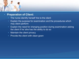
• Preparation ofClient:
• The nurse identify herself his to the client
• Explain the purpose for examination and the procedures which
may client perform
• Explain the need for changing position during examination asking
the client if he she has the ability to do so
• Maintain the client privacy
• Provide the client with clean gown
6.
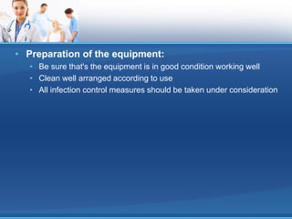
• Preparation ofthe equipment:
• Be sure that's the equipment is in good condition working well
• Clean well arranged according to use
• All infection control measures should be taken under consideration
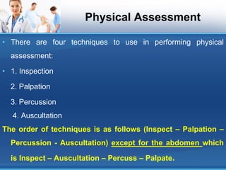
Physical Assessment
• Thereare four techniques to use in performing physical
assessment:
• 1. Inspection
2. Palpation
3. Percussion
4. Auscultation
The order of techniques is as follows (Inspect – Palpation –
Percussion - Auscultation) except for the abdomen which
is Inspect – Auscultation – Percuss – Palpate.
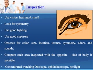
Inspection
• Use vision,hearing & smell
• Look for symmetry
• Use good lighting
• Use good exposure
• Observe for color, size, location, texture, symmetry, odors, and
sounds.
• Compare each area inspected with the opposite side of body if
possible.
• Concentrated watching Otoscope, ophthalmoscope, penlight
11.
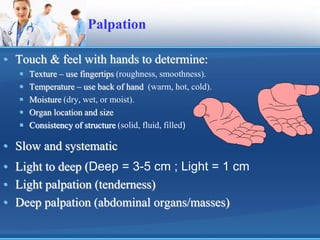
Palpation
• Touch &feel with hands to determine:
Texture – use fingertips (roughness, smoothness).
Temperature – use back of hand (warm, hot, cold).
Moisture (dry, wet, or moist).
Organ location and size
Consistency of structure (solid, fluid, filled)
• Slow and systematic
• Light to deep (Deep = 3-5 cm ; Light = 1 cm
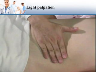
• Light palpation (tenderness)
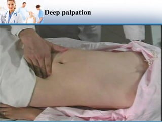
• Deep palpation (abdominal organs/masses)
12.
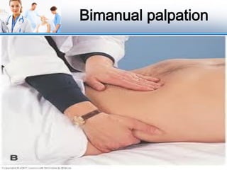
Types of palpation: -
• 1) Light palpation ..
• 2) Deep palpation .
• 3) Bimanual palpation
12
13.
Principles for AccuratePalpation
• Examiner finger nails should be short.
• Light Palpation precedes deep palpation.
• Start with light then deep palpation
• Tender area are palpated last
• Tell client to take slow deep breath to enhance muscle relaxation.
• Examine condition of the abdominal organs
• Depressed areas must be approximately “2cm”
• Assess turgor of skin measured by lightly grasping the body part
with finger tips.
13
Percussion
Tap aportion of the body to elicit tenderness that varies with
the density of underlying structures.
Percussion denotes location, size and density of
underlying structures, percussion requires dexterity.
Types of Percussion
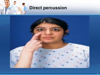
1) Direct (immediate) : involving striking the body surface
directly with one or two fingers.
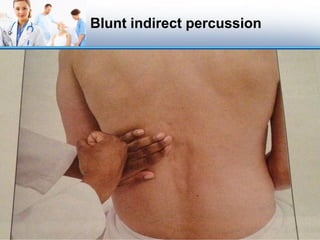
2) Blunt : used to detected tenderness over organs (e.g.
kidneys).
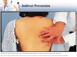
3) Indirect (mediate) : by tapping produce sound or tone that
varies with density of the underlying structure . Use a
quick & sharp stroke.
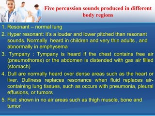
Five percussion soundsproduced in different
body regions
1. Resonant – normal lung
2. Hyper resonant: it’s a louder and lower pitched than resonant
sounds. Normally heard in children and very thin adults , and
abnormally in emphysema
3. Tympany : Tympany is heard if the chest contains free air
(pneumothorax) or the abdomen is distended with gas air filled
(stomach)
4. Dull are normally heard over dense areas such as the heart or
liver. Dullness replaces resonance when fluid replaces air-
containing lung tissues, such as occurs with pneumonia, pleural
effusions, or tumors
5. Flat: shown in no air areas such as thigh muscle, bone and
tumor
22.
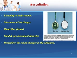
Auscultation
• Listening tobody sounds.
• Movement of air (lungs).
• Blood flow (heart).
• Fluid & gas movement (bowels).
• Remember the sound changes in the abdomen.
22
23.
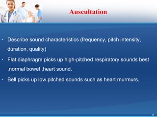
Auscultation
• Describe soundcharacteristics (frequency, pitch intensity,
duration, quality)
• Flat diaphragm picks up high-pitched respiratory sounds best
,normal bowel ,heart sound.
• Bell picks up low pitched sounds such as heart murmurs.
23
24.
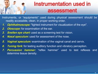
Instruments, or “equipments”used during physical assessment should be
readily accessible, clean, in proper working order.
1. Ophthalmoscope: "lighted instrument for visualization of the eye".
2. Otoscope: for examination of the ear.
3. Snellen eye chart: used as a screening test for vision.
4. Nasal speculum: used for assessment of the nose.
5. Vaginal speculum: examination of the vaginal canal and cervix.
6. Tuning fork: for testing auditory function and vibratory perception.
7. Percussion hammer: “reflex hammer” used to test reflexes and
determine tissue density.
24
Instrumentation used in
assessment


![✅ CCleaner Pro Free Crack 6.34 + Activation Key [APRIL-2025]](https://cdn.slidesharecdn.com/ss_thumbnails/physicalexamination-250415070608-e7301bc1-250415080446-ea0ab5fb-thumbnail.jpg?width=640&height=640&fit=bounds)
![✅ Download CCleaner Pro Key 2025 with Crack [Latest]](https://cdn.slidesharecdn.com/ss_thumbnails/physicalexamination-250415070608-e7301bc1-250415082125-6d1be3de-thumbnail.jpg?width=640&height=640&fit=bounds)


