Pelvic organ prolapse - Diagnosis and treatment
•
7 likes•1,780 views

Pelvic organ prolapse - Diagnosis and treatment

Recommended
More Related Content
What's hot
What's hot (20)
Viewers also liked
Viewers also liked (20)
Similar to Pelvic organ prolapse - Diagnosis and treatment
Similar to Pelvic organ prolapse - Diagnosis and treatment (20)
More from Tevfik Yoldemir
More from Tevfik Yoldemir (20)
Recently uploaded
Recently uploaded (20)
Pelvic organ prolapse - Diagnosis and treatment
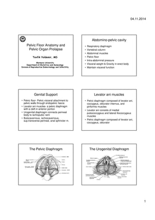
- 1. 04.11.2014 1 Pelvic Floor Anatomy and Pelvic Organ Prolapse Tevfik Yoldemir, MD Marmara University Department of Obstetrics and Gynecology Division of Reproductive Endocrinology and Infertility Abdomino-pelvic cavity • Respiratory diaphragm • Vertebral column • Abdominal muscles • Pelvic floor • Intra-abdominal pressure • Visceral weight & Gravity in erect body • Maintain visceral function Genital Support • Pelvic floor- Pelvic visceral attachment to pelvic walls through endopelvic fascia • Levator ani muscles- a pelvic diaphragm with a cleft in anterior portion • Urogenital diaphragm connects perineal body to ischiopubic rami • Bulocavernous, ischiocavernous, sup.transverse perineal, anal sphincter m. Levator ani muscles • Pelvic diaphragm composed of levator ani, coccygeus, obturator internus, and piriformis muscles • Levator ani consists of medial pubococcygeus and lateral iliococcygeus muscles • Pelvic diaphragm composed of levator ani, coccygeus, obturator The Pelvic Diaphragm The Urogenital Diaphragm
- 2. 04.11.2014 2 The Function of Pelvic Floor • Support pelvic and abdominal organs during stress of increased abdominal pressure • Allow for opening of the pelvic floor to accommodate excretory functions and parturition • Endopelvic fascia and visceral ligaments contains smooth muscles The Pelvic Floor Attachments • Pelvic floor support depends on its connection to the pelvic bones • An evolutionary solution for support of visceral organs • Pelvic floor muscles oppose gravity and increased abdominal pressures Prevention of Prolapse Attachments of Pelvic Floor • Tendinous arch of pelvic fascia-pubocervical fascia arises • Tendinous arch of levator ani- levator ani muscles arise • Pubourethral ligaments • Pubovesical ligaments Attachments of Pelvic Floor Pelvic Floor Dysfunction • A variety of fascial and anatomic defects • Cystocele, rectocele, uterine prolapse, enterocele, vault prolapse • Adequate diagnosis and staging of pelvic floor dysfunction is essential
- 3. 04.11.2014 3 Diagnosis of Pelvic Floor Dysfunction • Detailed physical examination • Pelvic ultrasound • Fluoroscopy of rectum & bladder • Magnetic resonance imaging (MRI) Normal Anatomy The axes of pelvic support • Three support axes • Upper vertical axis (cardinal-uterosacral ligament complex) • Horizontal axis leads to lateral and paravaginal supports – Two platforms pubocervical fascia and rectovaginal septum • Lower vertical axis supports the lower third of the vagina, urethra and anal canal DeLancey’s three levels of vaginal support • Apical suspension – Upper paracolpium suspends apex to pelvic walls and sacrum – Damage results in prolapse of vaginal apex • Midvaginal lateral attachment – Vaginal attachment to arcus tendineus fascia and levator ani muscle fascia – Pubocervical and rectovaginal fasciae support bladder and anterior rectum – Avulsion results in cystocele or rectocele • Distal perineal fusion – Fusion of vagina to perineal membrane, body and levators – Damage results in deficient perineal body or urethrocele Normal anterior anatomy Anterior compartment defects • Urethral hypermobility – Distal 4 cm of anterior vaginal wall – Cotton swab test – If describes an arc greater than 30 degrees from horizontal with valsalva – Results in genuine stress incontinence • Cystocele
- 4. 04.11.2014 4 Cystocele • Main support of urethra and bladder is the pubo-vesical-cervical fascia • Essentially a hernia in the anterior vaginal wall due to weakness or defect in this fascia – Midline weakness allows bladder to descend causing central cystocele – Tearing of endopelvic fascial connections from lateral sulci to arcus tendinii causes lateral or displacement cystocele – Detachment of pubocervical fascia from pericervical ring causes a transverse or apical cystocele • Symptoms include pelvic pressure and bulge or mass in the vagina Cystocele • Classified as Grade I, II, or III • Grade III is prolapse outside the introitus • Surgical repair is treatment of choice – Anterior Colporrhaphy – Paravaginal repair – Colpocleisis • Vaginal pessary Evaluation of a cystourethrocele Anterior defect Cystocele Bladder neck descent
- 5. 04.11.2014 5 Bladder neck descent Cystocele • Most Gr 1 and 2 cystoceles are asymptomatic • High grade cystoceles are associated with vaginal buldging, vaginal pressure, dyspareunia, UTI, obstructive voiding, urinary retention • A high grade cystocele may mask urethral hypermobility and stress incontinence Physical examination of Cystocele Enterocele • Simple enterocele • Complex enterocele- associated with vault prolapse and anterior or posterior vaginal prolapse • Cause vaginal pressure, dyspareunia, low back pain, constipation, symptoms of bowel obstruction Physical examination of Vaginal Cuff Prolapse Posterior compartment defects • Rectocele • Perineal deficiency – Bulbocavernous and superficial transverse muscle heads retracted • Perineal descent – Sagging and funneling of the levator ani around the perineum such that anus becomes most dependent – Difficulty with defecation
- 6. 04.11.2014 6 Rectocele • Chiefly a hernia in the posterior vaginal wall secondary to weakness or defect in the rectovaginal septum or fascia of Denonvilliers • Symptoms include difficulty evacuating stool, a vaginal mass, and fullness sensation • Rectovaginal exam confirms diagnosis Rectocele • Damage generally due to excessive pushing in childbirth or chronic constipation • Surgical treatment if symptomatic – Posterior Colporrhaphy – Laxatives and stool softeners • Temporary relief • Pessary not helpful Evaluation of a rectocele Rectocele • Defect of prerectal and pararectal fascia,and rectovaginal septum • Present in 80% asymptomatic patients • Vaginal mass,vaginal pressure, dyspareunia,constipation Physical examination of Rectocele Uterine Prolapse • Laxity of uterosacral ligaments • May present with vaginal mass, dyspareunia, urinary retention, back pain • Grade 4 prolapse is associated with ureteral obstruction
- 7. 04.11.2014 7 Physical examination of Uterine Prolapse Complete Uterovaginal procidentia Bladder descent Apical defect Pelvic Floor Relaxation • Associated with damage to pubococcygeus muscle • The muscle is lax, atrophied, poor tone • Urinary stress incontinence • Genital prolapse • Sexual Problem • Rectal stasis Muscular Component of Pubococcygeus muscle • Large diameter slow twitch type I fibers predominant- provide static visceral support • Fast twitch type II fibers- assists in active closure of pelvic visceral organs • 40% of women have lost function or coordination of this muscle
- 8. 04.11.2014 8 The Structures supporting Bladder and Urethra • Arcus tendineus fascia pelvis • Levator ani (pubococcygeus muscles) • Pubovesical muscles or ligaments • Vaginal muscle attachments to fascia and levator ani Physical examination of Pelvic Floor Dysfunction • General examination- cancer screen, stool OB, urinalysis, physical examination • Neurological examination- paresthesia of dermatome, bulbocavernous reflex, voluntary contraction of anal sphincter • Pelvic examination- cystocele, rectocele,uterine prolapse, vault prolapse • Urinary incontinence by Valsalva maneuver or coughing Patient position for evaluating pelvic floor defects Staging of Pelvic Organ Prolapse • Stage 0 - no prolapse • Stage I - the most distal portion is >1cm above level of hymen • Stage II - The most distal portion is <1cm proximal or distal to plane of hymen • Stage III - The most distal portion is >1cm below plane of hymen, but < total vaginal length - 2 cm • Stage IV - complete eversion of total length of lower genital tract, the distal portion is > TVL-2 cm, i.e. cervix or vaginal cuff Evaluation of Pelvic Floor Muscle Function • Assessing patient’s ability to contract and relax pelvic muscles separately • Measuring the force of contraction • Palpation of thickness of pelvic floor musculatures • Electromyography • Pressure recording
- 9. 04.11.2014 9 Measurement of Bladder Base Descent (The Q-tip Test) Lower Urinary Tract Symptoms caused by Pelvic Organ Prolapse • Stress incontinence • Frequency, urgency, urge incontinence • Hesitancy, weak stream, incomplete empty • Manual reduction of prolapse for voiding • Positional change to start or complete voiding Incontinence Assesment Bowel Symptoms caused by Pelvic Organ Prolapse • Difficulty with defecation • Incontinence of flatus • Incontinence of liquid stool • Incontinence of solid stool • Fecal staining of underwear • Digital manipulation to complete defecation • Feeling of incomplete evacuation • Rectal protrusion during or after defecation Sexual symptoms caused by Pelvic Organ Prolapse • Vaginal coitus? • Frequency of vaginal coitus? • Painful coitus? • Satisfaction with sexual activity? • Change in orgasm? • Incontinence experienced during sexual activity?
- 10. 04.11.2014 10 Local symptoms caused by Pelvic Organ Prolapse • Vaginal pressure or heaviness • Vaginal or perineal pain • Sensation or awareness of tissue protrusion from vagina • Low back pain • Abdominal pressure or pain • Observation or palpation of a mass Principles of reconstructive pelvic surgery • Site-specific repair • Rebuild weakened endopelvic fascia, repair fascial tears, and reattach prolapsed tissues to stronger sites • Goal is a vagina of normal depth, width and axis • Denervation or muscle trauma cannot be corrected surgically Cystocele Repair Cystocele Repair Burch Lateral defects
- 11. 04.11.2014 11 PVR Burch + PDR Colposuspension
- 12. 04.11.2014 12 Rectocele Rectocele Rectocele Pelvic Muscle Exercises Medications