This document discusses a study that examined the structural properties and ability to protect against oxidation of complexes formed between covalent conjugates of sodium caseinate and maltodextrins, and either liposomes of soy phosphatidylcholine or micelles of soy lysophosphatidylcholine in an aqueous medium. The study found that the conjugates highly encapsulated both the phosphatidylcholine and lysophosphatidylcholine, forming highly soluble complex particles with higher density and thermodynamic affinity for water than the pure conjugates. Lysophosphatidylcholine was a more effective cross-linking agent and the conjugates provided high levels of protection against oxidation for both phospholipids. The
![Impact of the structure of polyunsaturated soy phospholipids on the
structural parameters and functionality of their complexes with
covalent conjugates combining sodium caseinate with maltodextrins
M.G. Semenova*
, D.V. Zelikina, A.S. Antipova, E.I. Martirosova, N.V. Grigorovich,
R.A. Obushaeva, E.A. Shumilina, N.S. Ozerova, N.P. Palmina, E.L. Maltseva, V.V. Kasparov,
N.G. Bogdanova, A.V. Krivandin
N. M. Emanuel Institute of Biochemical Physics of Russian Academy of Sciences, Kosygin str., 4, 119334 Moscow, Russian Federation
a r t i c l e i n f o
Article history:
Received 18 February 2015
Received in revised form
15 June 2015
Accepted 16 June 2015
Available online 24 June 2015
Keywords:
Polyunsaturated phospholipids
Sodium caseinate
Maltodextrins
Covalent conjugates
Structural properties
Protection against oxidation
a b s t r a c t
A number of structural (the weight-average molar weight, Mw; the radius of gyration, RG; the hydro-
dynamic radius, Rh; the structure-sensitive parameter, r ¼ RG/Rh; the density, d; the intrinsic viscosity,
[h]; the z-potential), and thermodynamic (the second virial coefficient, A2, reflecting the nature and
intensity of both the biopolymerebiopolymer and biopolymeresolvent pair interactions; the molar
enthalpy of the bilayer phase transition, △Htr) parameters have been measured for the complex particles
formed between covalent conjugates of sodium caseinate (SC) with maltodextrins (MD) (dextrose
equivalent (DE) ¼ 2 and 10, Rweight ¼ MD: SC ¼ 2) and either liposomes of soy phosphatidylcholine (PC)
or micelles of soy lysophosphatidylcholine (LPC) in an aqueous medium (pH ¼ 7.0, I ¼ 0.001 M). The high
extent (>95%) of the encapsulation of both PC and LPC by the conjugates was found that led to the
formation of the highly soluble complex particles, having both essentially higher density and thermo-
dynamic affinity for an aqueous medium, as compared with the pure conjugates. LPC behaved as more
effective both inter- and intra-molecular cross-linking agent for the conjugate particles as compared with
PC. The data of the differential scanning calorimetry, electron spin resonance spectroscopy and small
angle X-ray scattering testified the maintenance of the PC bilayers under the formation of the complex
particles. The conjugates provided rather high level of the protection against oxidation to both PC and
LPC. The lowest extent of the oxidation of the phospholipids in the complexes was found for the LPC
micelles.
© 2015 Elsevier Ltd. All rights reserved.
1. Introduction
The development of smart nano-sized natural delivery systems
for biologically active compounds, containing prophylactic and
therapeutic agents for the improvement of human health (so-called
nutraceuticals), has aroused considerable interest under the novel
functional food formulation nowadays (Dickinson, 2014, chap. 1;
McClements, 2014; Semenova & Dickinson, 2010, chaps. 1-3). The
plant (a-Linolenic acid (ALA) 6-3, linoleic acid (LA) u-6) and fish
(6-3: eicosapentaenoic acid (EPA) and docosahexaenoic acid
(DHA)) polyunsaturated fatty acids (PUFAs) are one of the most
important and essential biologically active compounds, which the
human body cannot synthesize from other food components,
which is recognized to be significant for preserving health (Lee &
Ying, 2008, chap. 15; McClements, 2014; Phang & Garg, 2014,
chap. 16). The need for a balanced supply in both 6-3 and 6-6
polyunsaturated fatty acids is now supported by dietary guidelines
(Dietary Guidelines for Americans, 2010; McClements, 2014;
Michalski et al., 2013).
These essential PUFAs are building blocks of a huge diversity of
more complex natural molecules such as triacylglycerols and
phospholipids, themselves organized in various supramolecular
structures that could be generally natural delivery systems for
PUFAs (McClements, 2014; Phang & Garg, 2014, chap. 16). Recent
advance in nutrition research revealed that the supramolecular
arrangements of the lipid molecules and their physico-chemical
properties in a food matrix can modulate PUFAs release and* Corresponding author.
E-mail address: mariagersem@mail.ru (M.G. Semenova).
Contents lists available at ScienceDirect
Food Hydrocolloids
journal homepage: www.elsevier.com/locate/foodhyd
http://dx.doi.org/10.1016/j.foodhyd.2015.06.011
0268-005X/© 2015 Elsevier Ltd. All rights reserved.
Food Hydrocolloids 52 (2016) 144e160](https://image.slidesharecdn.com/cffadf95-3528-4550-9876-8ab19a966394-161028092821/85/Paper_Food_Hydrocolloids_RSF_1-1-320.jpg)
![bioavailability during digestion and their final metabolic fate,
significantly modifying their health impact (McClements, 2014;
Michalski et al., 2013; Shaw et al., 2008, chap. 7; Tamjidi,
Shahedi, Varshosaz, & Nasirpour, 2013). In particular, liposomes
and micelles of polyunsaturated phospholipids attracted significant
interest not only as the sources of the essential PUFAs, but also as
potential carriers for different kinds of both water-soluble and
water-insoluble nutraceuticals (Gibis, Rahn, & Weiss, 2013; Gibis,
Zeeb, & Weiss, 2014; Leser, Sagalowicz, Michel, & Watzke, 2006;
Liu, Ye, Liu, Liu, & Singh, 2013; Lu, Li, & Jiang, 2011; Lu, Nielsen,
Timm-Heinrich, & Jacobsen, 2011; Maherani, Arab-Tehrany,
Mozafari, Gaiani, & Linder, 2011; McClements, 2014; McClements
& Decker, 2000; Mozafari, Johnson, Hatziantoniou, & Demetzos,
2008; Singh, Thompson, Liu, & Corredig, 2012, chap. 11; Taylor,
Davidson, Bruce, & Weiss, 2005; Torchilin, 2007; Velikov & Pelan,
2008). However, currently their high susceptibility to oxidative
degradation during product preparation, transport, and storage as
well as low solubility in water (in the case of liposomes) or in
aqueous body fluids, in addition, possible both leakage and un-
controllable release of the involved nutraceuticals, pose a real
challenge for their use (McClements, 2014; Semenova & Dickinson,
2010, chaps. 1-3).
One of the general ways of improving the chemical stability of
these bioactive lipids is their encapsulation within colloidal de-
livery systems that inhibit their oxidative degradation (Gibis et al.,
2013; Mayer, Weiss, & McClements, 2013; McClements, 2014;
McClements & Decker, 2000; Salminen, Aulbach, Leuenberger,
Tedeschi, & Weiss, 2014; Waraho, McClements, & Decker, 2011).
In particular, to date it is well shown that biomacromolecules
present a great potential in such encapsulation due to mainly their
generally amphipilic nature that, on the one hand, provides surface
activity to the biomacromolecules and, on the other hand, underlies
their susceptibility to self-assembly and co-assembly with different
in nature compounds in an aqueous medium (Gibis et al., 2013;
Livney, 2008, chap. 9; McClements, 2014; Menendez-Aguirre
et al., 2014; Semenova, Anokhina, Antipova, Belyakova,
Polikarpov, 2014b; Semenova, Belyakova, Polikarpov, Antipova,
Anokhina, 2008; Semenova Dickinson, 2010, chaps. 1-3;
Semenova et al.,2012, 2014a). For example, by now, the predicted
novel functional ability of casein molecules/associates to behave as
delivery nanovehicles for hydrophobic nutraceuticals has been
distinctly demonstrated for: (i) polyphenols (Esmaili et al., 2011;
Hasni et al., 2011; Rahimi Corredig, 2012; Sahu, Kasoju, Bora,
2008); (ii) a vitamin (D2) (Semo, Kesselman, Danino, Livney,
2007); (iii) a polyunsaturated soy phosphatidylcholine (Semenova
Dickinson, 2010, chaps. 1-3; Semenova et al.,2006, 2008, 2012,
2014a,b); and (iv) for the chemotherapeutic drugs (Sahu et al.,
2008; Shapira, Assaraf, Epstein, Livney, 2010), under the elabo-
ration of non-saturated fat or low-saturated fat functional food
products (Livney, 2010; Semenova Dickinson, 2010, chaps.1-3). In
addition, the promising abilities of nano-sized (sodium caseinate
(SC) þ maltodextrin (MD)) covalent conjugates as delivery vehicles
for hydrophobic nutraceuticals were also clearly demonstrated
(Grigorovich et al., 2012; Markman Livney, 2012; Semenova et al.,
2014c, chap. 6). Both the high protection against oxidation for the
nutraceuticals studied (phosphatidylcholine (PC) (Semenova et al.,
2014c, chap. 6); vitamin D and epigallocatechin gallate (Markman
Livney, 2012)) and their high solubility in the wide range of pH
(including the protein isoelectric point) in the complexes with the
conjugates in an aqueous medium were revealed. Moreover, it was
shown that the covalent conjugation of the maltodextrins to the
protein can be used in order to control the release of PC under the
action of the gastric and intestinal enzymes in the simulated con-
ditions of the gastro-intestinal (GI) tract in-vitro (Semenova et al.,
2014c, chap. 6).
The nanoscale dimensions of biomacromolecule-based nano-
vehicles, which are typical of biopolymer molecules/associates, can
offer additional advantages, because, as was suggested by Acosta
(Acosta, 2009), the size of the delivery systems, especially below
500 nm, has enabled the promise of tackling problems of low oral
bioavailability or inefficient delivery of poorly water-soluble
nutraceuticals/drugs. This is a consequence of the enhancements
based on the following factors (Acosta, 2009; Semenova
Dickinson, 2010, chaps. 1-3): (i) the apparent solubility of the
active ingredients; (ii) the rate of mass transfer; (iii) the gastro-
intestinal retention time in the mucus covering the intestinal
epithelium; (iv) the rate of release (due to large surface area); and
(v) the direct uptake of particles by the intestinal epithelium
(Augustin Hemar, 2009; Chen, Weiss, Shahidi, 2006; Horn
Rieger, 2001; Medina, Santos-Martinez, Radomski, Corrigan,
Radomski, 2007).
Despite the almost exponential increase in the research activ-
ities in the area of the elaboration of the smart nanoparticle de-
livery systems for micronutrients and nutraceuticals during the
past decade (Augustin Hemar, 2009; Dickinson, 2014, chap. 1;
McClements, 2014; Ransley, Donnelly, Read, 2001; Semenova
Dickinson, 2010, chaps. 1-3; Velikov Pelan, 2008) our under-
standing of creating of the biopolymer-based nanovehicles with
specific functional properties is still rather limited, and a more clear
insight into both the structure e function relationships and the
physico-chemical mechanisms underlying the nanovehicles for-
mation is required to design functional biopolymer-based smart
ones more systematically (Dickinson, 2014, chap. 1; McClements,
2014; Semenova Dickinson, 2010, chaps. 1-3).
The present work is focused on the elucidation of the structural
basis underlying the formation of the nano-sized complexes be-
tween covalent conjugates (SC þ MD) and either liposomes of soy
phosphatidylcholine (PC) or micelles of soy lysophosphatidylcho-
line (LPC) in an aqueous medium, as well as on the ability of the
conjugates to protect the polyunsaturated phospholipids against
oxidation. In parallel, a more penetrating insight is sought into the
generality and the differences in the impact of the specific structure
of the PC liposomes and LPC micelles on both the molecular
mechanisms of the formation of the complexes and their physico-
chemical properties. Moreover, the estimation of the role of the
extent of the maltodextrin polymerization, that is reflected by their
dextrose equivalent (DE ¼ 2 and 10), in both the complex formation
and the complex behavior was also one of the objectives of our
investigation.
2. Materials and methods
2.1. Materials
The sample of sodium caseinate was purchased from Sigma (New
Zealand) and used as received. The samples of maltodextrins Paselli
SA-2 and MD-10 were kindly supplied by AVEBE (Netherlands) and
used as received. These maltodextrins were prepared by the enzy-
matic hydrolysis of a potato starch and had the DE ¼ 2 (SA2) and 10
(MD10). The molar masses of the individual molecules of malto-
dextrins SA-2 and MD-10 are 9 kDa and 1.8 kDa, respectively
(Harkema, 1998). Phosphatidylcholine Lipoid S 100 and Lysophos-
phatidylcholine Lipoid LPC were purchased from Lipoid GmbH,
Germany (PC: phospholipids [g/100 g]: phosphatidylcholine (by
anhydrous weight) ¼ 94; N-acyl-phosphatidylethanolamine ¼ 0.5;
phosphatidylethanolamine ¼ 0.1; phosphatidylinositol ¼ 0.1; lyso-
phosphatidylcholine ¼ 3.0. Nonpolar lipids [g/100 g]:
triglycerides ¼ 2.0; free fatty acids ¼ 0.5; DL-a-tocopherol ¼ 0.15 ÷
0.25. Typical fatty acid composition in % to total fatty acids: palmitic
acid ¼ 12 ÷ 17; stearic acid ¼ 2 ÷ 5; oleic acid ¼ 11 ÷ 15; linoleic acid
M.G. Semenova et al. / Food Hydrocolloids 52 (2016) 144e160 145](https://image.slidesharecdn.com/cffadf95-3528-4550-9876-8ab19a966394-161028092821/85/Paper_Food_Hydrocolloids_RSF_1-2-320.jpg)
![(u-6) ¼ 59 ÷ 70; linolenic acid (u-3) ¼ 3 ÷ 7).
LPC was produced from soybean lecithin by phospholipase A2.
LPC: phospholipids [g/100 g]: lysophosphatidylcholine ¼ 98.0;
phosphatidylcholine ¼ 0.2; phosphatidylethanolamine ¼ 0.1; un-
identified components ¼ 0.5; triglycerides ¼ 1.0; free fatty
acids ¼ 0.3; DL-a- tocopherol ¼ 0.1÷0.2. Typical fatty acid compo-
sition in % to total fatty acids: palmitic acid ¼ 24 ÷ 28; stearic
acid ¼ 4 ÷ 7; oleic acid ¼ 15 ÷ 17; linoleic acid (u-6) ¼ 44 ÷ 48;
linolenic acid (u-3) ¼ 1). DL-a-phosphatidylcholine dipalmitoyl
(DPPC) (Sigma (USA), P-5911, synthetic and 99% of purity) was used
as a model phospholipid for the differential scanning calorimetry
(DSC) measurements. The phosphate buffer solutions were pre-
pared using the analytical grade reagents (Laverna, Russia) (99.9%
of purity). They were filtered through a Millipore membrane
(nominal pore size ¼ 0.2 mm) to remove dust. The pH was adjusted
with HCl/NaOH. Analytically grade (99.9% of purity) sodium azide
(Laverna, Russia) (0.02 wt/v %) was added to the buffers as an anti-
microbial agent. Analytically grade ethanol (95.6% of purity) was
used for the PC dissolution. Analytically grade (99.5% of purity)
CuSO4 was used for the purpose of the acceleration of both the PC
and LPC oxidation (Laverna, Russia). Analytically grade (99.5% of
purity) trichloroacetic acid and 2-thiobarbituric acid (TBA) (Lav-
erna, Russia) were used in the TBA test for the oxidation mea-
surements of the phospholipids. Reagents of SENTINEL
DIAGNOSTICS (Italy) were used for the direct enzymatic-
colorimetric determinations of the phospholipids. All solutions
were prepared using a double-distilled water.
2.2. Preparation of the aqueous solutions of the protein,
maltodextrins, and their covalent conjugates
Solutions of the protein (SC), the maltodextrins (MD (either SA-
2 or MD10)), and the conjugates (SC þ MD (either SA2 or MD10)
with required concentrations were prepared using a double-
edistilled water and a phosphate buffer (pH ¼ 7.0, ionic strength of
the buffer ¼ 0.001 M). In addition, both the maltodextrin and
conjugate's solutions were heated at 85 C for 5 min and thereafter
allowed to be cooled for the room temperature. Centrifugation
(4000 rpm, 30 min, at 20 C) of the all biopolymer solutions was
carried out to remove a small fraction of an insoluble material. The
concentration of the protein, the maltodextrins, and the conjugates
in the solutions after centrifugation was checked using a refrac-
tometer (Shimadzu, Japan), with reference to the determined pre-
vious values of the protein, maltodextrin, and conjugate's refractive
indexes, n, equalled, within the 10% experimental error, to
0.20 Â 10À3
m3
/kg, 0.15 Â 10À3
m3
/kg, and 0.17 Â 10À3
m3
/kg,
respectively. Besides, in order to control the concentration of the
biopolymers defined by the refractometric method we have addi-
tionally checked it by drying the biopolymer solutions up to the
constant weight and using the appropriate buffer as the blank
sample. Mixed (SC þ SA2) and (SC þ MD10) aqueous solutions for
the formation of the conjugates, having the weight ratio of the
maltodextrins to the protein (Rweight ¼ 2), were prepared from the
individual biopolymer solutions.
2.3. Preparation of the covalent conjugates (sodium
caseinate þ maltodextrins)
(SC þ MD (either SA-2 or MD10)) conjugates were prepared
according to the method described in the work (Shepherd,
Robertson, Ofman, 2000). The lyophilized mixed (SC þ MD
(either SA-2 or MD10)) solutions were heated at 60С in a
controlled humidity environment (79% relative humidity) for 72 h.
It was previously shown that the chosen duration of heating under
the mentioned above environmental conditions was enough for
achieving of the excellent solubility, emulsification activity and
stability of the covalent (SC þ MD) conjugates even at pH ¼ 4.8, i.e.,
about the protein isoelectric point (pI) (O'Regan Mulvihill, 2009;
Shepherd et al., 2000).
2.4. Estimation of the covalent binding between the sodium
caseinate and maltodextrins in the conjugates
The extent of the covalent binding between the SC and malto-
dextrins was estimated according to O'Regan and Mulvihill (2009)
using the trinitrobenzenesulphonate method (TNBS). The calibra-
tion curve was constructed using the pure lysine stock solution
(2.7 Â 10À4
M) by measuring the absorbance of the resultant yellow
solutions at 340 nm using a bi-distillated water treated blank as a
reference. The concentration of the available amino groups in both
pure SC and (SC þ MD) conjugates was measured by reference to
the lysine standard curve. The change in the concentration of
available amino groups in the (SC þ MD) conjugates relative to the
unreacted SC (assumed representing 100% of available amino
groups) was calculated per gram of soluble protein (determined by
the Biuret Protein Assay) and expressed as a percentage loss of
available amino groups compared to SC. Thereafter the average
number of the loss of the available originally lysine groups per
average casein molecules (24,000 Da) (Semenova Dickinson,
2010, chaps. 1-3; Swaisgood, 2003) was calculated. As this took
place, both the primary sequences of amino acids in the individual
caseins (13 lysine residues (Lys) for aS1-casein; 21 Lys for aS2-
casein; 12 Lys for b-casein; and 9 Lys for k-casein) (Manson,
Carolan, Annan, 1977; Swaisgood, 2003) and their proportions
in SC (38% aS1-casein, 10% aS2-casein, 36% b-casein and 13% k-
casein) (Manson et al., 1977) were taken into account under the
calculation of the originally present available lysine groups: 0.38
(13) þ 0.10 (21) þ 0.36 (12) þ 0.13 (9) ¼ 12.53.
2.5. Preparation of both PC and DPPC liposomes as well as LPC
micelles
The required amount of PC was dissolved in the pure ethanol.
The aqueous solutions of both PC (10-3
M, containing 10 v/v % of
ethanol), LPC (10-3
M) and DPPC (10-3
M) were prepared under
mixing and shaking for an hour of the PC solution in ethanol, and
both LPC and DPPC in a dry state, with an appropriate amount of the
pure buffer solution, respectively. The LPC critical micelle concen-
tration (CMC), determined by static light scattering (See the Section
2.8.), was equal to 10-3
M under the experimental conditions used
(рН ¼ 7.0, ionic strength of the buffer ¼ 0.001 M, t ¼ 25
C and
40
C). In order to remove ethanol from the PC solutions we have
used the equilibrium dialysis (Visking Dialysis Tubing (Type 36/32),
Serva; the ratio of the volumes of the outer vessel to the inner one
equalled to 20, the duration time of the dialysis was 24 h) against a
pure phosphate buffer (pH ¼ 7.0, ionic strength ¼ 0.001 M). Pre-
liminary estimation showed no transmission of the PC molecules
through the dialysing tubes. The residual amount of ethanol in the
PC containing solutions was equal to 0.5 v/v %.
2.6. Formation of the complex particles (covalent
conjugates þ phospholipids)
The buffered solutions of the conjugates with the required
concentration were mixed and shaken with the solutions of the
phospholipids (in the pure ethanol for PC and in the buffered
aqueous medium for DPPC and LPC) at 40 C for an hour in order to
get mixed solutions, containing finally 10 v/v % of ethanol in the
case of PC, and where the ultimate concentrations of the compo-
nents were the following: 1.0 wt/v % of SC, 2.0 wt/v% of MD (either
M.G. Semenova et al. / Food Hydrocolloids 52 (2016) 144e160146](https://image.slidesharecdn.com/cffadf95-3528-4550-9876-8ab19a966394-161028092821/85/Paper_Food_Hydrocolloids_RSF_1-3-320.jpg)
![estimation of the total contributions from the other kinds of the
interactions (electrostatic (Ael
2 ), hydrogen bonding (A
h=b
2 ) and
hydrophobic (Ah
2)) as follows:
A*
2 À Aexc
2 ¼ Ael
2 þ A
h=b
2 þ Ah
2 (2)
For the spherical particles found generally in our experiments
(1 r 2) (See Tables 2, 4 and 5) we have used the simplest case of
the interacting solid spheres and the following equation for the
calculation of the Aexc
2 (Tanford, 1961):
Aexc
2 ¼ 10À3
4pNA
.
3 ð2RÞ3
(3)
The parameter R in the Equation (3) is the radius of the equiv-
alent hard sphere representing the biopolymer particle. The
equivalent hard sphere corresponds to the space occupied in the
aqueous medium by a single biopolymer particle, which is
completely inaccessible to both other biopolymer particles and
solvent molecules. In our calculations we have used the Rh as the
radius of the equivalent hard sphere.
The critical micelle concentration (CMC) of the LPC was deter-
mined by measuring the intensity of the scattering light (I90) for the
LPC aqueous solutions having the LPC concentration in the range
(10À7
M ÷ 10-1
M), using an LS-01 apparatus (Scientific Instruments,
St. Petersburg, Russia). The sharp increase in the I90 was referred to
the CMC.
2.9. The viscosimetry in diluted solutions
The intrinsic viscosity, [h], of the buffered aqueous solutions of a
pure SC, the conjugates (SC þ MD), and their complexes with the
phospholipids was measured at 25 C using an Ubbelohde
viscometer having the capillary diameter ¼ 0.54 mm. The experi-
mental error in the [h] determination, which was estimated on the
basis of not less than 2 experimental repetitions, did not exceeded
15%.
2.10. The z-potential measurements
Electrophoretic mobilities of the biopolymer samples: a pure SC,
the conjugates (SC þ MD), and their complexes with the phos-
pholipids were determined with a Zetasizer Nano ZS Malvern (UK)
calibrated against a standard latex dispersion. The experimental
error in the z-potential determination, which was estimated on the
basis of not less than 10 experimental repetitions, did not exceeded
10%.
2.11. DSC measurements
DSC measurements of the inherent phase transition for the
DPPC liposome bilayers were performed by the high sensitivity
differential scanning calorimetry using a DASM-4M model, Differ-
ential Scanning Calorimeter (Pushino, Russia). The baseline ther-
mogram was obtained using the appropriate buffer (0.5 ml) in all
two cells including the reference cell to normalize cell to cell dif-
ferences. The measurements were carried out in the temperature
range from 10 to 110 C at the constant pressure 2.5 atm. The
heating scan rate was maintained at 0.5 C/min for the all experi-
ments. The thermograms for the investigated samples were ob-
tained by subtracting the respective baseline thermogram from the
sample thermogram using the software provided by the manu-
facturer. The peak position in the plot of the “excess heat capacity”,
DCp, versus temperature on a heating scan was taken as the tem-
perature, ttr, of the solid-like gel to the fluid liquid-crystalline phase
transition for DPPC liposome bilayers in an aqueous medium. The
thermodynamic parameters (ttr, Dt, hpeak), and function (DHtr) of
the phase transition were also computed using the same software
as reported. The experimental error in their determination, which
was estimated on the basis of not less than 2 experimental repe-
titions, did not exceeded 5%.
2.12. Electron spin resonance (ESR) study
The structural state of both PC liposomes and LPC micelles in
their pure form and in the complexes with the conjugates (1.0 wt/v
% of the conjugates with either 2 Â 10-3
M of PC (0.156 wt/v %), or
2 Â 10-3
M of LPC (0.102 wt/v %)) was studied by the electron spin
resonance spectroscopy at 25 C. ESR spectra were recorded on a
Bruker EMX spectrometer (Germany). 16-doxylstearic acid radical
(16-DSA, Sigma) was used as a spin probe. 16-DSA localizes in the
hydrophobic region of the phospholipid particles at a depth of 20 Å
of a lipid layer.
The microviscosity of the phospholipid layers was estimated by
the rotational correlation time (tс) of the 16-DSA using the equation
for a fast motion of the nitroxyl radical:
tс ¼ 6; 65$DНþ
ffiffiffiffiffi
Iþ
IÀ
s
À 1
!
$10À10
; (4)
where △Hþ represents the resonance width of the low-field
component, Iþ and IÀ are the resonance heights of the low and
the high-field components of the ESR spectra, respectively.
The experimental error of the method did not exceeded 2%.
Both the calculation of the parameters and the correction of the
ESR spectra were performed using the computer program Bruker
WIN-EPR with the subprogram SYMFONIA.
2.13. Small-angle X-ray scattering (SAXS)
The SAXS measurements were performed with an X-ray
diffractometer of a local design at room temperature. The X-ray
radiation from the fine focus Cu X-ray tube was Ni-filtered and
focused with the glass mirror collimator. X-ray scattering patterns
Table 1
The amount of maltodextrin molecules covalently attached to the individual mass-averaged casein molecule (24 kDa) comprising original sodium caseinate (SC) particles in an
aqueous medium.
Rweight Percent of the loss of the available
amino groups of lysine in the SC
Rm
a
The quantity of the maltodextrin molecules covalently
bound to the individual averaged casein molecules
The quantity of the free maltodextrin
molecules
Conjugate (SC þ SA2)
2 30 5 4 1
Conjugate (SC þ MD10)
2 45 27 6 21
a
Under the calculation of the molar ratio Rm we have taken into account the molar masses of the individual molecules of maltodextrins (9 kDa and 1.8 kDa for the
maltodextrins SA2 and MD10, respectively) (Harkema, 1998) and the individual mass-averaged casein molecules (24 kDa) (Swaisgood, 2003) comprising the original sodium
caseinate particles.
M.G. Semenova et al. / Food Hydrocolloids 52 (2016) 144e160148](https://image.slidesharecdn.com/cffadf95-3528-4550-9876-8ab19a966394-161028092821/85/Paper_Food_Hydrocolloids_RSF_1-5-320.jpg)
z-potential(mV)
SC11,9304.412.423.6À11.21591.511.269À31.0
Conjugate(SCþSA2)60053.82.80.91.91251.751.244À22.0
Conjugate(SCþMD10)47063.10.62.6À2.01401.390.777À27.0
M.G. Semenova et al. / Food Hydrocolloids 52 (2016) 144e160 149](https://image.slidesharecdn.com/cffadf95-3528-4550-9876-8ab19a966394-161028092821/85/Paper_Food_Hydrocolloids_RSF_1-6-320.jpg)
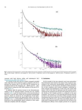
![Table 5
Schematic representation of the nanoscale structures and the experimental data relating to the assembly of covalent conjugate (SC þ MD10) induced by the interactions of the conjugate with either PC liposomes or LPC micelles in
an aqueous medium (pH ¼ 7.0, ionic strength of the buffer ¼ 0.001 M, 25 C).
Conjugate (SC þ MD10) þ LPC LPC Conjugate (SC þ MD10) PC Conjugate (SC þ MD10) þ PC
1
2
3
4
System Mw (kDa) A2 Â 105
(m3
mol kgÀ2
) A*
2 (m3
molÀ1
) Aexc
2 (m3
molÀ1
) Ael
2 þ A
h=b
2 þ Ah
2 (m3
molÀ1
) RG (nm) r ¼ RG/Rh d (mg/ml) 1/[h] (mg/ml) z-potential (mV)
Conjugate (SC þ MD10) þ LPC 15,000 1.4 22.0 1.9 20.1 157 1.70 1.5 248 À26.4
Conjugate (SC þ MD10) þ PC 5900 1.6 1.1 0.5 0.6 114 1.98 1.6 83 À25.6
Table 4
Schematic representation of the nanoscale structures and the experimental data relating to the assembly of the covalent conjugate (SC þ SA2) induced by the interactions of the conjugate with either PC liposomes or LPC micelles
in an aqueous medium (pH ¼ 7.0, ionic strength of the buffer ¼ 0.001 M, 25 C).
Conjugate (SC þ SA2) þ LPC LPC Conjugate (SC þ SA2) PC Conjugate (SC þ SA2) þ PC
System Mw (kDa) A2 Â 105
(m3
mol kgÀ2
) A*
2 (m3
molÀ1
) Aexc
2 (m3
molÀ1
) Ael
2 þ A
h=b
2 þ Ah
2 (m3
molÀ1
) RG (nm) r ¼ RG/Rh d (mg/ml) 1/[h] (mg/ml) z-potential (mV)
Conjugate (SC þ SA2) þ LPC 9400 2.4 4.3 0.44 3.9 109 2.0 2.9 86 À23.4
Conjugate (SC þ SA2) þ PC 6800 2.6 2.4 0.6 1.8 112 2.0 1.9 60 À22.8
M.G.Semenovaetal./FoodHydrocolloids52(2016)144e160151](https://image.slidesharecdn.com/cffadf95-3528-4550-9876-8ab19a966394-161028092821/85/Paper_Food_Hydrocolloids_RSF_1-8-320.jpg)
![conjugate particles formed in an aqueous medium. It manifests
itself as either less negative for the case of the conjugate
(SC þ MD10) or, by contrast, positive for the conjugate (SC þ SA2)
contributions of the additive term (Ael
2 þ A
h=b
2 þ Ah
2) into the value
of the A*
2 (Eq. (2)) as compared to the pure SC. It was also found that
the lower is the molar mass of the studied biopolymer particles the
poorer is their thermodynamic affinity for an aqueous medium, as
if a greater amount of hydrophobic patches of the casein mole-
cules, combined in the conjugates, become exposing into the
aqueous medium, rather than are hidden in the interior of the
particles. That is most pronounced in the case of the particles of
the (SC þ MD10) conjugate.
In addition, Table 2 shows that the formed conjugates have the
smaller size (RG) as compared with the pure protein, keep both the
spherical shape (1 r 2) and negative, but lower in the absolute
value than the protein has the z-potential, which is reflecting the
negative total charge of the particles. It is probable that such
decrease in the absolute value of the z-potential is attributable to
the partial protein charge neutralization as a result of the facilita-
tion of the interactions between the opposite charges of the caseins
under the new form of the self-assembly of casein molecules
modified by the covalently attached maltodextrins.
In turn, the densities d of the conjugate particles (Eq. (1)) have
changed as a result of the decrease in both their molar mass, Mw,
and size, RG. Hence, there is no alteration of the density value in
the case of the conjugate (SC þ SA2) in comparison with that of
the pure SC particles, and, in turn, there is an essential decrease in
the d for the case of the conjugate (SC þ MD10) (from d ¼ 1.2 mg/
ml for SC to d ¼ 0.7 mg/ml for the conjugate). This result can be
attributed to the largest decrease in the absolute value of Mw of
the conjugate (SC þ MD10) particles, as compared with the pure
SC, that does not followed by the same extent of the decrease in
the RG value.
In order to confirm the alteration of the structural and ther-
modynamic parameters of the SC particles as a result of the
conjugation with the maltodextrins, observed by light scattering
method, we have measured the intrinsic viscosity [h] of both SC
and the conjugates in diluted aqueous solutions (Table 2). The [h]
is the most convenient parameter to this end because its value is
sensitive to changes in the size, shape, and solvation of polymer
particles (Tanford, 1961). Thus an easy comparison of the inverse
values of the [h] obtained for both SC and the conjugate particles
with the values of the densities of these biopolymer particles,
calculated from the light scattering data (Eq. (1), Table 2), dem-
onstrates a number of discrepancies in the trend of their alteration.
We have found the same densities of the conjugate (SC þ SA2) and
the SC particles in contrast to the 1.6 times decrease in the value of
the 1/[h] for this conjugate in comparison to that of the pure
protein. In turn, in the case of the conjugate (SC þ MD10), there is
1.8 times increase in the value of the 1/[h] against the 1.7 times
decrease in the values of d as compared with those parameters of
the pure SC particles. These discrepancies indicate that one should
take into account that the value of [h] of the particles is controlled
not only by their density d but also by their shape and degree of
solvation (Tanford, 1961). In particular the solvation correlates
with the thermodynamic affinity of the biopolymer particles for
solvent. It is well known that the greater extent of the solvation of
the particle as a whole leads to the increase in the value of [h] and
hence to the decrease in its reciprocal value 1/[h] due to the
retention of the solvent inside the biopolymer particle moving
together with it. Such particle may be approximated by the
equivalent solid sphere (Tanford, 1961). The protein particles seem
to accord well with this approximation. On the contrary, if the
thermodynamic quality of the solvent for the biopolymer particle
is poorer, as was found for the conjugate (SC þ MD10) particles
(the lowest positive values of both A2 and A*
2), for example,
(Table 2), that is, when the solvent is not retained much by the
biopolymer particle, the model of a more permeable for the sol-
vent biopolymer particle may be more acceptable for the
description of its hydrodynamic properties (Tanford, 1961). Hence,
it may be proposed that the higher permeability of the conjugate
(SC þ MD10) particles for the solvent is responsible for the lower
value of the [h] and hence the higher value of the 1/[h] as
compared to both the protein and the conjugate (SC þ SA2) par-
ticles even though the value of d of the conjugate (SC þ MD10)
particles is essentially lower (Table 2). In addition, with account
taken of the contribution of the different shapes of polymer par-
ticles to the value of [h], one may also provide an additional
explanation for the discrepancies mentioned above. Thus, a more
asymmetric shape of polymer particles (r 1) may be also
responsible for a higher value of [h] and consequently for a lower
value of its reciprocal value (Tanford, 1961). Really, the lowest
value of 1/[h] was found for the more asymmetric particles of the
conjugate (SC þ SA2) (r ¼ 1.75), having an intermediate thermo-
dynamic affinity for the solvent, as compared with both the pro-
tein and the conjugate (SC þ MD10) particles.
The measured structural and thermodynamic parameters of
both the pure protein and the conjugate particles allow visualizing
them by the schematic representation based on the dual binding
model of the casein ‘micelles’ as built up from the individual caseins
(aS1, aS2, b and k) and suggested by Horne (1998) (Table 2).
3.3. Encapsulation ability of the covalent conjugates relatively the
polyunsaturated phospholipids (PC and LPC)
In order to get the conjugate-based delivery systems for the
phospholipids the co-assembly of the conjugates (1 wt/v% of SC þ 2
wt/v% MD (either SA2 or MD10)) with both PC liposomes (10-3
M;
0.078 wt/v%) and LPC micelles (10-3
M; 0.051 wt/v%) was performed
under pH ¼ 7.0, at the ionic strength of the phoshate buffer
equalled to 0.001 M, and temperature (40 C).
The high extent (95%) of the encapsulation of both PC lipo-
somes and LPC micelles by the conjugates was observed in the
aqueous medium for the weight ratio of the conjugates to the
phospholipid particles used. Relying on both the literature
(Antunes, Marques, Miguel, Lindman, 2009; Bai, Nichifor,
Bastos, 2010; Schulz, Olubummo, Binder, 2012; Str€omstedt,
Ringstad, Schmidtchen, Malmsten, 2010) and our experimental
data (Istarova et al., 2005; Semenova et al., 2012, 2014a) we can
suggest the contribution of the different kinds of the interactions
(electrostatic attraction between opposite charges, hydrogen
bonding and predominantly hydrophobic attraction) into the
complex formation between the phospholipids and the conjugates.
It is significant that, as this takes place, the formed complexes show
the same high level of the solubility in an aqueous medium that is
inherent to the pure covalent conjugates (Grigorovich et al., 2012).
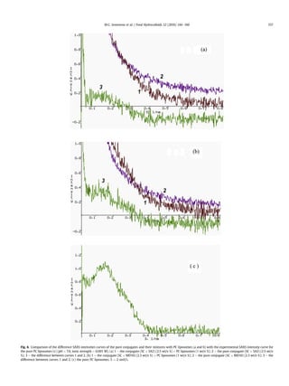
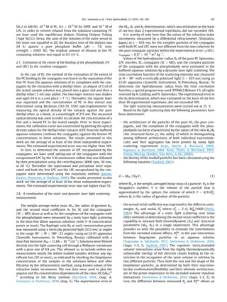
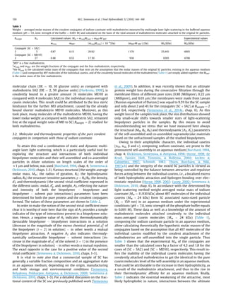
The appearance of the new peaks of the complex particles at the
size distribution diagrams obtained by dynamic light scattering
(Fig. 1) for the studied particles of pure PC liposomes, LPC micelles,
conjugates, and their complexes, give an additional evidence of the
involvement of both the conjugates and the phospholipid particles
into the complex formation. The lower polydispersity in the size
(more narrow size distributions) seems to be the common feature
of the complex particles involving both PC liposomes and LPC mi-
celles as compared with the polydispersity of the pure conjugates
(Fig. 1).
M.G. Semenova et al. / Food Hydrocolloids 52 (2016) 144e160152](https://image.slidesharecdn.com/cffadf95-3528-4550-9876-8ab19a966394-161028092821/85/Paper_Food_Hydrocolloids_RSF_1-9-320.jpg)
![3.4. Comparison of the structural and thermodynamic parameters
of the complex particles combining the covalent conjugates with
either PC liposomes or LPC micelles
Tables 4 and 5 show a comparison of the structural and ther-
modynamic parameters of the complex particles based on the
conjugates ((SC þ SA2) Table 4; (SC þ MD10) Table 5) and involving
either PC liposomes or LPC micelles. From this comparison the
generalities and distinctions in the impact of the structural orga-
nization of the phospholipids (PC liposomes or LPC micelles) on the
properties of the complex particles can be distinguished.
Firstly, it is worthy of note here that the architecture of the
random coil (1 r 2) is the general characteristic for the all
complex particles formed (Tables 4 and 5).
Secondly, the common similarity is the significant increase in
the density d of the complex particles as compared to that of the
pure conjugates (Table 2). This result could be generally attributed
to both the decrease in the size (RG) of the complex particles
accompanied with the simultaneous increase in their weight
average molar masses (Mw) (Eq. (1), Tables 2, 4 and 5). In the case of
the conjugate (SC þ MD10) the increase in the density of the
complex particles involving micelles of LPC was predominantly
caused by the greatest, namely threefold, increase in the Mw, since
the slight 1.12 times increase in the RG was found in this case
(Tables 2 and 5). It is vital to note here that both the greatest values
of the density caused by either the most contraction of the complex
particles, as in the case of the conjugate (SC þ SA2), (Tables 2 and 4)
or the most increase in their Mw, as in the case of the conjugate
(SC þ MD10) (Tables 2 and 5) were found as a result of the
encapsulation of the LPC micelles.
In support of the found increase in the density of the complex
particles, as compared to that of the pure conjugates, the similar
trend in the increase in the values of the reciprocal intrinsic vis-
cosity 1/[h] was revealed for the all complexes (Tables 2, 4 and 5).
This result indicates that the values of the intrinsic viscosity [h] of
the complex particles are mainly controlled by their densities d.
On the strength of these data it may be deduced that among the
zwitterionic phospholipids studied the LPC micelles seem to act as
more effective both inter- and intra-molecular cross-linking agents
for the conjugate particles in an aqueous medium.
The third common feature of the complex particles, including
the conjugates with either PC liposomes or LPC micelles, is the
general decrease in the values of their second virial coefficients A2
(in the weight units) characterizing the thermodynamic quality of
the solvent for the weight unity of the complex particles, whereas
there is the general rise in the positive values of their second virial
coefficients A*
2 (in the molal units), reflecting the thermodynamic
quality of the solvent for the complex particles as a whole
(Semenova Dickinson, 2010, chaps. 1-3; Tanford, 1961). The
exception is the case of the complexes of the conjugate (SC þ SA2)
with PC liposomes. As this takes place the complex particles
involving LPC micelles have the higher thermodynamic affinity for
an aqueous medium (more positive value of A*
2) than the complexes
of the conjugates with PC liposomes (Tables 4 and 5). That is most
Fig. 1. The size distributions of the pure phospholipids (dot), pure conjugates (dash) and their complexes (solid) in the buffered aqueous medium (pH ¼ 7.0, ionic
strength ¼ 0.001 M): for the system of the conjugate (SC þ SA2) with either LPC micelles (a) or PC liposomes (b); for the system of the conjugate (SC þ MD10) with either LPC
micelles (c) or PC liposomes (d).
M.G. Semenova et al. / Food Hydrocolloids 52 (2016) 144e160 153](https://image.slidesharecdn.com/cffadf95-3528-4550-9876-8ab19a966394-161028092821/85/Paper_Food_Hydrocolloids_RSF_1-10-320.jpg)