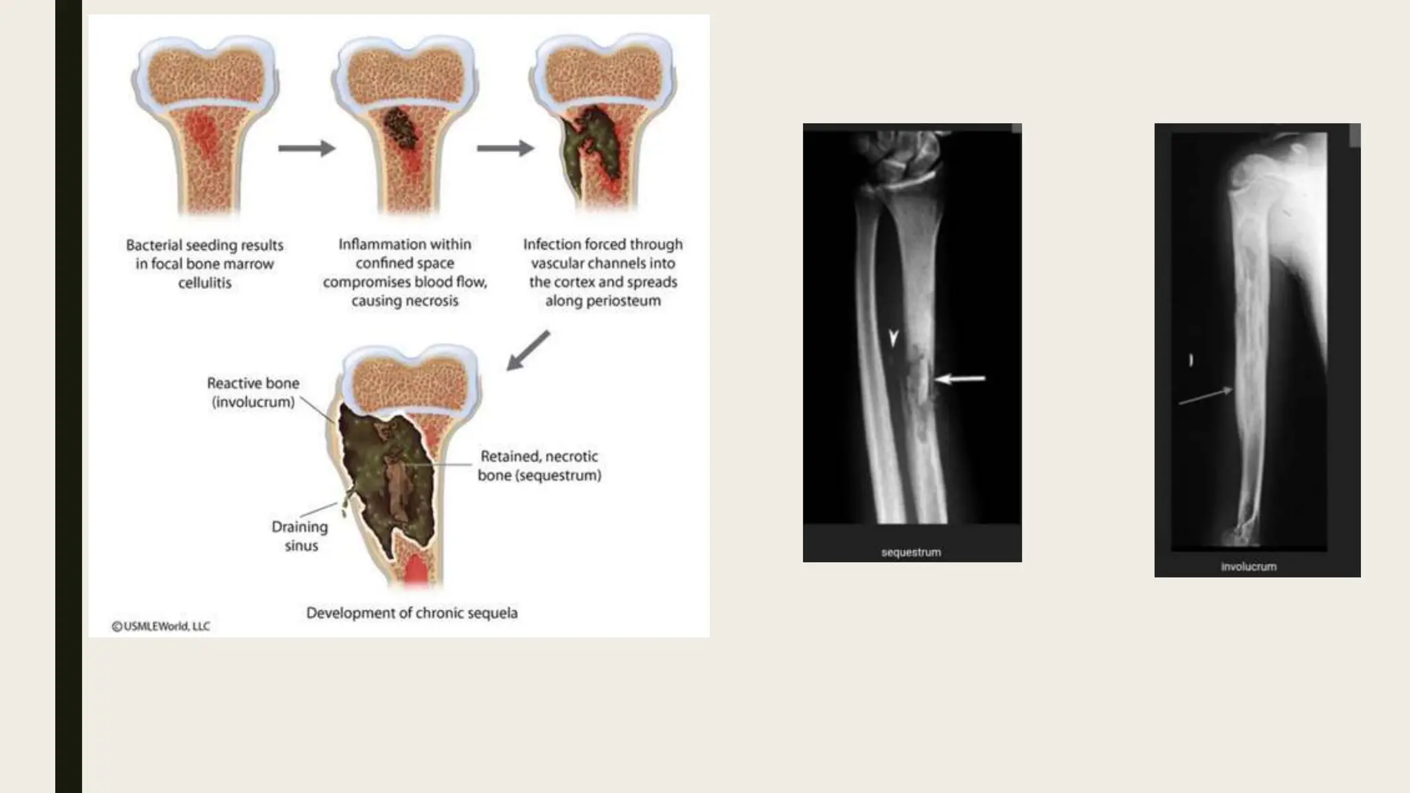
OSTEOMYELITIS
Leelawathy A/P Pandian
Definition
Inflammation or swelling of bone and marrow that is
cause by an infecting organism
Epidemiology
1. Location
Dialysis patient  spine and ribs
IV drug user  medial or lateral clavicle
Diabetics  foot and decubitus ulcer
■ neonates: metaphysis and/or epiphysis
■ children: metaphysis
■ adults: epiphyses and subchondral regions
Risk Factors
■ Recent trauma or surgery
■ Immunocompromised patients
■ IV drug user
■ Poor vascular supply
■ Peripheral neuropathy
■ Systemic conditions (diabetes & sickle cell)
Etiology
Mechanism of spread
1. Hematogenous
■ originated or transported by blood
■ may be due to bacterial or viral systemic illness
■ most common etiology in children
■ vertebrae are the most common hematogenous site in adults
■ S. aureus is the most common organism
2. Contiguous-spread
■ associated with previous surgery, trauma, wounds, or poor vascularity
■ can be bacterial (most common), mycobacterial, or fungal in nature
3. Direct-inoculation
■ penetrating injuries
■ open fractures
■ surgical contamination
Associated conditions
1. Orthopedic manifestations
■ septic arthritis
■ Abscess
2. Medical conditions
■ immunosuppression
■ dialysis
■ IV drug use
■ diabetes
■ poor nutrition
■ vascular disease
Classification
■ Waldvogel classification
■ Cierny-Mader classification
Presentation
History
■ duration
■ prior treatments
■ characterize host (Immunocompromised)
Symptoms
■ pain
■ fever
-more common in acute osteomyelitis
Physical exam
1. Vital signs
■ fever, tachycardia, and hypotension (sepsis)
2. Inspection
■ erythema, tenderness, and edema are commonly seen
■ draining sinus tract
- more common in chronic osteomyelitis
- if able to probe bone through sinus, chronic osteomyelitis is present
3. Motion
■ limp and/or pain inhibition with weight-bearing or motion may be present
■ assess the joints above and below the area of concern
4. Neurovascular
■ assessment of vascular insufficiency locally or systemically
Imaging
Xray
■ Osteomyelitis must extend at least 1 cm and compromise 30 to 50% of bone mineral
content to produce noticeable changes on plain radiographs.
■ Early findings may be subtle, and changes may not be obvious until 5 to 7 days from the
onset in children and 10 to 14 days in adults.
■ Chronic
-bone lucency, sclerotic rim, osteopenia, periosteal reaction (codmans triangle)
-sequestrum: devitalized bone that serves as a nidus for infection
-involucrum: formation of new bone around an area of bony necrosis
-cloaca: gap in the cortex of a bone that allows the drainage of pus or other material
from the bone into the adjacent tissues.
CT Scan
Indications
■ assist in diagnosis and surgical planning by identifying necrotic bone
■ sensitivity and specificity may be affected by hardware artifact and scatter
MRI
Indications
■ assists in the diagnosis and surgical planning
■ best test for diagnosing early osteomyelitis and localizing infection
Views
■ T2 sequences will show bone and soft tissue edema
Findings
■ penumbra sign (T1 - dark central abscess with bright internal wall and dark external sclerotic rim)
Sensitivity and specificity
■ if negative rules out osteomyelitis
■ if positive may overestimate the extent of osteomyelitis
Laboratory analysis
1. Leukocyte count (WBC)
■ only elevated in 1/3 of acute osteomyelitis
2. Erythrocyte sedimentation rate (ESR)
■ usually elevated in both acute and chronic osteomyelitis (90%)
■ a decrease in ESR after treatment is a favorable prognostic indicator
3. C-reactive protein
■ most sensitive test with elevation in 97% of cases
■ decreases faster than ESR in successfully treated patients
4. blood cultures
■ often negative, but may be used to guide therapy for hematogenous osteomyelitis
5. Microbiology
■ sinus tract cultures
- not reliable for guiding antibiotic therapy
■ culture of bone
-gold-standard for guiding antibiotic therapy
Treatment
Success in the treatment is dependent on various factors
1. Patient factors
■ immunocompetence of patient
■ nutritional status
2. Injury factors
■ the severity of the injury as demonstrated by segmental bone loss
3. Infection location
■ metaphyseal infections heal better than mid-diaphyseal infections
4. Other factors affecting prognosis and treatment include:
■ residual foreign materials and/or ischemic and necrotic tissues
■ inappropriate antibiotic coverage
■ lack of patient cooperation or desire
Non operative treatments
■ Suppressive antibiotics
antibiotics should be tailored to a specific organism, preferably after a bone biopsy is obtained
chronic suppressive antibiotics may be useful in patients who are immunocompromised or in whom surgery is
not feasible
high rates of recurrence if suppressive antibiotics are discontinued
Indications
■ when operative intervention is not feasible
■ Hyperbaric oxygen therapy
Indications
■ can be used as addition in refractory osteomyelitis
Operative treatments
Irrigation and debridement followed by organism specific antibiotics
Indications
■ acute osteomyelitis that fails to improve on IV antibiotics
■ subacute osteomyelitis
■ abscess formation
■ chronic osteomyelitis
■ draining sinus
Amputation
■ amputation at the level that will eradicate infected tissue to healing tissue with capacity to heal
Indications
■ chronic infection with pervasive wound or bone damage that is unable to be salvaged
Complications
1. Persistence or extension of infection
2. Amputation
3. Sepsis
4. Malignant transformation
■ 1% in chronic osteomyelitis
■ most commonly squamous cell carcinoma (Marjolin's ulcer)
■ risk factors
-chronic draining sinus
■ treatment
-wide surgical resection

Osteomyelitis Ortho Slides Hospital.pptx

  • 1.
  • 2.
    Definition Inflammation or swellingof bone and marrow that is cause by an infecting organism
  • 3.
    Epidemiology 1. Location Dialysis patient spine and ribs IV drug user  medial or lateral clavicle Diabetics  foot and decubitus ulcer ■ neonates: metaphysis and/or epiphysis ■ children: metaphysis ■ adults: epiphyses and subchondral regions
  • 4.
    Risk Factors ■ Recenttrauma or surgery ■ Immunocompromised patients ■ IV drug user ■ Poor vascular supply ■ Peripheral neuropathy ■ Systemic conditions (diabetes & sickle cell)
  • 5.
    Etiology Mechanism of spread 1.Hematogenous ■ originated or transported by blood ■ may be due to bacterial or viral systemic illness ■ most common etiology in children ■ vertebrae are the most common hematogenous site in adults ■ S. aureus is the most common organism 2. Contiguous-spread ■ associated with previous surgery, trauma, wounds, or poor vascularity ■ can be bacterial (most common), mycobacterial, or fungal in nature 3. Direct-inoculation ■ penetrating injuries ■ open fractures ■ surgical contamination
  • 7.
    Associated conditions 1. Orthopedicmanifestations ■ septic arthritis ■ Abscess 2. Medical conditions ■ immunosuppression ■ dialysis ■ IV drug use ■ diabetes ■ poor nutrition ■ vascular disease
  • 8.
  • 9.
  • 10.
    Presentation History ■ duration ■ priortreatments ■ characterize host (Immunocompromised) Symptoms ■ pain ■ fever -more common in acute osteomyelitis
  • 11.
    Physical exam 1. Vitalsigns ■ fever, tachycardia, and hypotension (sepsis) 2. Inspection ■ erythema, tenderness, and edema are commonly seen ■ draining sinus tract - more common in chronic osteomyelitis - if able to probe bone through sinus, chronic osteomyelitis is present 3. Motion ■ limp and/or pain inhibition with weight-bearing or motion may be present ■ assess the joints above and below the area of concern 4. Neurovascular ■ assessment of vascular insufficiency locally or systemically
  • 12.
    Imaging Xray ■ Osteomyelitis mustextend at least 1 cm and compromise 30 to 50% of bone mineral content to produce noticeable changes on plain radiographs. ■ Early findings may be subtle, and changes may not be obvious until 5 to 7 days from the onset in children and 10 to 14 days in adults. ■ Chronic -bone lucency, sclerotic rim, osteopenia, periosteal reaction (codmans triangle) -sequestrum: devitalized bone that serves as a nidus for infection -involucrum: formation of new bone around an area of bony necrosis -cloaca: gap in the cortex of a bone that allows the drainage of pus or other material from the bone into the adjacent tissues.
  • 15.
    CT Scan Indications ■ assistin diagnosis and surgical planning by identifying necrotic bone ■ sensitivity and specificity may be affected by hardware artifact and scatter MRI Indications ■ assists in the diagnosis and surgical planning ■ best test for diagnosing early osteomyelitis and localizing infection Views ■ T2 sequences will show bone and soft tissue edema Findings ■ penumbra sign (T1 - dark central abscess with bright internal wall and dark external sclerotic rim) Sensitivity and specificity ■ if negative rules out osteomyelitis ■ if positive may overestimate the extent of osteomyelitis
  • 16.
    Laboratory analysis 1. Leukocytecount (WBC) ■ only elevated in 1/3 of acute osteomyelitis 2. Erythrocyte sedimentation rate (ESR) ■ usually elevated in both acute and chronic osteomyelitis (90%) ■ a decrease in ESR after treatment is a favorable prognostic indicator 3. C-reactive protein ■ most sensitive test with elevation in 97% of cases ■ decreases faster than ESR in successfully treated patients 4. blood cultures ■ often negative, but may be used to guide therapy for hematogenous osteomyelitis 5. Microbiology ■ sinus tract cultures - not reliable for guiding antibiotic therapy ■ culture of bone -gold-standard for guiding antibiotic therapy
  • 17.
    Treatment Success in thetreatment is dependent on various factors 1. Patient factors ■ immunocompetence of patient ■ nutritional status 2. Injury factors ■ the severity of the injury as demonstrated by segmental bone loss 3. Infection location ■ metaphyseal infections heal better than mid-diaphyseal infections 4. Other factors affecting prognosis and treatment include: ■ residual foreign materials and/or ischemic and necrotic tissues ■ inappropriate antibiotic coverage ■ lack of patient cooperation or desire
  • 18.
    Non operative treatments ■Suppressive antibiotics antibiotics should be tailored to a specific organism, preferably after a bone biopsy is obtained chronic suppressive antibiotics may be useful in patients who are immunocompromised or in whom surgery is not feasible high rates of recurrence if suppressive antibiotics are discontinued Indications ■ when operative intervention is not feasible ■ Hyperbaric oxygen therapy Indications ■ can be used as addition in refractory osteomyelitis
  • 20.
    Operative treatments Irrigation anddebridement followed by organism specific antibiotics Indications ■ acute osteomyelitis that fails to improve on IV antibiotics ■ subacute osteomyelitis ■ abscess formation ■ chronic osteomyelitis ■ draining sinus Amputation ■ amputation at the level that will eradicate infected tissue to healing tissue with capacity to heal Indications ■ chronic infection with pervasive wound or bone damage that is unable to be salvaged
  • 21.
    Complications 1. Persistence orextension of infection 2. Amputation 3. Sepsis 4. Malignant transformation ■ 1% in chronic osteomyelitis ■ most commonly squamous cell carcinoma (Marjolin's ulcer) ■ risk factors -chronic draining sinus ■ treatment -wide surgical resection

Editor's Notes

  • #7 Staphylococcus aureus Haemophillus influenza Ac acromioclavicular joint Sc sternoclavicular joint
  • #8 Vascular disesase Peripherary artery disease Varicose vein Chronic venous insufficiency carotid artery disease Buergers disease
  • #9 acute -within 2 weeks subacute -within one to several months chronic -after several months Acute: <2 weeks · Subacute: 2-6 weeks · Chronic: >6 weeks;
  • #13 Nidus place for bacteria to develop and multiply
  • #19 Hyperbarbaric oxygen therapy Breathing oxygen in pressurized chamber Allows lungs to gather three times more oxygen Help body grow new skin blood vessel and connective tissue Refractory osteomyletis Tht does not respond or returns after appropritate treatment
  • #20 Anaerobes organism Clostridium Bacteroider fragilis Lactobacillus Peptostreptococus
  • #21 all devitalized and necrotic tissue should be removed extensive debridement is essential to eradicate the infection sequestrum must be eliminated from the body, or infection is likely to recur dead space management goal is to replace dead bone and scar tissue with vascularized tissue options include vascularized bone grafts local tissue flaps or free flaps antibiotic-impregnated acrylic beads (PMMA)  vacuum-assisted closure improves wound healing and dead space closure in multiple ways   remove interstitial fluids eliminate superficial purulence or slime allow arterioles to dilate, which allows granulation tissue to proliferate decrease in capillary afterload to promote inflow of blood bony stability is required for successful eradication of infection external fixation preferred to internal fixation