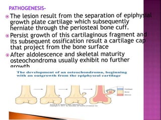
Roll no. 1823
Chandrakala meena
•the first hint of a tumor presence in bone is -
pathologic fractures.
Benign bone tumors-
 Predominant tissue-
 1.bone forming

 2.Cartilage forming
 3.Unknown origin
 Benign tumors-
 1.osteoma
 Osteoblastoma& osteiod
osteoma
 2.chondroma
 Osteochondroma
 Chondroblastoma
 Chondromyxoid fibrom
 3.giant cell tumor
 Osteochondroma is a bony exostosis projecting
from the external surface of a bone.
 It is usually has a hyline lined cartilagious cap.
 It is the most common benign bone tumor .
 Arise from- metaphysis of distal femur and
proximal tibia.
 Osteochondroma present as slow growing masses
which can be painful if the stalk is fractured or
they imping on a nerve.
 male female ratio 2:1
 75% occure before the age of 20
Causes due to-
 Abnormality in the growth plate.
 genetic disorder cause- heredietry multiple
osteochondroma result of mutation in gene
EXT1& EXT2.
1. Mashroom shaped
 2.Size about 1cm to 20 cm .
3. Outer layer of osteochondroma
composed of benign hyline cartilage
4. Newly formed bone forms the
inner portion of the head &
stalk,cortex of the stalk omerges
with cortex of host bone.

1.covered by thin layer of cartilage
2.cap made up of hyline cartilage
3.binucleate chondrocyte present
in
lacuna
4.contain hyline cartilage & bony
cartilage
histology-
Gross-
--Radiograph of an
osteochondroma from
the distal femur.
-osteochondroma are
sessile and
pedunculated.
1.Fracture
2.Bony deformity
3. Vascular injury
4.mechanical irritation
5.Malignancy - 5% to 20% multiple
heridietry exostosis progress to osteosarcoma.
6. Bursae formation.
osteochondroma.pptx

osteochondroma.pptx

  • 1.
  • 2.
    •the first hintof a tumor presence in bone is - pathologic fractures. Benign bone tumors-
  • 3.
     Predominant tissue- 1.bone forming   2.Cartilage forming  3.Unknown origin  Benign tumors-  1.osteoma  Osteoblastoma& osteiod osteoma  2.chondroma  Osteochondroma  Chondroblastoma  Chondromyxoid fibrom  3.giant cell tumor
  • 4.
     Osteochondroma isa bony exostosis projecting from the external surface of a bone.  It is usually has a hyline lined cartilagious cap.  It is the most common benign bone tumor .  Arise from- metaphysis of distal femur and proximal tibia.  Osteochondroma present as slow growing masses which can be painful if the stalk is fractured or they imping on a nerve.
  • 5.
     male femaleratio 2:1  75% occure before the age of 20 Causes due to-  Abnormality in the growth plate.  genetic disorder cause- heredietry multiple osteochondroma result of mutation in gene EXT1& EXT2.
  • 7.
    1. Mashroom shaped 2.Size about 1cm to 20 cm . 3. Outer layer of osteochondroma composed of benign hyline cartilage 4. Newly formed bone forms the inner portion of the head & stalk,cortex of the stalk omerges with cortex of host bone.  1.covered by thin layer of cartilage 2.cap made up of hyline cartilage 3.binucleate chondrocyte present in lacuna 4.contain hyline cartilage & bony cartilage histology- Gross-
  • 8.
    --Radiograph of an osteochondromafrom the distal femur. -osteochondroma are sessile and pedunculated.
  • 9.
    1.Fracture 2.Bony deformity 3. Vascularinjury 4.mechanical irritation 5.Malignancy - 5% to 20% multiple heridietry exostosis progress to osteosarcoma. 6. Bursae formation.