1. B Fall 2015 C
Optometry
FALL 2015
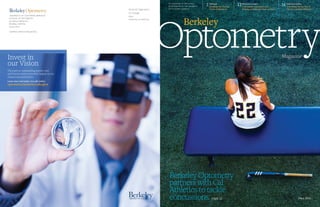
Berkeley Optometry
partners with Cal
Athletics to tackle
concussions. page 12
Berkeley
THE MAGAZINE OF THE SCHOOL
OF OPTOMETRY AT THE UNIVERSITY
OF CALIFORNIA, BERKELEY
Djibouti
Breaking the Vicious
Cycle of Blindness
2 Return to Learn
Concussion evaluation and
helping students get back to class
12 Infection Killer
Unlocking the secrets of
the eye’s natural defenses
16
UNIVERSITY OF CALIFORNIA, BERKELEY
SCHOOL OF OPTOMETRY
302 Minor Hall #2020
Berkeley, California
94720-2020
ADDRESS SERVICE REQUESTED
Nonprofit Organization
U.S. Postage
PAID
University of California
Invest in
our Vision
The path to outstanding patient care
and vision science research begins in our
classrooms and clinics.
Learn more and make your gift online.
optometry.berkeley.edu/give
Magazine
3. 2 Fall 2015 3
IN VIEW
OptometryNews
Branding, renovation
projects, and our
extraordinary clinic
By now you’ve
probably noticed that
our magazine looks
very different from
past editions. The
updated design is the
first step in a greater
plan to bring our
school’s publications
—both printed and
web-based—in
line with the new
branding established
for the Berkeley
campus. And while
the design is new, the
content will continue to focus on the groundbreaking research,
teaching and clinical care that is the foundation of what we do
here at Berkeley Optometry. I hope you enjoy it and welcome
your feedback.
Many of you will already know that a priority for the School
moving forward is to renovate and extend our clinic. The clinic
is the life-blood of our school; it is the foundation upon which
we build our research and our education. As always, there is great
news to share. Over the last year, we’ve seen 70,000 patients!
The strength of the clinic is our extraordinary faculty and staff,
talented interns, dedicated residents and the unparalleled level of
patient care that they provide—that is what sets us apart; this is
what makes us Berkeley Optometry. I look forward to you joining
me as we embark on an era of fundraising to build a facility
worthy of the great works that happen here every single day.
Lastly, we have been working hard to improve the physical
appearance of our school. I believe that the way our building
looks—especially high traffic areas such as Minor Hall’s ground
floor and entranceway—establishes a tone for the institution and
plays an important role in attracting top level students, faculty
and staff. We are the top rated Optometry school in the country,
and it’s important that we do everything we can to look the
part. With this in mind, the ground floor of Minor Hall has been
restored and renovated and now properly reflects our institution’s
preeminence, and the building’s amazing history (think
Manhattan project and the development of modern day contact
lenses). Please come by and take a look!
—John Flanagan
Shown is an image of human retinal cells taken with adaptive optics
scanning laser ophthalmoscopy, an instrument that allows doctors
to examine the back of a patient's eye. Real-time eye tracking allows
researchers to optically stimulate individual photoreceptors, as
illustrated by the green focused beam in the main figure and in the
inset, which shows a magnified mosaic of cones, the cells in the
retina responsible for color vision. The dark branched structures are
shadows of blood vessels. (Image courtesy of Lawrence Sincich and
Kady Bruce, University of Alabama Birmingham.)
DEAN'S MESSAGE
For most of us, sight is how we connect to the world
around us. But for the 285 million people around the
globe that are blind or visually impaired, the joys and
benefits of sight are unattainable. Now, a bold new
initiative to restore vision by regenerating neurons
and neural connections in the visual system that have
been lost or damaged, is underway. In the first round of
funded projects, the National Eye Institute (NEI) has
committed $17.9 million toward building and testing
innovative new ophthalmic imaging systems that will
be essential for evaluating new treatments as they are
developed. Of those funds, $3.2 million are going to
a retinal mapping project led by Austin Roorda, a UC
Berkeley professor of optometry and vision science.
The Audacious Goals Initiative brings together scien-
tists from around the country who, specializing in diverse
fields of vision science, will work simultaneously toward
more than one solution.
“These ambitious projects will give us a window into
the visual system,” said NEI Director Paul A. Sieving, M.D.,
Ph.D. in an NEI press announcement. “Tools developed
will enhance the study of functional changes in the retina
and optic nerve, in real-time and at the cellular level, and
will be indispensable when evaluating new regenerative
therapies for eye diseases.”
Dr. Roorda is the principal investigator of the retinal
mapping project. He will be working with E.J. Chichilnisky
and Daniel Palanker, both professors of ophthalmology at
An Audacious Goal:
Restoring Sight to the Blind
Artist's rendering
of neural activity
in the retina.
Light that enters
the eye activates
rod and cone
photoreceptors,
which in turn
activate retinal
ganglion cells.
Signals travel
to the brain via
retinal ganglion
cell axons.
Welcomes
As we celebrate the start of a new
academic year we’d like to welcome
some new additions to the Berkeley
Optometry family
We are delighted to introduce
our new librarian Jeff Loo, and
Eric Craypo, our new Director
of Communications. Please also
welcome new clinic faculty Drs.
Ashley Craven, Alexandra (Sasha)
Cross, Sarah Kochik, Jing Zeng,
Jennifer Lim and Melissa Valdellon,
and recent appointees Drs. Neda
Ghanbari, Stephanie Chen Joo, Anne
Tasaki, Jackie Theis and Tan Truong.
Please give them all a warm welcome
to Berkeley Optometry!
PHOTOGRAPHYNATIONALEYEINSTITUTE.
Stanford University, and B. Hyle Park, an assistant profes-
sor of bioengineering at UC Riverside.
According to NEI, “the mapping project will enable
scientists to stimulate individual neurons and observe
other cells as they become active in response. Mapping
these intricate signaling patterns will help explain how
the retina processes visual information before it is sent
to the brain, and will be an important tool for monitoring
function in regenerated cells.”
News from the School of
Optometry and beyond.
4. 4 Fall 2015 5
IN VIEW
OptometryNews
Here’s the catch: a person with Diabetic Retinopathy
—the leading cause of blindness in working age adults
around the world—can still see well when they are in
the early stages of the disease. But if they wait until
visual symptoms appear to seek help, it's often too late;
and the treatment is unlikely to work. Then as vision
becomes further impaired, they blame the treatment for
the blindness, which discourages others from seeking
treatment. It’s a vicious cycle of blindness.
The solution, explains Berkeley Optometry’s Dr. Jorge
Cuadros is early detection and patient engagement: “If
you treat it early, 90% of people can maintain good vision.
You can avoid vision impairment.” But in many places
around the world—including underserved communities
in this country—the screening of diabetic patients is
hampered by both a dearth of equipment and clinicians
trained to do the assessments. Djibouti, for example, has
only two ophthalmologists for the entire country.
With this in mind, Dr. Cuadros traveled to Djibouti, an
east African country of 800,000 located on the Red Sea,
late last year to train a group of nurses and other clinicians
to operate a digital retinal camera and to detect Diabetic
Retinopathy using a free and non-proprietary software
called EyePACS—a program developed by Dr. Cuadros
and Dr. Wyatt Tellis of UCSF.
Dr. Ethan Chorin, founder and director of Perim Asso-
ciates, the Berkeley-based international policy consultancy
that helped introduce EyePACS in Djibouti, says “EyePACS
is a great example of technology in service to development.
It is simple to use, responds to a widespread need, and
empowers local clinicians to do their own screening and
treatments, rather than relying on external aid.”
During the initial screening of 140 people, the group
found that 64 had some diabetic retinal disease and will
need to be counseled and monitored closely to avoid
vision impairment in the future, and 20 had severe diabetic
retinal disease requiring immediate treatment. For these
84 people, the screenings have likely prevented blindness.
But it was too late for 5 of those 20 patients—the disease
had progressed so far that treatment is unlikely to help. A
devastating reminder that early detection is critical.
In the next phase of the project, Cuadros anticipates
that newly trained health care professionals—traveling
around the country—will be able to screen 50% of Dji-
bouti’s diabetic population. That’s about 40,000 people.
He thinks it can be done by the end of 2017. The program
will follow the success of similar endeavors that Cuadros
initiated in the U.S., Canada and Mexico. All told, over
340,000 people with diabetes have been screened using
the EyePACS system, saving thousands from blindness.
Dr. Cuadros recently told the Huffington Post, “If we can
test early and widely, we can save many from this fate. The
testing technology is now there, it's simple, portable, and
it works really well.”
Bono Aims to
Rock Blindness
Bono, lead singer for the rock band U2, and sunglasses
manufacturer Revo have partnered to create the “Buy
Vision, Give Sight” program. For each pair of Revo
sunglasses sold, $10—up to a total of $10 million—will
be donated by Revo to the “Buy Vision, Give Sight”
initiative. The goal of the initiative is to help prevent
vision impairment and blindness in more than 5 million
people by 2020.
Bono was diagnosed with glaucoma 20 years ago, and
although he has had access to excellent treatment, the
experience inspired him to seek ways increase access to
basic eye care for others. “Thanks to good medical care
my eyes are okay, but tens of millions of people around
the world with sight problems don’t have access to
glasses, or even a basic eye test. Poor eyesight may not be
life-threatening, but it dramatically affects your life and
your livelihood if you aren’t able to fix it,” said Bono in a
Revo press release announcing the partnership.
Revo and Bono are partnering with the non-profit
Brien Holden Vision Institute, who believe that sight is
a fundamental right for all humans. The group’s mission
is to provide sustainable solutions for eye care and end
avoidable blindness and vision impairment in under-re-
sourced communities. According to Revo, the funds col-
lected “will help pay for basic eye care services, such as
eye tests and prescription glasses, and build stronger eye
care services in target communities for the longer term
by training local people to provide eye care and detect
eye diseases in their communities.”
Revo’s line of sunglasses are now available at the
Berkeley Optometry eye clinic.
You are studying for your optics and
anatomy classes in the beautiful, cozy, and
sunlit Pamela & Kenneth Fong Optometry
Library, but your eyes wander every now
and then, and you catch a glimpse of two
large and colorful paintings of Snoopy,
the beloved Peanuts character created by
Charles Schulz. You think to yourself, what
is Snoopy doing in our Optometry library?
It’s a good question—these are no ordinary
paintings!
The two lithographs were donated
in 2006 by Dr. Pamela Fong ’77, whose
generous donation in 2000 helped establish
the Fong Library. They were created by artist
UP NEXT:
Online
Education!
Change is afoot at
Berkeley Optometry,
and one of our
most exciting new
endeavors is online
education. Soon
optometrists around
the state, country—
and even the world!—
will have access to
interactive cases and
stimulating courses
via the Berkeley
Optometry Online
Education program.
Earning Continuing
Education credits
just got a whole lot
easier—and more
interesting. Coming
to a laptop near
you in 2016!
This Dean’s
a LiferThe College of Optometrists has invited John Flanagan,
Dean of UC Berkeley’s School of Optometry, to become
a Life Fellow of the College in recognition of the
outstanding contribution he has made to the profession
of optometry. Specifically, the award recognizes Dean
Flanagan’s contribution to research into glaucoma
and diabetic eye disease, and to the development of
optometric education in the UK and Canada.
Before coming to UC Berkeley in 2014, Dean Flanagan
held faculty positions at the School of Optometry
and Vision Science, University of Waterloo, and the
Department of Ophthalmology and Vision Sciences,
University of Toronto. He was Director of the Glaucoma
Research Unit, Toronto Western Research Institute,
and a Senior Scientist at the Toronto Western Hospital,
University Health Network.
The College of Optometrists is the professional,
scientific and examining body for optometry in the UK,
working for the public benefit.
Breaking the Vicious Cycle
of Blindness in Djibouti
A patient in the Djibouti General Hospital awaits
the results of retinal photography. Fortunately,
she didn't have retinal disease.
>OVERHEARD “I spent hours at the Oakland
Zoo, often surrounded by school kids on
field trips, to observe the different animals.
Sure enough, when goats, antelope and other
grazing prey animals put their head down to
eat, their eyes rotated to maintain the pupils’
horizontal alignment with the ground.”
Vision scientist and UC Berkeley professor of optometry Martin
Banks on research that suggests pupil shape reveals whether one
is hunter or hunted. For grazing animals, seeing panoramically—
even when eating—is key to detecting approaching predators.
Opto Dog?
Tom Everhart, the only fine artist legally
authorized to use characters from the
Peanuts comic strip. Everhart met Schulz
in 1980 and they instantly forged a bond
that would become a lifelong friendship.
Everhart’s paintings and lithographs have
been exhibited worldwide at the Louvre
in Paris, Los Angeles, New York, Montreal,
Tokyo, Rome, Venice—and at the Fong
library! BY JANE POUVARANUKOAH
5. 6 Fall 2015 7
Top10
NAVIGON North America $59.99
MORE THAN NAVIGATION: LANE GUIDANCE, JUNCTION VIEWS, SPEED INFO,
PEDESTRIAN NAVIGATION, AND MUCH MORE!
Navigon is worth its price, even next to free apps like Google Maps.
You'll get a more extensive feature set, innovative optional plug-ins,
and a more informative display.
Bigger and Brighter
$Free
NEED A MAGNIFIER BUT ON
A BUDGET...BIGGER AND
BRIGHTER TO THE RESCUE!
This application helps you to
read small letters by making
the images bigger, brighter
and clearer with scientific
color changing methods.
Glucose
Buddy
$Free
IF YOU ARE DIABETIC WITH LOW
VISION, THIS APP IS WAITING TO
HELP YOU STAY HEALTHY!
Glucose Buddy is a data storage
utility for people with diabetes.
Users can manually enter
glucose numbers, carbohydrate
consumption, insulin dosages,
and activities. Then, you can
view all of your data on a free
glucosebuddy.com online account.
Dragon Dictation $Free
STOP TYPING AND START SPEAKING
Use your voice to dictate a text
message or email, create Facebook
status updates or a Tweet, and
anything in between—simply
speak and see your text
content appear. Dragon
Dictation recognizes and
transcribes your words
quickly and easily.
MedHelper $Free
YOUR PERSONAL
HEALTHCARE ASSISTANT
Track your prescription medications,
treatment, and appointment
schedules. Set alarm reminders and
a log of all your past doses. It’s easy
to install and MedHelper is ready to
become your 24/7 healthcare assistant.
Vision Sim $Free
HAVING LOW VISION CAN BE FRUSTRATING,
ESPECIALLY WHEN FRIENDS AND FAMILY
CANNOT RELATE!
For those suffering from macular
degeneration, diabetic retinopathy, glaucoma, or
cataracts, this app shows people the world through
your eyes. VisionSim turns on the camera of the
iPhone and filters the lens with distortion that
mimics the selected eye condition.
5
4
6
8
CamFind $Free
SEARCH THE PHYSICAL WORLD!
This mobile visual search engine allows
you to search for anything from your
mobile phone just by taking a picture.
KNFB Reader$99.99
HAVING TROUBLE SEEING DOCUMENTS?
HAVE KNFB READER DO THE WORK FOR YOU!
It's not always convenient to use a powerful magnifying glass like the one being
displayed by Dr. Katsikos in the photo above. The KNFB app converts printed text into
high quality speech to provide accurate, fast, and efficient access to both single and
multiple page documents with the tap of a button on the iPhone. So easy!1>
2
7
9
TapTapSee $Free
CAN’T IDENTIFY THE
OBJECTS AROUND YOU?
TAPTAPSEE CAN!
TapTapSee is a mobile
camera application designed
specifically for the blind and
visually impaired. The app
utilizes the iDevice’s camera
and VoiceOver functions
to photograph objects and
identify them out loud for you!
3
Ashley’s ListOur Ian L. Bailey Low Vision Resident,
Dr. Ashley Katsikos, finds that her patients are reaping huge benefits
from both iPhone and Android apps designed to make basic tasks
such as reading, getting places, or counting money a whole lot
easier. There’s even an app that will show family and friends what
it feels like to have low vision! Here’s Dr. Katsikos’ top ten list.
10 LookTel $9.99
COUNT YOUR MONEY THE NEW-FASHIONED WAY!
LookTel Money Reader instantly recognizes
currency and speaks the denomination, enabling people with visual
impairments or blindness to quickly and easily identify and count
bills. Several currencies are supported including the US Dollar,
Euro, British Pound, Canadian Dollar, and Australian Dollar.
6. 8 Fall 2015 9
See more of our good times on
instagram @BerkeleyOptometry
ThroughourEyes Experience the life and times of
Berkeley Optometry students
through their (smartphone) lens!
STUDENTS
Maria Jen | CLASS OF 2017Jane Pouvaranukoah | CLASS OF 2017
Yay done with finals, practicals, quals, and hopefully
second year!! #twomoreyears #kidsatheart
Optobears at the Big Game: Cal vs. Stanford
#GOBEARS
Just ran our first 10K together! Team #EyesEyesBaby Two months ago I saw my first Team Care patient
with Dr. Chu, and today, I saw my LAST patient
for the semester! This semester is passing by so
quickly... #berkeleyoptometry
Optometry rafting trip! Lots of lasting
friendships were made this day.
The selfie game at Berkeley Optometry is strong! Whachu know about gonioscopy? #berkeleyoptometry
Keratometry...yo mama tree.
Richard Phan | CLASS OF 2018Milan Lockhart | CLASS OF 2018
7. STUDENTS
QuickFacts
A look at the class of 2019, and what they
will experience over the course of their
four years at Berkeley Optometry.
Applicants Academics Student Profile Student Experience
Class of 2019
284Applications
124Interviews
69Students
matriculated
3.49Average GPA in Bio, Chem and Physics
4.00-2.75Overall GPA range
351Academic Average
on the OAT
3.57Undergraduate GPA
52In-State
60Women
9Men
23Average age
440hours
Preclinical Laboratory Training
2nd year in program
When students start seeing
real patients
100%Number of students who learn
how to do a full eye exam by
end of first semester
2,500Individual patient
encounters by graduation
40%Of grads go into coveted
residency slots throughout the US
12Out-of-State
1 from China,
1 from Iran,
and 3 from
Canada
>
>
>
>
Fall 2015 11
8. Fall 2015 13
J
osephine Devanbu was a sophomore neuroscience
major at Brown University when she hit her head
at a dance party two years ago. “I knew I’d hit my
head too hard, but I didn’t expect there to be serious
consequences,” she says. When she described her
symptoms to student health services over the phone
a couple of days later, they wanted to rush her to the
hospital in an ambulance. “From the beginning, there
was a lot of tension between different assessments
of what I could and couldn’t do, about what I was
and wasn’t up to,” she says now. “I didn’t want to go to the hospital
at all. I had a lot of studying to do. Little did I know that I wasn’t
going to be doing much coursework for months,” she adds.
Hits to the head—on the athletic field, in the dorm, and on
the street—are a common form of student injury. About 500
concussions are diagnosed by the UC Berkeley student health
center each year (less than one-tenth of those are suffered by
athletes). And yet the injury is notoriously tricky to diagnose and
treat. Two UC Berkeley doctors, one a sports physician and the
other an optometrist, are deploying the tools of optometry to help
bring greater clarity to the field and to ease the way for all students,
whether athletes or not, back into their academic saddles.
Concussion symptoms are due to a complex pathophysiologic
change in the brain, and at times these symptoms may be subtle,
says Dr. Casey Batten, MD, UC Berkeley Head Team Physician, and
co-investigator of a new study examining the use of optometry
measurements to evaluate mild head injury patients. “Concussions
do not entail structural damage, but only functional disturbance,”
Batten says. If you’re experiencing profound visual changes, or
have other signs or symptoms suggestive of possible swelling or
bleeding inside your skull, you may not be concussed but may
have another, more serious traumatic brain injury. However, if
none of those more salient signs of damage are present after a
hit to the head, and yet you are experiencing amnesia, say, or
dizziness, nausea, headaches, exhaustion, or confusion, then you
may have a concussion, says Batten.
Currently, diagnosis is based largely on subjective patient reports
and these range all over the map, often depending on both the extent
and the context of the injury, says the project’s other co-investigator,
Dr. Jacqueline Theis, OD, FAAO, a Clinical Instructor and previous
resident in the UC Berkeley Binocular Vision and Neuro-Optometry
clinic. An athlete chomping at the bit to return to play, for example,
may minimize his symptoms, while a cyclist hit by a car may feel an
equivalent injury more keenly. With this project, the researchers are
hoping to employ optometric measurements, currently not a key
part of concussion diagnosis, to create a standardized, quantifiable,
and portable way to diagnose the shape-shifting disturbance. If the
researchers are right, the work will be a major new contribution by
optometry to the study and treatment of concussion, a territory
pretty much uncharted for optometrists except those specifically
treating vision-related concussion symptoms. A standardized
vision-based evaluation protocol would be extremely valuable to any
medic, sports doctor, military field doctor, or coach who regularly
needs to diagnose, evaluate, treat, or advocate for those who have hit
their heads, says Batten.
A Partnership Between Berkeley
Optometry and Cal Athletics
Aims to Streamline Concussion
Evaluation and Help Students
Get Back to Their Studies
Dr. Jacqueline Theis and Cal
Field Hockey player Keats
Iwanaga. A collaboration
between Cal Athletics and the
School of Optometry will get
baseline data on as many as
500 Division 1 athletes.
Return
toLearnAfter a Hit on the Head
BY GORDY SLACK
9. 14 Fall 2015 15
Batten should know. As a team physician, he often
evaluates players who have hit their heads on the field. He
would love to have more objective measurement tools to help
identify when it’s okay to send such a player back into play,
when she should be benched, and when she should be retired
from the game.
Optometry may be especially well positioned to detect
concussions because the network of muscular and neuronal
systems that govern the movement of eyes is so thoroughly
distributed throughout the brain. “There are so many pathways
related to vision,” says Theis, “a comprehensive eye movement
examination can correlate pathways across every lobe of the
brain.” Even subtle transient dysfunction may show up in
vision and eye movement.
“Fast eye movements employ coordination of nuclei and
gaze centers in the frontal lobe, midbrain, and brainstem.
Not to mention the subject would have to ‘see’ the object,
recognize the object that needs to be tracked, which requires
the afferent visual pathway from the eyes traversing back
to the occipital lobe, and onwards for further higher-order
processing in parietal and temporal lobes. If anything is wrong
in any of those pathways, it will show up in the patient’s visual
and eye movement assessment,” says Theis. Depending on the
kinds of problems you detect, it may be possible to infer the
focus and the degree of the injury. Just as importantly, it may
be possible to tell when recovery is achieved. “We hope to use
the eyes to objectively say, ‘You are done! Your concussion
is over. This was precisely when it ended.’ Although we are a
long way away from that goal,” says Theis.
Today, it is impossible to say precisely how a hit on the
head effects each of the visual pathways involved, but it is
clear that the effects are real, says Theis. And they certainly
should be measurable. So the first challenge to overcome is a
paucity of baseline data about how an individual’s eyes move
before they incur a concussion.
The Need for Baseline Data
The first step is to get reliable baseline data on normal eye
movement patterns, says Theis. Currently, there are a lot
of promising studies noting post-concussive changes in
vision, but without baseline data, it is hard to verify that
these changes are directly from the concussion. At their
new, well-scrubbed UC Berkeley Sports Vision Institute,
housed in the Simpson Center for Student Athlete High
Performance, adjacent to Memorial Stadium, Theis has
already conducted baseline screenings on 100 of UC
Berkeley’s Division 1 athletes. In coming months she is
hoping to screen 300 to 400 more.
The baseline screening, composed of ten simple tests,
takes only 15-20 minutes. Theis first checks visual acuity
to make sure each subject has 20/20 vision. Most student
athletes do. Next, she performs a series of tests that check
each eye’s ability to focus—including the gross amount,
accuracy, and ability to repeatedly engage and relax focus.
After concussion, Theis expects “a fatigue component”
to be introduced, which would explain why concussed
students can get exhausted in class just looking back and
forth from their notes to the blackboard.
Some studies suggest that the ability to bring your
eyes together, known as convergence, is decreased after a
concussion. Using a Bernell Convergence/Accommodation
ruler, Theis moves a small target closer and closer to the
subject’s face, recording how much the eyes can converge,
and then how quickly they can recover from their break in
convergence.
Theis also tests oculomotor coordination by
investigating how the two eyes work together when looking
at both distant and near objects. Perceiving single vision
with two eyes is a complex accomplishment requiring
coordination of nuclei, nerves, and muscles. If this system
fails, a patient will have double vision. “If you have injury or
swelling in the brain, it is likely this system could be affected,”
says Theis.
The researchers will also test visual tracking. “To look at
something and keep your eyes fixated in one place, a lot of
different neurons have to fire,” says Theis. Just by asking a
subject to track a visual target moving from side to side she
can test multiple pathways in the brain. The neural pathway
governing tracking from the midline to the right is different
than the one going to the left, or up from the midline, or
down, says Theis.
Finally, administering the Developmental Eye Movement
Test, Theis first measures how quickly subjects can read
numbers listed in vertical columns. Then she times them
reading number sequences arranged horizontally, but irregularly
interspaced as words would be in a book. “If the subject has
trouble with their saccades [rapid eye movements,] or has
trouble with their larger regression eye movements,” she says,
“it will take disproportionately longer for them to do this
test than to read the vertical columns. That would indicate
binocular vision related eye-tracking problems.”
“We know that, unfortunately, some small percentage of
our athletes are going to get concussions while engaged in
sports,” says Theis. “If an athlete’s tracking ability changes
post-concussion compared to their baseline measurements,
it would support that eye-tracking movements are impaired
by the concussion. If this is the case, one could argue that it
would be unjust to mandate someone with a concussion to be
expected to participate with the same scholastic rigor as they
did pre-concussion.”
“Currently, optometry doesn’t play a role in concussion
management,” Batten says. “Yet the literature shows that a
multidisciplinary approach in evaluation is the way to go. You
can’t put all your stock into symptom scores only, or balance
tests only, or cognitive tests only. You need to assess multiple
domains, and standardized visual tests could prove to be
vital,” he says.
In addition to developing techniques for better identifying
and quantifying the severity of concussions, Theis and Batten
also hope to develop techniques and practices for helping
athletes, and other students, make the smoothest possible
transition back to their studies.
“In the past, the majority of focus has been on clearing
student athletes to play sports,” says Batten. “But we’re
finding that concussion can also have a significant negative
affect on their academic performance. A lot of people who’ve
had concussions can often quickly return to training without
symptoms. But when they sit down to study or to read a book
they run into trouble.”
“One thing brains do is filter out irrelevant information
by dampening certain neuronal signals,” says Theis. “After a
concussion it seems that a lot less signal dampening is going
on. After a head injury, people often complain that they are
visually overwhelmed. Memory is affected, too. Without
filtering, it’s harder to distinguish what’s important and what’s
not, as well as maintain focus, which is key to productive
studying. People also tend to be very light sensitive, and
have trouble keeping their place while reading, or reading for
prolonged periods of time,” says Theis. Combined, all of these
symptoms can have a big impact on academic performance.
Impact on Studying
When Josephine Devanbu, the neuroscience student, tried
to study in the days and weeks after her head injury, her
persistent but low-grade headache intensified, she felt like
her body was in a fog, and like there was pressure building
in her skull, she remembers. Whereas she had put in long
hours of studying all through high school and college, after
her concussion she couldn’t focus for any length of time. She
felt discouraged, guilty, and increasingly anxious about the
workload she would face when she was finally better.
“There was this constant double take,” Devanbu says.
“Was I supposed to be worried about my health or was I
supposed to be worried about my coursework?”
Some of her professors “totally got it,” she says, and urged
her to focus on recovery. Others were skeptical and required
convincing by her doctor that she was not exaggerating her injury.
“Unfortunately, the scientific literature is equivocal on the
effectiveness of rehab,” says Theis. “Some studies show that if
you rehabilitate the visual problems they get better faster.
Other studies seem to show the opposite, that simple rest is the
best,” she says. “Nobody knows which it is. Or if maybe it’s
different in different cases. But if we can measure the effects
of rehabilitation on visual signs and symptoms, that could go
a long way.”
At academic institutions like Berkeley and Brown, the
pressure to get students back into class can be as great as the
pressure to get them back onto the field. Evidence suggests
that returning too soon to either activity can delay the healing
process, says Batten. If that’s so, it would be important to know,
he adds. Not least so that doctors can help advocate for academic
accommodations with the professors of concussed students like
Devanbu. Extensions on assignments and exams, for example,
can help make time for students to recover without unnecessary
stress. But before granting such accommodations, some
professors ask for quantitative science to validate the request.
Theis’s and Batten’s work, should produce that kind of evidence.
In recent years, concussion has demanded more and more
attention, both from sport and military doctors. Helping to
positively identify and measure the condition using objective
optometric measurements will not only help doctors of students,
soldiers, and others who have hit their heads know how best
to proceed, but it will also contribute key information to the
intensifying national discussion about concussion. Debates
based on vague definitions and assumptions are notoriously
difficult to resolve; this study could help to clarify the terms.
“We have a unique situation,” says Batten. “At Berkeley
not only do we have arguably the best optometry school in the
country, and a great intercollegiate athletics program, but our
offices are located right across the street from each other. The
ball is rolling now. If we can keep it moving down the field, this
will grow into something big.”
Optometry may be especially
well positioned to help detect
concussions because the network
of muscular and neuronal systems
that govern the movement of
eyes is so thoroughly distributed
throughout the brain.
Dr. Jacqueline Theis
takes the field while
Cal club lacrosse
players Emily
Brown and Danielle
Lecher practice
their stick work.
10. 16 Fall 2015 17
by Robin Meadows
Unlocking the
secrets of the eye’s
natural defenses
Infection
Killer
A
fter decades of trying,
Suzanne Fleiszig finally has
contact lenses for her mice—
her lab mice, that is. Now
she’s poised to learn how
contact lenses increase the
risk of eye infections and how
to reduce that risk.
When she began her PhD in
the mid-1980s, extended-wear
lenses had just been introduced. “The infection rate went
crazy,” says Fleiszig, Professor of Optometry and Vision
Science at the UC Berkeley School of Optometry. “I’ve
been working on it ever since.”
More than 40 million people in the U.S. wear contact
lenses and, according to a 2015 survey from the Centers
for Disease Control (CDC), nearly all of them engage
in behaviors that could increase their risk of eye
infection. These include keeping contact lens cases
too long, reusing contact lens solution, and sleeping
in contact lenses. “Infections are usually from people
doing things they’re not supposed to do,” says Pam
Satjawatcharaphong, Assistant Clinical Professor at the
UC Berkeley School of Optometry.
The students she sees at UC Berkeley eye clinics are
no exception. “They study so hard that they sleep in their
lenses a lot. But even when that’s FDA approved, there’s
a higher risk of infection,” says Satjawatcharaphong, who
is chief mentor of the UC Berkeley Cornea & Contact
Lens Residency. “You only have two eyes. Why risk it?”
The CDC survey also found that a third
of contact lenses wearers have gone to
the doctor for red or painful eyes. While
serious eye infections are not common, the
outcome can be severe. Infection can turn
corneas from clear to cloudy and can even
cause permanent vision loss.
Fleiszig studies bacteria called
Pseudomonas aeruginosa, the most common
cause of eye infections in people who
wear contact lenses. Most of the time, eyes
are remarkably infection-free. “You can put
ridiculously large numbers of bacteria
in them and nothing happens,” Fleiszig
says. “Why?” She thinks the answer is
key to thwarting infections caused by
contact lenses.
Her work could also have implications
beyond the eye. That’s because all of our
body surfaces—including our corneas, skin and digestive
tract—share an outer layer of epithelial cells that resist
bacteria. “We walk around all day long and are exposed
to bacteria but we don't get infected,” she notes. “We
want to identify the factors that maintain eye health.”
Understanding how eyes resist infection could help us
ward off infections in other parts of our bodies as well.
But learning how eyes stay sterile is a challenge. “It’s
difficult to study because nothing happens,” Fleiszig says.
In other words, how do you study the absence of infection?
Her approach includes figuring out how to recreate the
eye's resistance to infection in epithelial cells from human
corneas that are grown in the lab. These cultured cells are
defenseless. “Bacteria go crazy and win,” she says.
So far, she knows that bacteria can be attracted to
our corneas and want to invade them. But their efforts
are usually thwarted thanks to the eye’s defenses. For
example, the cells bristle with peptides—short amino
acids chains—that are antimicrobial. “Bacteria don’t swim
to the epithelial cell surface, they try to stay a distance
away,” Fleiszig says. But in the eye they can’t get very
far away. The tear fluid on our eyes is so thin—about a
tenth of the thickness of a hair—that the bacteria have
nowhere to go. “They’re squeezed between the cornea
and the eyelids,” she says. “It’s a hostile environment."
Like corneas, the insides of the eyelids are lined by
antimicrobial epithelial cells.
Fleiszig thinks contact lenses make this environment
friendlier by giving bacteria another place to live. If she's
right, contact lenses serve as launching pads where
bacteria figure out how get around the defenses of the
cornea’s outer layer. “They’re far enough away not to
be affected by the antimicrobial peptides, but could
be close enough to read and adapt to them,” she says.
“Pseudomonas aeruginosa is a master of adaptation—it’s a
huge problem in hospitals, it‘s a terrible, deadly bug.”
If bacteria adapt to the peptides, they could penetrate
the surface of the cornea and enter the
vulnerable layer underneath, which is called
the stroma. “That has the potential to destroy
the cornea, so it's better to prevent infection
rather than treat it once its ongoing,” she says.
In the past year, Fleiszig’s lab has
developed a microscope that can track
infections in eyes. “We can see individual
bacteria in the eyes of living mice,” she says.
And, even more recently, she got another
critical tool. “We just got a batch of contact
lenses for mice,” she says. “They’re tiny—a
mouse eye is only about two millimeters.” She
has to use the little lenses carefully because
the company only made a couple hundred,
and she's not sure if she’ll ever get any more.
The new contact lenses for mice will help
Fleiszig explore how eyes defend themselves
against infection, how contacts help bacteria
breach that defense, and how Pseudomonas aeruginosa
invades corneas. “I have my hypotheses and now I finally
have the tools,” she says. “For the first time, we’re in a
position to see what’s going on as infections develop.”
Ultimately, Fleiszig’s work could give contact lens
wearers a natural alternative to the drugs now used to
treat eye infections.
BY ROBIN MEADOWS
“Under-
standing
how eyes
resist infection
could help
us ward off
infections
in other
parts of
our bodies
as well.”
Facing page: Dr.
Suzanne Fleiszig in the
lab. Above: a mouse
contact lens—in
foreground—and a
human contact lens
rest on tip of a finger.
11. 18 Fall 2015 19
What inspired you to make the
transition from seeing patients
to designing frames?
A I was the junior partner with
two other optometrists where
in addition to patient care, each
of us took on the responsibility
of overseeing one area of our
practice. I was responsible for
overseeing our opticians, optical
boutique, and the buying for our
dispensary. I very soon found that
I enjoyed being in the “front of the house” assisting patients with
their eyewear selections and working with frames more than being
in the dark exam rooms all day.
Q Where does your interest in design come from?
A I’ve always had an interest in design—mainly interior and
graphic design. At UCLA, I was the Art Director for the campus
yearbook and also did an internship at an interior design firm
my senior year. In fact, they offered me a full time position upon
graduation so I actually was faced with the decision to matriculate
to Berkeley Optometry or go down a different career path as an
interior designer. After a lot of consideration, I decided to pursue
optometry but always kept an active interest in interior design.
Q Did you have early influences as a child growing up in LA?
A Both my mother and grandmother are artists so I was always
exposed to the arts. While I did grow up in a very “Ozzie &
Harriet” suburb of LA, my grandparents lived overseas—Bolivia,
Jakarta, Yemen, Saigon, and Bangkok—so I was fortunate to still
get exposure to a variety of cultural influences. I definitely got the
travel bug from my grandparents.
Q Transitioning from seeing patients to designing frames is a
big change. What gave you the confidence that you could pull
it off, and the courage to actually try it?
A Ha! I had absolutely no idea if I could! I was in completely new
territory! It was one of those “at-a-crossroad” type of decisions
that I needed to make. While I loved optometry, I knew that I
needed to explore a more creative outlet and frame design seemed
like a natural extension of my career path rather than a totally
divergent one. I knew though that if it wasn't right for me, I still
had my OD degree and could return to private practice if I wanted
to. It’s always easier to make a decision when you know you have a
fall back position!
I had the very good fortune of having a mentor in Dick Haft, the
owner of Liz Claiborne Optics, who gave me my first opportunity
as its Creative Director to delve into the world of frame design. He
taught me the technical aspects of frame design and production,
and I relied on my previous experience in graphic and interior
design to tackle the aesthetics. While very different, there still is a
sense of balance, proportion, and color that extends to all areas of
design—including frame design—that I was able to tap into.
Q When you spoke at last Spring’s Professional Development
Day here at Berkeley Optometry, you told the students that they
should “sweat the small stuff.” Can you elaborate on that here
for our readers?
A There is an amazing book written by Chris Hadfield titled “An
Astronaut's Guide to Life on Earth.” If you ever want to feel like a total
underachiever, read it! He explains that it's often the little things that
ultimately make a big difference in the success of a mission or in life.
When I was practicing, being in a dark room everyday seemed at first
like a very little thing, but it became a big issue for me. I'm not sure
why I didn't come to that realization earlier, but it was only after being
in practice a few years that I did. Not being able to be spontaneous
with my schedule was another. I was in a very busy practice, and we
had a backlog of about a month which meant that I couldn’t just
spontaneously take any day off—it had to be scheduled months in
advance. So, my enjoyment of optometry as a career really had less to do
with the actual practice of seeing patients and being a primary eyecare
provider, it was the little things—like being in a dark room—that made
me consider whether I had made the right career decision for me.
Q We’ve read that you are inspired by architecture. How does that
inspiration manifest in your frames?
A To me, architecture is a wonderful source of inspiration and reference
for frame design. Like architecture, we need to focus not only on the
aesthetics, but on the engineering and ergonomics of what we are
designing. A pair of glasses needs to not only look good, but fit and
function properly. One architectural reference in my own collection is
the use of 45 degree miters which allows the break in the temple to be
completely hidden in the end piece.
Q Looking back on your time at Berkeley Optometry, can you see
the beginnings of the thought processes that ultimately led you to
become interested in designing frames?
A After I made the decision to go to Berkeley Optometry rather than
pursue interior design, I really concentrated on the academic and
clinical curriculum and didn’t give design a lot of serious thought.
Having said that, I was editor of our senior yearbook so I got to stretch
some of my creative muscles and was voted “Most Likely to Prescribe
Gucci Contact Lenses” so my classmates must have known something
that I didn’t!
Q Now that you are reflecting back, if you could go back to campus
and take any Berkeley Optometry faculty member to dinner, who
would it be, why would you chose them, and what’s on the menu?
A Can I invite two? I'd love to have dinner with Karla Zadnick and
Debbie Chew. Both of them were my clinical instructors, and I learned
so much from both of them. And, more importantly, they made me
laugh and made clinic enjoyable. I think that dinner would be a hoot!
I think that's important in life in general. To laugh and have a good
time. I think it would be a really casual dinner of beer and pizza since
that was the meal of choice when I was in clinic (although I'm sure
Deb would be horrified at that thought!).
Q What are you most proud of?
A This sounds really corny, but relative to Berkeley Optometry, I’m
most proud of the fact that as a class (’86) we’ve remained close friends
and in many cases, our friendship has gotten stronger as we've gone
down the path of life. Next year it'll be 30 years since we graduated, but
it seems like almost no time has passed at all!
How to Frame
a Career
“[I] was voted ‘Most Likely to Prescribe Gucci Contact Lenses’
so my classmates must have known something that I didn’t!”
QQ & A WITH BLAKE KUWAHARA, ’86
Blake talks about creative inspiration, the
importance of paying attention to the
little things, making the transition from
clinician to frame designer, and who to
take to dinner.
A celebrity favorite, Kuwahara’s designs
are worn by Brad Pitt, Sandra Bullock,
Catherine Zeta-Jones, Halle Berry, Robert
Downey, Jr., Will Smith, Samuel L. Jackson,
Casey Affleck, Slash, and Charlize Theron
among many others.
12. Fall 2015 2120
Voted American’s
Finest Optical Retailer!
Tanya Gill ’99
WORK: Owner, Oakland Vision Center
HOME: Oakland, CA
WEB: oaklandvisioncenter.com
Dr. Gill’s Oakland Vision Center—an innovative,
stylish and busy vision care business—was
voted America’s Finest Optical Retailer for 2015
by Invision Magazine! The practice’s motto “We
Love Eyes” is "not just a cute phrase to sell
glasses,” Dr Gill told Invision Magazine. They
really mean it!
Advice for current students: “If you want
to pursue private practice take or audit as many
classes at the Haas School of Business that you
possibly can! They will prove invaluable.”
Ophthalmology Medical
Science Liaison
Avanti Ghanekar ’11
WORK: Medical Science Liaison, Genentech
HOME: Oakland, CA
WEB: gene.com
Dr. Ghanekar works as a Medical Science Liaison at
Genentech, providing medical support to pipeline
products, phase 3 and post-market trials, and
investigator-initiated trials. She also works on projects
looking at genetics, imaging, and functional endpoints
for dry and wet AMD, diabetic eye disease, and retinal
vein occlusions..
Advice for current students: “Keep trying different
things until you find something that sticks. Knowing
what makes you miserable is just as important as
knowing what makes you happy!"
Developing the Next
Generation of Display Panels
David Hoffman,
PhD in Vision Science ’10
WORK: Samsung Display America Lab
HOME: Fremont, CA
WEB: samsung.com/us/labs
David worked with Professor Martin Banks to
develop technology needed to create volumetric
3D displays and explored how these systems
change the way we perceive depth and how it
impacts our comfort when viewing 3D imagery.
At Samsung he researches core technologies
for display panels that produce appreciable
improvement in visual quality—ultimately leading
to new and exciting user experiences.
Advice for current students: “It’s easy to feel
like you are developing a specialized skill set, but
more often than not, in the process you have
built up strong competencies in a broad swath of
different areas. You never know until much later
just which of these areas will be valuable.”
Serving the South Bay
Sonia Menchavez ’13
WORK: Optometry Director, Ravenswood Family Health Center
HOME: San Jose, CA
WEB: ravenswoodfhc.org
After receiving her MPH in 2014, Dr. Menchavez joined Ravenswood—
a community health center operating in the mostly uninsured and
underserved community of East Palo Alto—where she sees patients full
time and directs the optometry program.
Advice for current students: “Figure out what it is you are
passionate about in optometry and pursue it. Try not to compare
yourself to others in optometry school. Everyone is trying their best and
learning at their own speeds.”
Providing Free Healthcare
Around the World
Kelly Kao ’09
WORK: CEO, See the Lord
HOME: Santa Clara, CA
WEB: seethelord.org
After receiving her degree, Dr. Kao hit the
ground running; first seeing patients in the
Berkeley Optometry eye clinic, doing research
for Google Glass, and now running See the Lord,
a non-profit Catholic ministry that provides free
comprehensive eye exams and patient education
to underserved populations in Taiwan, the
Philippines and here in the States. Soon they will
expand their services to Latin America!
Advice for current students: “Your professors
have a wealth of knowledge and experience. Pick
their brains as much as you can.”
Our young alums are
doing big things! We’re
so proud of them that we
had to brag. Here are a
few of their stories.
WhereAreTheyNowLOOKING BACK
13. Fall 2015 2322
1 10
2 5 9
3
4 6
87
LOOKING BACK
AlumniNotes Our Alumni do amazing things—in and out of the
clinic! Here's a sampling of what they're up to.
1952
Saul Levine says “the years keep flying by while enjoying
the best of all professions—what a ride it’s been!” He has
reduced his workload to two mornings per week, and is
still trying to perfect his golf game.
1968
1 | Ernie Takahashi and his wife Jenny recently traveled
to Machu Picchu. Dr. Takahashi was the California
Optometric Association’s 2014 Optometrist of the Year!
1973
Clark Abramson and Gary Nelson went fly fishing
together on the Missouri River in Craig, Montana.
(A long way from Berkeley) Dr. Nelson caught
the biggest fish!!
1975
2 | Jerry Chan runs a full service optometry practice in
Grass Valley, CA. Dr. Chan is still accepting new patients,
as you can see in the photo.
1981
3 | Dr. Julie Helmus, daughter of Dr. Mark Helmus, and
Dr. Joann Helmus, ’86, has joined the practice of Helmus
+ Baker Optometry in Davis, CA. Julie graduated with
distinction from Pacific University College of Optometry
in May.
1982
4 | David Brew, & Anita Scheifler, ’85 retired from
Lenscrafters in 2005 and now enjoy traveling to places
such as Istanbul, where they visited in May of 2015.
1986
5 | Drs. Jeff Azus, Paul Jensen, Blake Kuwahara,
Paul Peng, and Rick Robison (Class of 1986) together
with Drs. Alice Azus and Jane Ogawa-Tsuetaki
(Class of 1987) took part in a Lions in Sight mission in
Aguascalientes, Mexico.
1988
6 | In March, a team of all-Cal Optometry grads along
with Deborah Steinberg, headed to Bolivia to do vision
screenings on school children in Coroico and surrounding
communities with a grant from Rotary International,
where they had a 12% referral rate. Phase 2 of the project
provided extended care for the children that failed the
screening and low cost glasses. Phase 3 of the project
brings Rotarians from Italy to train Bolivians on making
glasses and providing new lab equipment. While in Bolivia
they screened 1178 children from 30 different schools.
The screening team was Wayne Nishio,’81, Devinder
Grewal, ’10, Ross Redding, ’87, Deborah Steinberg, 88,
and Tony Giannotti, ’79.
2007
7 | Introducing the newest member of the Kanai family!
Kuniyoshi Kanai presents Leo (left) born in November
2014, and Alisa (right), 4 years old.
2008
8 | This year Debora Lee Chen, and husband Terry
welcomed their first daughter, Mia Chen, into their family.
Mia was born on April 7, 2015. Debora is working in the
Binocular Vision Clinic at Berkeley Optometry.
2009
9 | Justin Kwan married Lora Kim on April 11, 2015 on
board the historic and majestic Queen Mary in Long
Beach, CA. It was a super fun day celebrating with
friends and family. Dr. Kwan currently is in his fifth
year teaching full time at Southern California College
of Optometry at Marshall B. Ketchum University in
cornea and contact lens. He also heads the dry eye and
refractive surgery clinic.
2012
10 | Melanie Akau completed a 2-year post-residency
optometry research fellowship at the Boston VA Hospital
and married Matthew Taliaferro in August, whom she met
during her last year of optometry school. They were each
other’s first and only E-Harmony date! The couple reside
in the greater Boston area and Melanie will be joining a
private MD/OD practice in Nashua, NH this Fall.
Hey Alumni!
Do you have a story to tell?
About your career or you life?
We'd love to hear from you!
Send us pics and details.
optoalumni@berkeley.edu
14. Fall 2015 2524
What You Supported
Where did support come from?
TheYearinNumbers
Berkeley Optometry alumni and friends have never been more generous with their time and talent!
Over the past year more than 300 Berkeley Optometry alumni volunteered at or on behalf of the school.
We hosted a whopping 3000 participants in Alumni, CE, Career Management, student and patient care
events. The voices of Berkeley Optometry alumni are loud and clear, and our ears are open!
Total Unrestricted Giving
2014 2013
$316,825
$474,144
$1,103,661.55
+$157,319
Total
Giving
Student
Donors
89
462Alumni
Donors
792Number of Donors
$29,484.39
New Donors
Total
$ from
New
Donors:
249
42%
Research
43%
Dean’s Initiatives
(Annual Fund)
Alumni
Friends
Students, Faculty & Staff
Corporations, Foundations,
and Other Organizations
Trusts
58%
LOOKING BACK
17%
14%
9%
2%
10%
Learning Environment
(Facilities)
5%
Student Scholarship
(PSSF)