
Recommended
More Related Content
What's hot
What's hot (20)
Similar to OPTICAL COHERENCE TOMOGRAPHY SCAN
Similar to OPTICAL COHERENCE TOMOGRAPHY SCAN (20)
Recently uploaded
Recently uploaded (20)
OPTICAL COHERENCE TOMOGRAPHY SCAN
- 1. Optical Coherence Chua, Chelsi Aryll Flores, Novy Faith Tomography scan
- 3. What Is Optical Coherence Tomography? • Introduced in 1991 by NEEC & MIT • Similar to ultrasound imaging • Uses near infrared laser instead of sound waves • Characterizes scatter variation
- 4. What Is Optical Coherence Tomography? ▪It uses light waves to take cross-section pictures of the retina. ▪It has become the standard of care for the assessment and treatment of most retinal diseases. ▪ It is similar to a CT scan which is used to image internal organs inside the body. ▪It uses an array of light to rapidly scan the eye.
- 5. Retina ▪A layer at the back of the eyeball containing cells that are sensitive to light and that trigger nerve impulses that pass via the optic nerve to the brain, where a visual image is formed.
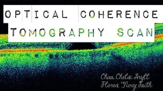
- 6. OCT Image of a Normal Eye
- 7. Preparation for OCT exam •To prepare you for an OCT exam, an ophthalmologist may put dilating eye drops in your eyes. These drops widen your pupil and make it easier to examine the retina.
- 8. Procedure ▪You will sit in front of the OCT machine and rest your head on a support to keep it motionless. The equipment will then scan your eye without touching it.
- 9. Duration of the OCT exam ▪Scanning takes about 5 – 10 minutes. ▪If your eyes were dilated, they may be sensitive to light for several hours after the exam.
- 10. What conditions can OCT help to diagnose?
- 11. MACULAR HOLE ¤ A tear or opening forms in your macula. ¤ As the hole forms, things in your central vision will look blurry, wavy or distorted. As the hole grows, a dark or blind spot appears in your central vision. ¤ A macular hole does not affect your peripheral (side) vision. OCT scan results for macular hole
- 12. | Wrinkles, creases or bulges form on your macula. | The macula must lie flat against the back of your eye to work properly. | When the macula wrinkles or bulges, the central vision is affected. MACULAR PUCKER
- 13. A swelling or thickening of the eye's macula, the part of your eye responsible for detailed, central vision. MACULAR EDEMA
- 14. » Happens when a part of the retina called the macula is damaged. » With AMD you lose your central vision. You cannot see fine details, whether you are looking at something close or far. But your peripheral (side) vision will still be normal. » For instance, imagine you are looking at a clock with hands. With AMD, you might see the clock’s numbers but not the hands. AGE-RELATED MACULAR DEGENERATION (AMD) OCT Results for age related macular degeneration
- 15. * a disease that damages the eye’s optic nerve. * It usually happens when fluid builds up in the front part of the eye. * That extra fluid increases the pressure in your eye, damaging the optic nerve. GLAUCOMA Chronic glaucoma
- 16. Central serous chorioretinopathy (CSR) OCT OF CSR
- 17. § This is when high blood sugar levels cause damage to blood vessels in the retina. § These blood vessels can swell and leak, or they can close, stopping blood from passing through. § Sometimes abnormal new blood vessels grow on the retina. § All of these changes can steal your vision. DIABETIC RETINOPATHY
- 18. ✓ A condition in which the vitreous gel has an abnormally strong adhesion to the retina. ✓Over time, the gel tends to pull forward and can cause vessel and retinal distortion causing retinal swelling and decreased vision. VITREOUS TRACTION OCT of vitreous traction
- 19. Advantages of OCT > Non-invasive > Minimal cooperation needed > Resolution ~ 10 μm (vs. US=150 μm; UBM=50μm) > Pick up earliest signs of disease > Quantitatively monitor disease/staging
- 20. Advantages of OCT >provides a faster and easier method for diagnosis. >chances of error are reduced >gives a better image of the test results. >provides broad and dynamic range of operations. >involves rapid data acquisition rate. >uses simple and inexpensive catheter / endoscope. >The device used in OCT is portable.
- 21. Disadvantages of OCT • Diminished penetration through retinal/subretinal hemorrhage • OCT has a limited penetration power. (Requires pupil diameter > 4 mm) • Femtosecond laser used in OCT is expensive. • During OCT, the transverse resolution needs to be similar to axial resolution.
- 22. Limitations of Retinal OCT •Dilation of the pupils may sometimes be necessary •Dioptric media must be somewhat transparent •Exploration typically limited to posterior pole •Good lacrimal film necessary
- 23. • https://www.williamsoneyeinstitute.com/other- diseases/vitreomacular-traction/ • https://www.aao.org/eye- health/treatments/what-is-optical-coherence- tomography - American academy of ophthalmology • http://visionsource- visionhealthinstitute.com/vision-care- products/advanced-diagnostic-testing/optical- coherence-tomography-oct/ - Vision Health Institute • http://www.c4sightcare.com/page87aa.html • https://globalnewsinsights.com/2017/07/10/adv antages-and-disadvantages-of-optical-coherence- tomography/