Call Girls Alandi Road Call Me 7737669865 Budget Friendly No Advance Booking
OCT-for-counsellors-ppt.pdf
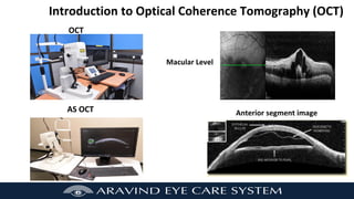
1. Introduction to Optical Coherence Tomography (OCT)
Macular Level
OCT
AS OCT Anterior segment image
2. Learning Objectives
Define OCT and AS OCT and its purpose
Explain the indications for OCT and AS OCT
Check the pre-requisites for the procedure in each patient
Recognize few retina, glaucoma and corneal diseases
3. Brief anatomy - Layers of the Retina
• The retina is a multi-layered structure located in
the inner surface of the eye
• It has 10 layers
• (RPE) retinal pigmented layer is towards the
choroid.
• It is a light-sensitive structure where an image of
the visual world is created through the optics of
the eye.
• Ganglion cell layer is absent in the fovea so there
is foveal depression
4. Activity
Instruct the trainees to see and label the retinal layers
Pigmented
Epithelium
Bacillary Layer
Inner Nuclear
Layer
Inner plexiform
Layer Ganglion Cell
Layer
Choroid
6. OCT - Introduction
• OCT is a non-invasive, detailed painless procedure
• OCT scans can delineate the retinal layers
• OCT shows the cross-section of retina
• OCT uses light to evaluate the retina
• OCT is useful to evaluate the patient both for
diagnosis and for follow-up of retinal diseases
7. Light waves
Coherent Light waves (the crest
meet the crest and the trough
meets the trough
The crest of one wave does not
meet the crest of other wave the
trough of a wave does not meet
the other some it is incoherent
A wave will propagate in the form of a waveform only up to a certain length and that length is called the
coherence length. Beyond coherence length the light will fade away.
8. Pre- requisites before performing OCT
• Name, age, MR.no. has to be verified before
seating the patient on OCT machine
• New vessels in Iris / AC angle needs to evaluate
before dilatation
• Dilatation is essential before performing OCT
• Some special features are specific to each OCT
machine. Hence each patient according to
retinal problem may require OCT on a particular
OCT machine.
9. Counseling the patient
Instruct the patient to remove spectacles or contact lenses to avoid incorrect classification of
result or additional results
Technician will take scan your eye to diagnose the disease
A light beam from the machine scans the optic nerve and retinal nerve fiber layer
This test is simple, quick and painless
This test is completely safe and done without touching the eye
You have to sit in front of the machine and follow the instructions properly
The test will take only few minutes
Be relaxed and follow all the instructions
Media Hazy (cataract, corneal opacity, Vitreous Hem) pt. will show low signal.
10. OCT – Indications (Retina)
Age Related Macular Degeneration
Macular hole
Cystoid Macular Oedema
Epi Retinal Membrane
Central Serous Retinopathy
Diabetic Retinopathy
Hypertensive Retinopathy
Polypoidal Choroidal Vasculopahy
Optic disc Pit
Best disease
Coloboma
11. OCT – Indications (Glaucoma)
Macula thickness
Retinal nerve fibre layer (RNFL)
(Anterior Segment (AS) Angle
Posterior Segment – RNFL)
Angle open distance
Angle
Opening
Distance
Trabecular
Iris Angle
Trabecular
ciliary distance
(TCPD)
Trabecular Iris angle (TIA)
Trabecular ciliary distance (TCPD)
Iris thickness (ID1
Iris concavity / convexity
AS OCT is used to image the defect in the anterior segment and the
posterior segment.
12. OCT – Indications (Cornea)
Cornea – AS OCT - is a non contact equipment
LASIK
DSAEK
Dystrophies and degenerations
Keratoconus
Keratoplasty - PKP, Degeneration
Investigating the distribution of the corneal thickness,
Intacs
Shape of the stromal interface after lamellar corneal surgery,
Association between host and corneal graft in keratoplasty,
Dimension of the anterior chamber, and
Lesions of the corneal diseases.
Descemets detachment
13. AS OCT - Contraindications
⮚ OCT cannot be seen in
Hazy Media
Vitreous Hemorrhage Infected Eye
⮚Under age (Below 3Yrs)
⮚ Non Co operative patient
⮚Infected eye
14. Assessment
1. How many layers are the in retina?
A. 12 layers B. 8 layers
C. 10 layers D. 15 layers
2. What are the important things to take care of
while obtaining an OCT scan?
A. Before doing OCT check M.R.No, Name
B Correct head & chin position
C. Exact looking fixation point
D. All of the above
4.
15. Mindmap
OCT
•OCT definition and the
abbreviation
•Purpose in Retina,
Glaucoma and Cornea
patients
•Pre requisite and process
flow in OCT
•OCT – Tips and Tricks
16. THANK YOU
Sign up to view all training resources for Introduction to Optical Coherence Tomography (OCT)
competency and more (It is free!)
Explore more training resources related to allied ophthalmic personnel