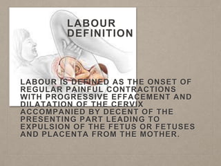
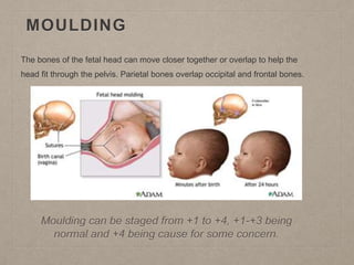
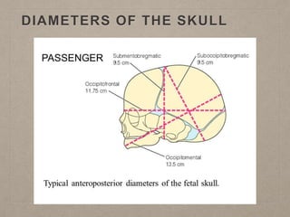
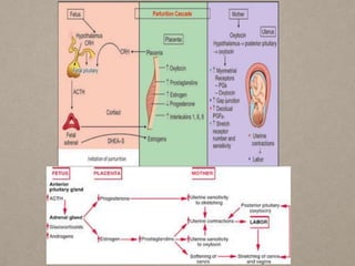
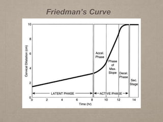
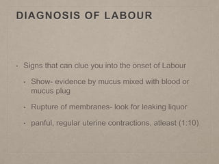
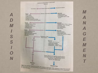
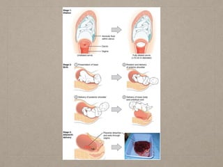
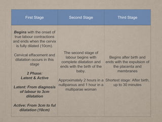
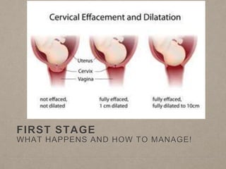
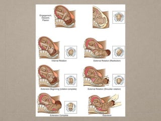
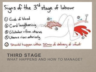
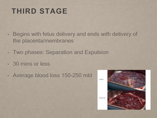
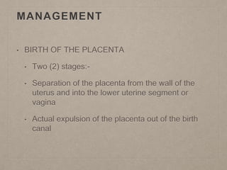
NORMAL LABOUR AND
DELIVERY
RHEA MARCANO
413003930
CONTENTS
• 1. Definition of normal Labour
• 2. Factors influencing progress of Labour
• 3. Diagnosis of Labour
• 4. Stages of Labour
• 5. Management of Labour
LABOUR
DEFINITION
LABOUR IS DEFINED AS THE ONSET OF
REGULAR PAINFUL CONTRACTIONS
WITH PROGRESSIVE EFFACEMENT AND
DILATATION OF THE CERVIX
ACCOMPANIED BY DECENT OF THE
PRESENTING PART LEADING TO
EXPULSION OF THE FETUS OR FETUSES
AND PLACENTA FROM THE MOTHER.
FACTORS TO HELP DETERMINE
IF LABOUR IS NORMAL
• Mature Fetus 37-42 weeks
• Spontaneous expulsion
• Vertex is the presenting part
• Vaginal Delivery
• Time ( not < 3hour but not >18 hours)
• Complications??
INFLUENTIAL FACTORS OF THE
PROGRESS OF LABOUR
• 3P’s
• Power
• Passenger
• Passage
FEMALE PELVIS
• Basic framework for the birth canal
• True Pelvis- Inlet, cavity and Outlet ( The fetus must
pass through all three in order for labour to be
sucessful)
• Types of Pelvis- Gynaecoid, Anthropoid, Android
and Platypelloid
THE FETAL SKULL
MOULDING
The bones of the fetal head can move closer together or overlap to help the
head fit through the pelvis. Parietal bones overlap occipital and frontal bones.
Moulding can be staged from +1 to +4, +1-+3 being
normal and +4 being cause for some concern.
DIAMETERS OF THE SKULL
INITIATION OF LABOUR
CAUSES OF THE ONSET OF
NORMAL LABOUR
• It is unknown but the following theories are proposed:
• Hormonal Factors
• Oestrogen Theory
• Progesterone withdrawal theory
• Prostaglandin Theory
• Oxytocin Theory
• Fetal Cortisol Theory
• Mechanical Factors
• Uterine Distension Theory
• Stretch of the lower uterine segment
Friedman’s Curve
DIAGNOSIS OF LABOUR
• Signs that can clue you into the onset of Labour
• Show- evidence by mucus mixed with blood or
mucus plug
• Rupture of membranes- look for leaking liquor
• panful, regular uterine contractions, atleast (1:10)
A
D
M
I
S
S
I
O
N
M
A
N
A
G
E
M
E
N
T
• ON ADMISSION:
Review antenatal record
Complete history if record isn't available
• GENERAL EXAMINATION OF MOTHER
General condition- pallor, oedema, abdominal scars,
maternal height
Vital signs- Blood pressure, Pulse, respiration, temperature
(measured and recorded)
Heart and Lungs
Urinalysis- protein, sugar, ketones
• Abdominal Examination:
Detail examination, determine fetal presentation,
position and engagement
Auscultate fetal heart sound
Evaluate uterine contractions
Attach Carditocography (CTG) for 20 min trace
• VAGINAL EXAMINATION
Confirm degree of dilatation and effacement
Identify the presenting part
Fetal head engagement if any doubt
Confirm or artificially rupture if necessary (ROM)
Exclude cord prolapse
• BLADDER/BOWEL CARE
Administer an Enema
allow to empty bladder ever 1 1/2 - 2 hours
• NUTRITION IN EARLY LABOUR
No food after labour is established to prevent regurgitation and
aspiration
Place IV to start administration of fluids
• POSITIONING OF LABOURING MOTHER
Once everything is well with mom and baby, patient may ambulate
or lay in bed as the feel comfortable
• MONITORING, PROGRESS OF LABOUR
• PAIN RELIEF
Opiate drugs- Pethidine given IM q4hrly
Epidural analgesia
PARTOGRAM
• A cartogram is a composite
graphical record of key data
(maternal & fetal) during
labour entered against time
on a single sheet of paper.
• Relevant measurements
such as cervical dilatation,
fetal heart rate, duration of
labour and vital signs
• Monitors progress of Labour
COMPONENTS OF A PARTOGRAM
• Patient Identification
• Time (recorded in 1hr intervals)
• Fetal Heart Rate
• State of Membranes
• Cervical Dilatation
• Uterine Contractions
• Drugs & Fluids
• BP (2hr intervals)
• Pulse Rate (30min intervals)
• Oxytocin
• Urinalysis
• Temperature
STAGES OF LABOUR
First Stage Second Stage Third Stage
Begins with the onset of
true labour contractions
and ends when the cervix
is fully dilated (10cm).
Cervical effacement and
dilatation occurs in this
stage
2 Phase:
Latent & Active
Latent: From diagnosis
of labour to 3cm
dilatation
Active: From 3cm to ful
dilatation (10cm)
The second stage of
labour begins with
complete dilatation and
ends with the birth of the
baby.
Approximately 2 hours in a
nulliparous and 1 hour in a
multiparae woman
Begins after birth and
ends with the expulsion of
the placenta and
membranes
Shortest stage: After birth,
up to 30 minutes
FIRST STAGE
WHAT HAPPENS AND HOW TO MANAGE!
• 1. Contractions
• Regular
• Increasing Frequency
• Stronger
• 2. Cervical Dilatation and Effacement
• 3. Engagement of the presenting part
MANAGEMENT
• Continuity of care
• Observation of progress of Labour
• Monitoring fetal & maternal well-being
• Adequate pain relief (according to mothers wishes)
• Adequate hydration to prevent Ketosis
Lactate ringer solution
SECOND STAGE
WHAT HAPPENS AND HOW TO MANAGE?
SECOND STAGE
• First sign of the second stage is the urge to push
• Full Dilatation to Delivery of the fetus
• Signs to look for:-
• (1) Distention of the perineum
• (2) Dilatation of the anus
• Satisfactory progress:- steady descent of the fetus
through the birth canal & onset of the expulsive phase
MANAGEMENT
• Continuous monitoring during this phase
• Maternal Position, usually semi-recumbent or
supported sitting position with thighs abducted but
any comfortable position expect supine for an
uncomplicated pregnancy
• Encourage to bear down with the contractions
MANAGEMENT (CONT’D)
• Maternal condition - BP and PR measured every 15-
30mins and after contractions
• Fetal Condition- Fetal heart rate, measured
continuously or after contractions
• Uterine Contractions- strength, length and frequency
continuously assessed
• Progress of descent- recorded every 30 mins
CONDUCTING THE DELIVERY
• position patient
• antiseptic solution to clean skin of lower abdomen, vulva, anus and upper
thigh, then drape
• DELIVERY OF THE HEAD
• Control delivery of the head
• Perform episiotomy if required
• Perform Ritgen’s Maneuver
• Clear the airways after delivery of the head
CONDUCTING THE DELIVERY
(CONT’D)
• DELIVERY OF THE
SHOULDERS
• Anterior shoulder assisted
by gentle downward
traction of the head
• Posterior shoulder is
delivered by elevating the
head.
CONDUCTING THE DELIVERY
• DELIVERY OF THE TRUNK
• Grasp baby around the chest after shoulders delivered to help with birth
of trunk
• Baby swept unto mother’s abdomen
• Note time of delivery
• CUTTING THE UMBILICAL CORD
• wait 15-20 seconds then clamp
• plastic crushing clip placed 1-2cm above umbilicus and cut 1cm beyond
the clamp
IMMEDIATE CARE OF THE NEWBORN
• Assess baby
• Health baby with spontaneous respiration place
on mother’s abdomen, dry& cover baby
• No spontaneous respiration or respiratory
problems then resuscitate baby
• APGAR scores
EVENTS OCCURRING DURING LABOUR
• Flexion and Descent
• Internal Rotation of the fetal head
• Crowning
• Extension
• Restitution
• Internal rotation of the shoulders
• External rotation of the fetal body
• Lateral flexion of the body
THIRD STAGE
WHAT HAPPENS AND HOW TO MANAGE?
THIRD STAGE
• Begins with fetus delivery and ends with delivery of
the placenta/membranes
• Two phases: Separation and Expulsion
• 30 mins or less
• Average blood loss 150-250 mld
MANAGEMENT
• BIRTH OF THE PLACENTA
• Two (2) stages:-
• Separation of the placenta from the wall of the
uterus and into the lower uterine segment or
vagina
• Actual expulsion of the placenta out of the birth
canal
TWO MECHANISMS OF SEPARATION
• Mathews-Duncan mechanism (raw surface exposed
when delivered)
• Schultz Mechanism (placenta inserted at fundus,
placenta inverts and covers the raw surface)
SIGNS OF SEPARATION
• Globular and hard uterus
• Sudden gush of blood
• Cord Lengthening (Most reliable clinical sign)
BIRTH OF THE PLACENTA
• Two methods:
• Passive Management (wait for spontaneous
expulsion of the placenta)
• Active Management
ACTIVE MANAGEMENT OF THE
THIRD STAGE
• Help prevent postpartum hemorrhage
• Includes:-
• Use of oxytocin (given around the time of the
anterior shoulder delivery, 10 units)
• Controlled cord traction
• Uterine massage
ACTIVE PLACENTA DELIVERY
• Brandt’s Andrew method
• Placenta separation
• Controlled cord traction
• Delivery of the membranes
• Examination of the Placenta:- placenta, membranes &
umbilical cord for completeness and anomalies
• EXAMINATION OF THE PERINEUM
• look for lacerations, also vulva outlet, vaginal
canal & cervix should be inspected
• Repair lacerations or episiotomies immediately
IMMEDIATE MANAGEMENT
AFTER THE DELIVERY
• EARLY POSTPARTUM MANAGEMENT
• Monitor for postpartum hemorrhage, keep for atlas 1 hour in delivery suite
(bleeding- ask to report any sudden gushes of blood, bp and pulse)
• Before discharging from delivery suite
• Check uterus frequently to ensure it is firm
• Remove intrauterine clots
• Look at introitus for NO hemorrhage
• Keep bladder empty
• Ensure baby is breathing well, pink and well flexed
REFERENCES
• Obstetrics ten teacher
• Various online resources
LEARNING RESOURCE
• http://intranet.tdmu.edu.ua/data/kafedra/internal/gine
cology2/classes_stud/en/nurse/adn/ptn/2/Nursing%2
0Care%20of%20Childbearing%20Family_Practicum
/04.%20Labor%20and%20birth%20process..htm
QUESTIONS??
Let’s Deliver Babies……………………
THANK YOU!!!
• Engagement: The fetus is engaged if the widest leading part (typically the widest
circumference of the head) is negotiating the inlet.
• Station: Relationship of the bony presenting part of the fetus to the maternal ischial
spines. If at the level of the spines it is at “0 (zero)” station, if it passed it by 2cm it is
at “+2” station.
• Attitude: Relationship of fetal head to spine: flexed, neutral (“military”), or extended
attitudes are possible.
• Position: Relationship of presenting part to maternal pelvis, i.e. ROP=right occiput
posterior, or LOA=left occiput anterior.
• Presentation: Relationship between the leading fetal part and the pelvic inlet:
cephalic, breech (complete, incomplete, frank or footling), face, brow, mentum or
shoulder presentation.
• Lie: Relationship between the longitudinal axis of fetus and long axis of the uterus:
longitudinal, oblique, and transverse.
• Caput or Caput succedaneum: oedema typically formed by the tissue overlying the
GLOSSARY
Pelvic types
Traditional obstetrics characterizes four types of pelvises:
• Gynecoid: Ideal shape, with round to slightly oval (obstetrical inlet slightly less
transverse) inlet: best chances for normal vaginal delivery.
• Android: triangular inlet, and prominent ischial spines, more angulated pubic arch.
• Anthropoid: the widest transverse diameter is less than the anteroposterior
(obstetrical) diameter.
• Platypelloid: Flat inlet with shortened obstetrical diameter.

normallabouranddeliveryppt-170622025809 (1).pdf

  • 1.
  • 2.
    CONTENTS • 1. Definitionof normal Labour • 2. Factors influencing progress of Labour • 3. Diagnosis of Labour • 4. Stages of Labour • 5. Management of Labour
  • 3.
    LABOUR DEFINITION LABOUR IS DEFINEDAS THE ONSET OF REGULAR PAINFUL CONTRACTIONS WITH PROGRESSIVE EFFACEMENT AND DILATATION OF THE CERVIX ACCOMPANIED BY DECENT OF THE PRESENTING PART LEADING TO EXPULSION OF THE FETUS OR FETUSES AND PLACENTA FROM THE MOTHER.
  • 4.
    FACTORS TO HELPDETERMINE IF LABOUR IS NORMAL • Mature Fetus 37-42 weeks • Spontaneous expulsion • Vertex is the presenting part • Vaginal Delivery • Time ( not < 3hour but not >18 hours) • Complications??
  • 5.
    INFLUENTIAL FACTORS OFTHE PROGRESS OF LABOUR • 3P’s • Power • Passenger • Passage
  • 6.
    FEMALE PELVIS • Basicframework for the birth canal • True Pelvis- Inlet, cavity and Outlet ( The fetus must pass through all three in order for labour to be sucessful) • Types of Pelvis- Gynaecoid, Anthropoid, Android and Platypelloid
  • 8.
  • 9.
    MOULDING The bones ofthe fetal head can move closer together or overlap to help the head fit through the pelvis. Parietal bones overlap occipital and frontal bones. Moulding can be staged from +1 to +4, +1-+3 being normal and +4 being cause for some concern.
  • 10.
  • 11.
  • 12.
    CAUSES OF THEONSET OF NORMAL LABOUR • It is unknown but the following theories are proposed: • Hormonal Factors • Oestrogen Theory • Progesterone withdrawal theory • Prostaglandin Theory • Oxytocin Theory • Fetal Cortisol Theory • Mechanical Factors • Uterine Distension Theory • Stretch of the lower uterine segment
  • 15.
  • 16.
    DIAGNOSIS OF LABOUR •Signs that can clue you into the onset of Labour • Show- evidence by mucus mixed with blood or mucus plug • Rupture of membranes- look for leaking liquor • panful, regular uterine contractions, atleast (1:10)
  • 17.
  • 18.
    • ON ADMISSION: Reviewantenatal record Complete history if record isn't available • GENERAL EXAMINATION OF MOTHER General condition- pallor, oedema, abdominal scars, maternal height Vital signs- Blood pressure, Pulse, respiration, temperature (measured and recorded) Heart and Lungs Urinalysis- protein, sugar, ketones
  • 19.
    • Abdominal Examination: Detailexamination, determine fetal presentation, position and engagement Auscultate fetal heart sound Evaluate uterine contractions Attach Carditocography (CTG) for 20 min trace
  • 20.
    • VAGINAL EXAMINATION Confirmdegree of dilatation and effacement Identify the presenting part Fetal head engagement if any doubt Confirm or artificially rupture if necessary (ROM) Exclude cord prolapse • BLADDER/BOWEL CARE Administer an Enema allow to empty bladder ever 1 1/2 - 2 hours
  • 21.
    • NUTRITION INEARLY LABOUR No food after labour is established to prevent regurgitation and aspiration Place IV to start administration of fluids • POSITIONING OF LABOURING MOTHER Once everything is well with mom and baby, patient may ambulate or lay in bed as the feel comfortable • MONITORING, PROGRESS OF LABOUR • PAIN RELIEF Opiate drugs- Pethidine given IM q4hrly Epidural analgesia
  • 22.
    PARTOGRAM • A cartogramis a composite graphical record of key data (maternal & fetal) during labour entered against time on a single sheet of paper. • Relevant measurements such as cervical dilatation, fetal heart rate, duration of labour and vital signs • Monitors progress of Labour
  • 24.
    COMPONENTS OF APARTOGRAM • Patient Identification • Time (recorded in 1hr intervals) • Fetal Heart Rate • State of Membranes • Cervical Dilatation • Uterine Contractions • Drugs & Fluids • BP (2hr intervals) • Pulse Rate (30min intervals) • Oxytocin • Urinalysis • Temperature
  • 25.
  • 27.
    First Stage SecondStage Third Stage Begins with the onset of true labour contractions and ends when the cervix is fully dilated (10cm). Cervical effacement and dilatation occurs in this stage 2 Phase: Latent & Active Latent: From diagnosis of labour to 3cm dilatation Active: From 3cm to ful dilatation (10cm) The second stage of labour begins with complete dilatation and ends with the birth of the baby. Approximately 2 hours in a nulliparous and 1 hour in a multiparae woman Begins after birth and ends with the expulsion of the placenta and membranes Shortest stage: After birth, up to 30 minutes
  • 28.
    FIRST STAGE WHAT HAPPENSAND HOW TO MANAGE!
  • 29.
    • 1. Contractions •Regular • Increasing Frequency • Stronger • 2. Cervical Dilatation and Effacement • 3. Engagement of the presenting part
  • 30.
    MANAGEMENT • Continuity ofcare • Observation of progress of Labour • Monitoring fetal & maternal well-being • Adequate pain relief (according to mothers wishes) • Adequate hydration to prevent Ketosis Lactate ringer solution
  • 31.
    SECOND STAGE WHAT HAPPENSAND HOW TO MANAGE?
  • 32.
    SECOND STAGE • Firstsign of the second stage is the urge to push • Full Dilatation to Delivery of the fetus • Signs to look for:- • (1) Distention of the perineum • (2) Dilatation of the anus • Satisfactory progress:- steady descent of the fetus through the birth canal & onset of the expulsive phase
  • 33.
    MANAGEMENT • Continuous monitoringduring this phase • Maternal Position, usually semi-recumbent or supported sitting position with thighs abducted but any comfortable position expect supine for an uncomplicated pregnancy • Encourage to bear down with the contractions
  • 34.
    MANAGEMENT (CONT’D) • Maternalcondition - BP and PR measured every 15- 30mins and after contractions • Fetal Condition- Fetal heart rate, measured continuously or after contractions • Uterine Contractions- strength, length and frequency continuously assessed • Progress of descent- recorded every 30 mins
  • 35.
    CONDUCTING THE DELIVERY •position patient • antiseptic solution to clean skin of lower abdomen, vulva, anus and upper thigh, then drape • DELIVERY OF THE HEAD • Control delivery of the head • Perform episiotomy if required • Perform Ritgen’s Maneuver • Clear the airways after delivery of the head
  • 36.
    CONDUCTING THE DELIVERY (CONT’D) •DELIVERY OF THE SHOULDERS • Anterior shoulder assisted by gentle downward traction of the head • Posterior shoulder is delivered by elevating the head.
  • 37.
    CONDUCTING THE DELIVERY •DELIVERY OF THE TRUNK • Grasp baby around the chest after shoulders delivered to help with birth of trunk • Baby swept unto mother’s abdomen • Note time of delivery • CUTTING THE UMBILICAL CORD • wait 15-20 seconds then clamp • plastic crushing clip placed 1-2cm above umbilicus and cut 1cm beyond the clamp
  • 38.
    IMMEDIATE CARE OFTHE NEWBORN • Assess baby • Health baby with spontaneous respiration place on mother’s abdomen, dry& cover baby • No spontaneous respiration or respiratory problems then resuscitate baby • APGAR scores
  • 39.
    EVENTS OCCURRING DURINGLABOUR • Flexion and Descent • Internal Rotation of the fetal head • Crowning • Extension • Restitution • Internal rotation of the shoulders • External rotation of the fetal body • Lateral flexion of the body
  • 41.
    THIRD STAGE WHAT HAPPENSAND HOW TO MANAGE?
  • 42.
    THIRD STAGE • Beginswith fetus delivery and ends with delivery of the placenta/membranes • Two phases: Separation and Expulsion • 30 mins or less • Average blood loss 150-250 mld
  • 43.
    MANAGEMENT • BIRTH OFTHE PLACENTA • Two (2) stages:- • Separation of the placenta from the wall of the uterus and into the lower uterine segment or vagina • Actual expulsion of the placenta out of the birth canal
  • 44.
    TWO MECHANISMS OFSEPARATION • Mathews-Duncan mechanism (raw surface exposed when delivered) • Schultz Mechanism (placenta inserted at fundus, placenta inverts and covers the raw surface)
  • 45.
    SIGNS OF SEPARATION •Globular and hard uterus • Sudden gush of blood • Cord Lengthening (Most reliable clinical sign)
  • 46.
    BIRTH OF THEPLACENTA • Two methods: • Passive Management (wait for spontaneous expulsion of the placenta) • Active Management
  • 47.
    ACTIVE MANAGEMENT OFTHE THIRD STAGE • Help prevent postpartum hemorrhage • Includes:- • Use of oxytocin (given around the time of the anterior shoulder delivery, 10 units) • Controlled cord traction • Uterine massage
  • 48.
    ACTIVE PLACENTA DELIVERY •Brandt’s Andrew method • Placenta separation • Controlled cord traction • Delivery of the membranes • Examination of the Placenta:- placenta, membranes & umbilical cord for completeness and anomalies
  • 49.
    • EXAMINATION OFTHE PERINEUM • look for lacerations, also vulva outlet, vaginal canal & cervix should be inspected • Repair lacerations or episiotomies immediately
  • 50.
    IMMEDIATE MANAGEMENT AFTER THEDELIVERY • EARLY POSTPARTUM MANAGEMENT • Monitor for postpartum hemorrhage, keep for atlas 1 hour in delivery suite (bleeding- ask to report any sudden gushes of blood, bp and pulse) • Before discharging from delivery suite • Check uterus frequently to ensure it is firm • Remove intrauterine clots • Look at introitus for NO hemorrhage • Keep bladder empty • Ensure baby is breathing well, pink and well flexed
  • 51.
    REFERENCES • Obstetrics tenteacher • Various online resources
  • 52.
  • 53.
  • 54.
    • Engagement: Thefetus is engaged if the widest leading part (typically the widest circumference of the head) is negotiating the inlet. • Station: Relationship of the bony presenting part of the fetus to the maternal ischial spines. If at the level of the spines it is at “0 (zero)” station, if it passed it by 2cm it is at “+2” station. • Attitude: Relationship of fetal head to spine: flexed, neutral (“military”), or extended attitudes are possible. • Position: Relationship of presenting part to maternal pelvis, i.e. ROP=right occiput posterior, or LOA=left occiput anterior. • Presentation: Relationship between the leading fetal part and the pelvic inlet: cephalic, breech (complete, incomplete, frank or footling), face, brow, mentum or shoulder presentation. • Lie: Relationship between the longitudinal axis of fetus and long axis of the uterus: longitudinal, oblique, and transverse. • Caput or Caput succedaneum: oedema typically formed by the tissue overlying the GLOSSARY
  • 55.
    Pelvic types Traditional obstetricscharacterizes four types of pelvises: • Gynecoid: Ideal shape, with round to slightly oval (obstetrical inlet slightly less transverse) inlet: best chances for normal vaginal delivery. • Android: triangular inlet, and prominent ischial spines, more angulated pubic arch. • Anthropoid: the widest transverse diameter is less than the anteroposterior (obstetrical) diameter. • Platypelloid: Flat inlet with shortened obstetrical diameter.