
Recommended
More Related Content
What's hot
What's hot (20)
Similar to Large Left Pneumothorax X-Ray Images
Similar to Large Left Pneumothorax X-Ray Images (20)
More from Walif Chbeir
More from Walif Chbeir (12)
Recently uploaded
Recently uploaded (20)
Large Left Pneumothorax X-Ray Images
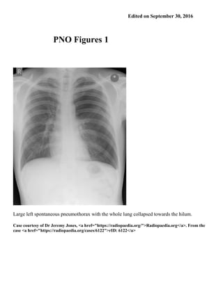
- 1. Edited on September 30, 2016 PNO Figures 1 Large left spontaneous pneumothorax with the whole lung collapsed towards the hilum. Case courtesy of Dr Jeremy Jones, <a href="https://radiopaedia.org/">Radiopaedia.org</a>. From the case <a href="https://radiopaedia.org/cases/6122">rID: 6122</a>
- 2. Large left pneumothorax with the whole lung collapsed towards the hilum. By not copyrightable - https://en.wikipedia.org/wiki/File:Rt_sided_pneumoD.jpg, Public Domain, https://en.wikipedia.org/w/index.php?curid=40565535 https://en.wikipedia.org/wiki/Pneumothorax
- 3. By Photographed by User Clinical Cases 00:42, 7 November 2006 - Originally from tension pneumthorax page on clinicalcases.orgTransferred from en.wikipedia to Commons.; description page is/was here; uploader to en Wiki was Clinical Cases at en.wikipedia, CC BY-SA 2.5, https://commons.wikimedia.org/w/index.php?curid=2235891
- 4. Frontal Supine. In the supine projection note the visible left visceral pleural edge and a relative transradiancy of the involved hemithorax. Case courtesy of Dr Nikos Karapasias, <a href="https://radiopaedia.org/">Radiopaedia.org</a>. From the case <a href="https://radiopaedia.org/cases/25667">rID: 25667</a>
- 5. Erect projection. In the erect position the lung falls inferiorly and air is seen at the apex of the chest showing the familiar appearance of the lung edge parallel to but separated from the chest wall. Note the visible visceral pleural edge and a relative transradiancy of the involved hemithorax. Case courtesy of Dr Nikos Karapasias, <a href="https://radiopaedia.org/">Radiopaedia.org</a>. From the case <a href="https://radiopaedia.org/cases/25667">rID: 25667</a>
- 6. Right Pneumothorax due to rib fractures p.a. x-ray of the chest in upright position reveals rib fractures and a pneumothorax on the right side. Case courtesy of Dr Jens Christian Fischer, <a href="https://radiopaedia.org/">Radiopaedia.org</a>. From the case <a href="https://radiopaedia.org/cases/12821">rID: 12821</a>
- 7. Pneumothorax The zoomed view clearly demonstrates the thin pleural line and the lack of the pulmonary vessels in the right apex. Case courtesy of Dr Jens Christian Fischer, <a href="https://radiopaedia.org/">Radiopaedia.org</a>. From the case <a href="https://radiopaedia.org/cases/12821">rID: 12821</a>
- 8. Right-sided pneumothorax and fracture of ribs 8-10. Case courtesy of Dr Sajoscha Sorrentino, <a href="https://radiopaedia.org/">Radiopaedia.org</a>. From the case <a href="https://radiopaedia.org/cases/14780">rID: 14780</a>
- 9. Right pneumothorax. Pleural reflection indicated by the arrows. Case courtesy of Dr M Osama Yonso, <a href="https://radiopaedia.org/">Radiopaedia.org</a>. From the case <a href="https://radiopaedia.org/cases/18975">rID: 18975</a>
- 10. Right pneumothorax with deep sulcus sign and collapse of the right lung. ICC projects over the right hemithorax. Case courtesy of Dr Paresh K Desai , <a href="https://radiopaedia.org/">Radiopaedia.org</a>. From the case <a href="https://radiopaedia.org/cases/10037">rID: 10037</a>
- 11. This is a supine film which demonstrates a deep sulcus sign at the base of the left hemithorax in
- 12. keeping with a small pneumothorax. Multiple rib fractures are present, on the left involving the 8th, 9th and 10 ribs. Case courtesy of Dr Paul Smith, <a href="https://radiopaedia.org/">Radiopaedia.org</a>. From the case <a href="https://radiopaedia.org/cases/10885">rID: 10885</a>
- 13. Right PNO. This is a nice example of the value of an expiratory film to improve the conspicuity of a pneumothorax. The image shows increase in apparent size of the pneumothorax on the expiratory view compared to the inspiratory view. Arrows show the pleura. Also seen is an acute clavicular fracture on the right. No rib fractures are seen though there may be an occult one. Case courtesy of Dr J. Ray Ballinger, <a href="https://radiopaedia.org/">Radiopaedia.org</a>. From the case <a href="https://radiopaedia.org/cases/23725">rID: 23725</a>
- 14. In the supine projection note the visible left visceral pleural edge and a relative transradiancy of the involved hemithorax.
- 15. In the erect projection note the difference: a large left pneumothorax , probably in tension. Note the visible visceral pleural edge and a relative transradiancy of the involved hemithorax.
- 16. In the supine position gravity causes the lung to fall dorsally allowing the air to collect anteromedially, laterally and basally. Note the visible visceral pleural edge and a relative transradiancy of the involved hemithorax.
- 17. In the erect position the lung falls inferiorly and air is seen at the apex of the chest showing the familiar appearance of the lung edge parallel to but separated from the chest wall. Case courtesy of Dr Nikos Karapasias, <a href="https://radiopaedia.org/">Radiopaedia.org</a>. From the case <a href="https://radiopaedia.org/cases/25667">rID: 25667</a>