
Recommended
Recommended
More Related Content
Similar to Normal Labral Variant Figures II - Walif Chbeir
Similar to Normal Labral Variant Figures II - Walif Chbeir (8)
More from Walif Chbeir
More from Walif Chbeir (6)
Recently uploaded
Recently uploaded (20)
Normal Labral Variant Figures II - Walif Chbeir
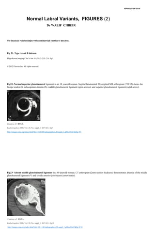
- 1. Edited 10-09-2016 Normal Labral Variants, FIGURES (2) Dr WALIF CHBEIR No financial relationships with commercial entities to disclose. Fig 21. Type A and B labrum. Magn Reson Imaging Clin N Am 20 (2012) 213–228; fig1. © 2012 Elsevier Inc. All rights reserved. Fig22- Normal superior glenohumeral ligament in an 18 yearold woman. Sagittal fatsaturated T1weighted MR arthrogram (750/15) shows the biceps tendon (t), subscapularis tendon (S), middle glenohumeral ligament (open arrows), and superior glenohumeral ligament (solid arrow). Courtesy of: RSNA. RadioGraphics 2000, Vol. 20, No. suppl_1: S67-S81; fig7. http://images.rsna.org/index.html?doi=10.1148/radiographics.20.suppl_1.g00oc03s67&fig=F7. Fig23- Absent middle glenohumeral ligament in a 40 yearold woman. CT arthrogram (2mm section thickness) demonstrates absence of the middle glenohumeral ligament (*) and a wide anterior joint recess (arrowheads). Courtesy of : RSNA. RadioGraphics 2000, Vol. 20, No. suppl_1: S67-S81; fig10. http://images.rsna.org/index.html?doi=10.1148/radiographics.20.suppl_1.g00oc03s67&fig=F10
- 2. Fig 24- Normal middle glenohumeral ligament in a 30 year old man. CT arthrogram (2mm section thickness) shows the middle glenohumeral ligament (arrowhead) attached to the anterior labrum (arrow). Courtesy of: RSNA. RadioGraphics 2000, Vol. 20, No. suppl_1: S67-S81; fig8. http://images.rsna.org/index.html?doi=10.1148/radiographics.20.suppl_1.g00oc03s67&fig=F8 Fig25- Normal middle glenohumeral ligament in an 18 year old woman. Transverse fatsaturated MR arthrogram (560/14) demonstrates the middle glenohumeral ligament attaching medially on the glenoid neck (arrow). Courtesy of: RSNA. RadioGraphics 2000, Vol. 20, No. suppl_1: S67-S81; fig9. http://images.rsna.org/index.html?doi=10.1148/radiographics.20.suppl_1.g00oc03s67&fig=F9 Fig 26- Normal inferior glenohumeral ligament in an 18 year old woman. Sagittal fatsaturated T1weighted MR arthrogram (750/15) demonstrates the biceps tendon (t), subscapularis tendon (S), and anterior and posterior bands of the inferior glenohumeral ligament (arrows). Courtesy of: RSNA. RadioGraphics 2000, Vol. 20, No. suppl_1: S67-S81; fig11. http://images.rsna.org/index.html?doi=10.1148/radiographics.20.suppl_1.g00oc03s67&fig=F11 Fig 27- Normal inferior glenohumeral ligament in a 57year old man. CT arthrogram (2mm section thickness) shows the anterior band of the inferior glenohumeral ligament in the axillary joint recess (arrow).
- 3. Courtesy of: RSNA RadioGraphics 2000, Vol. 20, No. suppl_1: S67-S81; fig12. http://images.rsna.org/index.html?doi=10.1148/radiographics.20.suppl_1.g00oc03s67&fig=F12 Fig 28- Tear of the anterior labrum. Photograph of a plastic model shows extensive fraying of the anterior labrum (arrows). Courtesy of: RSNA RadioGraphics 2000, Vol. 20, No. suppl_1: S67-S81; fig24. Courtesy of: http://images.rsna.org/index.html?doi=10.1148/radiographics.20.suppl_1.g00oc03s67&fig=F24 Fig29- Tear of the anterior labrum. Sagittal fatsaturated MR arthrogram (744/20) demonstrates absence of the labrum and residual irregularity of the anterior glenoid (arrows). Courtesy of: RSNA. RadioGraphics 2000, Vol. 20, No. suppl_1: S67-S81; fig25. http://images.rsna.org/index.html?doi=10.1148/radiographics.20.suppl_1.g00oc03s67&fig=F25 Fig30- Tear of the anterior labrum in a 26 year old man. Transverse CT arthrogram (2mm section thickness) shows injected contrast material extending into a tear of the anterior labrum (arrow).
- 4. Courtesy of: RSNA. RadioGraphics 2000, Vol. 20, No. suppl_1: S67-S81; fig26. http://images.rsna.org/index.html?doi=10.1148/radiographics.20.suppl_1.g00oc03s67&fig=F256 Fig31-Type II SLAP lesion. Photograph of a plastic model shows detachment of the superior labrum from the glenoid (arrows). Note the irregular fraying and hemorrhagic aspect of the free edge of the labrum. Courtesy of: RSNA. RadioGraphics 2000, Vol. 20, No. suppl_1: S67-S81; fig31. http://images.rsna.org/index.html?doi=10.1148/radiographics.20.suppl_1.g00oc03s67&fig=F31 Fig32- Type II SLAP lesion in a 37 year old man. Coronal MR arthrogram (520/14; flip angle, 40°) demonstrates contrast material between the superior labrum and the glenoid (arrow). Note also the slight irregularity of the labral margin (arrowhead). Courtesy of: RSNA. RadioGraphics 2000, Vol. 20, No. suppl_1: S67-S81; fig32. http://images.rsna.org/index.html?doi=10.1148/radiographics.20.suppl_1.g00oc03s67&fig=F32 Fig33- Type II SLAP lesion. Coronal fat saturated MR arthrogram (700/16) shows contrast material between the superior labrum and the glenoid (arrow). Note the lateral extension of the tear of the superior labrum.
- 5. Courtesy of: RSNA. RadioGraphics 2000, Vol. 20, No. suppl_1: S67-S81; fig33. http://images.rsna.org/index.html?doi=10.1148/radiographics.20.suppl_1.g00oc03s67&fig=F33 Fig34- Type II SLAP lesion in a 26year old man. CT arthrogram (1mm section thickness) shows contrast material between the superior labrum and the superior glenoid rim (arrow). The wide separation indicates a SLAP lesion. Courtesy of: RSNA RadioGraphics 2000, Vol. 20, No. suppl_1: S67-S81; fig34. http://images.rsna.org/index.html?doi=10.1148/radiographics.20.suppl_1.g00oc03s67&fig=F34 Figure 35. Type II SLAP lesion. On a photograph obtained during arthroscopy, the superior labrum (S) is separated from the superior glenoid rim. Note the irregular fraying of the free edge of the superior labrum (arrowheads). G = glenoid, H = humeral head. Courtesy of: RSNA. RadioGraphics 2000, Vol. 20, No. suppl_1: S67-S81; fig35. http://images.rsna.org/index.html?doi=10.1148/radiographics.20.suppl_1.g00oc03s67&fig=F35
- 6. Figure 36. Type II SLAP lesion versus superior sublabral recess. Drawings representing a coronal section through the labralbicipital attachment demonstrate a normal recess with a sharp free edge of the labrum (arrow in 1), a type II SLAP lesion with an irregular appearance of the free edge of the labrum (arrow in 2), a type II SLAP lesion with wide separation between the superior labrum and the glenoid (arrows in 3), and a type II SLAP lesion with lateral extension of the labral tear (arrowhead in 4). Courtesy of: RSNA. RadioGraphics 2000, Vol. 20, No. suppl_1: S67-S81; fig36. http://images.rsna.org/index.html?doi=10.1148/radiographics.20.suppl_1.g00oc03s67&fig=F36 Figure 37. Magn Reson Imaging Clin N Am 20 (2012) 213–228; fig3. © 2012 Elsevier Inc. All rights reserved. Fig38. Courtesy of: radedasia.com. GLENOID LABRUM MRI SIMPLIFIED 2: Are you normal or not? http://radedasia.com/glenoid-labrum-labral-variants-mri-simplified-2-are-you-normal-or-not/ Fig39- Courtesy of: radedasia.com . GLENOID LABRUM MRI SIMPLIFIED 3: Buford Complex http://radedasia.com/buford-complex-glenoid-labrum-mri-simplified-3/
- 7. Fig 40- Courtesy of: radedasia.com GLENOID LABRUM MRI SIMPLIFIED 4: Sublabral Foramen and Recess. http://radedasia.com/glenoid-labrum-mri-sublabral-foramen-and-recess/ Fig41- Courtesy of: radedasia.com http://radedasia.com/glenoidlabrummrisublabralforamenandrecess/ Fig42- Labral variants and Tears :
- 8. Courtesy of : The Radiology Assistant http://www.radiologyassistant.nl/en/p4f49ef79818c2/shouldermranatomy.html