Download as PPSX, PPTX
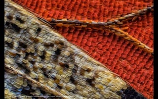
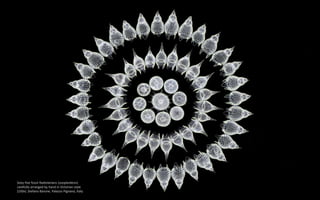
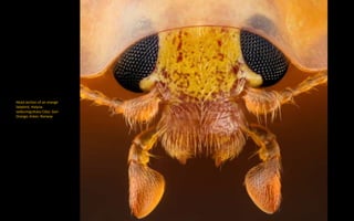
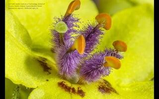
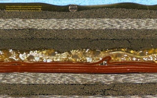
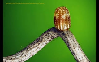
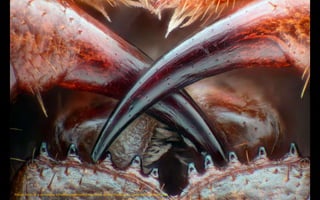
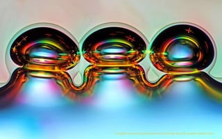
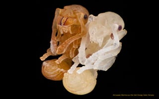
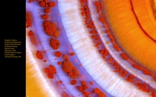
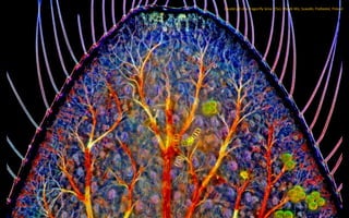
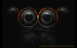
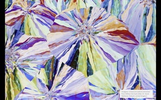
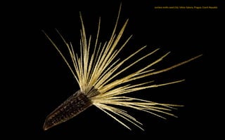
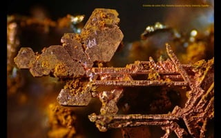
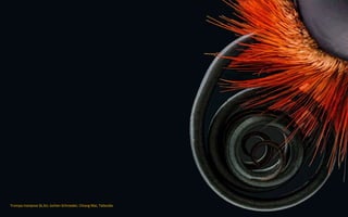
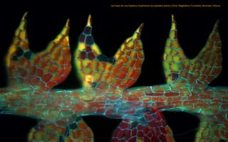
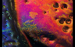
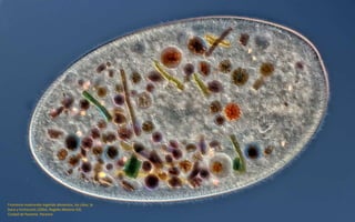
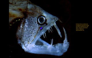
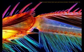

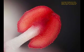
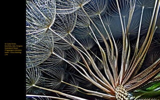
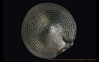
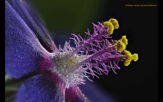
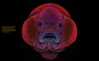
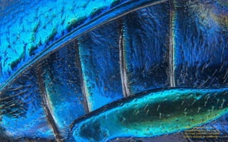
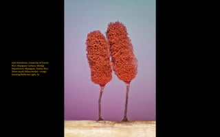
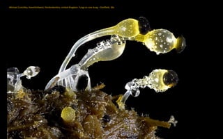
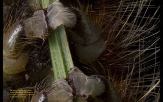
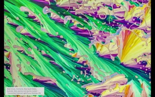
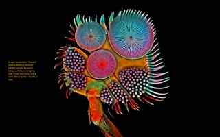
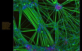
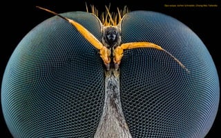
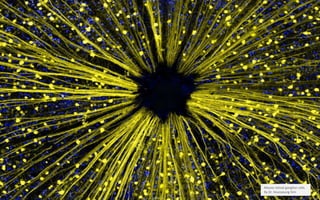
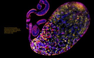
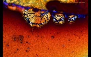
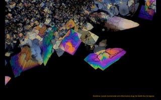
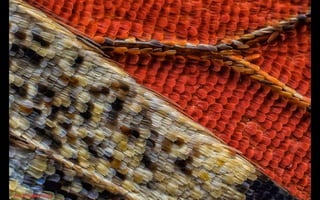

This document contains descriptions and images from microscopy submissions to the Microscopy UK website. It includes microscopic views of butterfly wings, fossil radiolarians, ladybug anatomy, mullein flowers, composite materials, butterfly eggs, centipede fangs, melted crystals, ant pupae, agate, dragonfly gills, spider eyes, willow bark extracts, seeds, insect parts, water fleas, flowers, fish anatomy, beetles, slime molds, fungi, insect legs, crystals, neurons, wasp eyes, retinal cells, fruit fly anatomy, coffee crystals, and diclofenac crystals at various magnifications. Contributors provided locations and affiliations.


