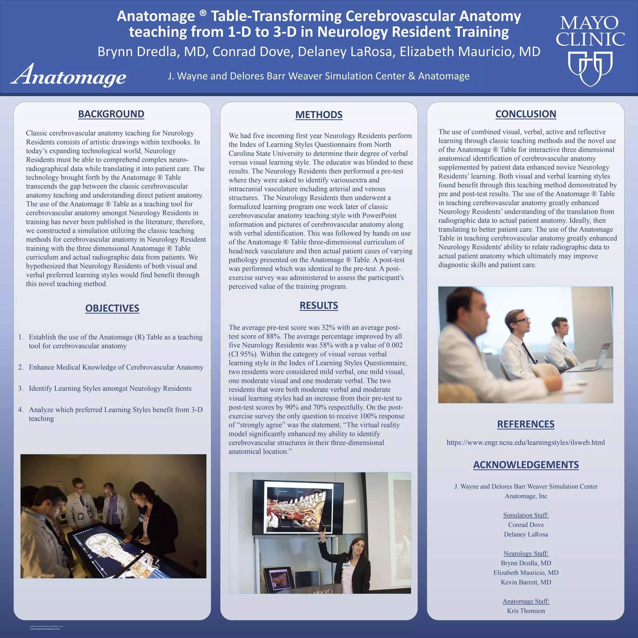
RESEARCH POSTER PRESENTATION DESIGN © 2015
www.PosterPresentations.com
1. Establish the use of the Anatomage (R) Table as a teaching
tool for cerebrovascular anatomy
2. Enhance Medical Knowledge of Cerebrovascular Anatomy
3. Identify Learning Styles amongst Neurology Residents
4. Analyze which preferred Learning Styles benefit from 3-D
teaching
BACKGROUND
Classic cerebrovascular anatomy teaching for Neurology
Residents consists of artistic drawings within textbooks. In
today’s expanding technological world, Neurology
Residents must be able to comprehend complex neuro-
radiographical data while translating it into patient care. The
technology brought forth by the Anatomage ® Table
transcends the gap between the classic cerebrovascular
anatomy teaching and understanding direct patient anatomy.
The use of the Anatomage ® Table as a teaching tool for
cerebrovascular anatomy amongst Neurology Residents in
training has never been published in the literature; therefore,
we constructed a simulation utilizing the classic teaching
methods for cerebrovascular anatomy in Neurology Resident
training with the three dimensional Anatomage ® Table
curriculum and actual radiographic data from patients. We
hypothesized that Neurology Residents of both visual and
verbal preferred learning styles would find benefit through
this novel teaching method.
OBJECTIVES
We had five incoming first year Neurology Residents perform
the Index of Learning Styles Questionnaire from North
Carolina State University to determine their degree of verbal
versus visual learning style. The educator was blinded to these
results. The Neurology Residents then performed a pre-test
where they were asked to identify variousextra and
intracranial vasculature including arterial and venous
structures. The Neurology Residents then underwent a
formalized learning program one week later of classic
cerebrovascular anatomy teaching style with PowerPoint
information and pictures of cerebrovascular anatomy along
with verbal identification. This was followed by hands on use
of the Anatomage ® Table three-dimensional curriculum of
head/neck vasculature and then actual patient cases of varying
pathology presented on the Anatomage ® Table. A post-test
was performed which was identical to the pre-test. A post-
exercise survey was administered to assess the participant's
perceived value of the training program.
METHODS
The average pre-test score was 32% with an average post-
test score of 88%. The average percentage improved by all
five Neurology Residents was 58% with a p value of 0.002
(CI 95%). Within the category of visual versus verbal
learning style in the Index of Learning Styles Questionnaire,
two residents were considered mild verbal, one mild visual,
one moderate visual and one moderate verbal. The two
residents that were both moderate verbal and moderate
visual learning styles had an increase from their pre-test to
post-test scores by 90% and 70% respectfully. On the post-
exercise survey the only question to receive 100% response
of “strongly agree” was the statement, “The virtual reality
model significantly enhanced my ability to identify
cerebrovascular structures in their three-dimensional
anatomical location.”
RESULTS
CONCLUSION
The use of combined visual, verbal, active and reflective
learning through classic teaching methods and the novel use
of the Anatomage ® Table for interactive three dimensional
anatomical identification of cerebrovascular anatomy
supplemented by patient data enhanced novice Neurology
Residents’ learning. Both visual and verbal learning styles
found benefit through this teaching method demonstrated by
pre and post-test results. The use of the Anatomage ® Table
in teaching cerebrovascular anatomy greatly enhanced
Neurology Residents’ understanding of the translation from
radiographic data to actual patient anatomy. Ideally, then
translating to better patient care. The use of the Anatomage
Table in teaching cerebrovascular anatomy greatly enhanced
Neurology Residents' ability to relate radiographic data to
actual patient anatomy which ultimately may improve
diagnostic skills and patient care.
REFERENCES
https://www.engr.ncsu.edu/learningstyles/ilsweb.html
ACKNOWLEDGEMENTS
J. Wayne and Delores Barr Weaver Simulation Center
Anatomage, Inc
Simulation Staff:
Conrad Dove
Delaney LaRosa
Neurology Staff:
Brynn Dredla, MD
Elizabeth Mauricio, MD
Kevin Barrett, MD
Anatomage Staff:
Kris Thomson
J. Wayne and Delores Barr Weaver Simulation Center & Anatomage
Brynn Dredla, MD, Conrad Dove, Delaney LaRosa, Elizabeth Mauricio, MD
Anatomage ® Table-Transforming Cerebrovascular Anatomy
teaching from 1-D to 3-D in Neurology Resident Training

Neuro poster 48x48

  • 1.
    RESEARCH POSTER PRESENTATIONDESIGN © 2015 www.PosterPresentations.com 1. Establish the use of the Anatomage (R) Table as a teaching tool for cerebrovascular anatomy 2. Enhance Medical Knowledge of Cerebrovascular Anatomy 3. Identify Learning Styles amongst Neurology Residents 4. Analyze which preferred Learning Styles benefit from 3-D teaching BACKGROUND Classic cerebrovascular anatomy teaching for Neurology Residents consists of artistic drawings within textbooks. In today’s expanding technological world, Neurology Residents must be able to comprehend complex neuro- radiographical data while translating it into patient care. The technology brought forth by the Anatomage ® Table transcends the gap between the classic cerebrovascular anatomy teaching and understanding direct patient anatomy. The use of the Anatomage ® Table as a teaching tool for cerebrovascular anatomy amongst Neurology Residents in training has never been published in the literature; therefore, we constructed a simulation utilizing the classic teaching methods for cerebrovascular anatomy in Neurology Resident training with the three dimensional Anatomage ® Table curriculum and actual radiographic data from patients. We hypothesized that Neurology Residents of both visual and verbal preferred learning styles would find benefit through this novel teaching method. OBJECTIVES We had five incoming first year Neurology Residents perform the Index of Learning Styles Questionnaire from North Carolina State University to determine their degree of verbal versus visual learning style. The educator was blinded to these results. The Neurology Residents then performed a pre-test where they were asked to identify variousextra and intracranial vasculature including arterial and venous structures. The Neurology Residents then underwent a formalized learning program one week later of classic cerebrovascular anatomy teaching style with PowerPoint information and pictures of cerebrovascular anatomy along with verbal identification. This was followed by hands on use of the Anatomage ® Table three-dimensional curriculum of head/neck vasculature and then actual patient cases of varying pathology presented on the Anatomage ® Table. A post-test was performed which was identical to the pre-test. A post- exercise survey was administered to assess the participant's perceived value of the training program. METHODS The average pre-test score was 32% with an average post- test score of 88%. The average percentage improved by all five Neurology Residents was 58% with a p value of 0.002 (CI 95%). Within the category of visual versus verbal learning style in the Index of Learning Styles Questionnaire, two residents were considered mild verbal, one mild visual, one moderate visual and one moderate verbal. The two residents that were both moderate verbal and moderate visual learning styles had an increase from their pre-test to post-test scores by 90% and 70% respectfully. On the post- exercise survey the only question to receive 100% response of “strongly agree” was the statement, “The virtual reality model significantly enhanced my ability to identify cerebrovascular structures in their three-dimensional anatomical location.” RESULTS CONCLUSION The use of combined visual, verbal, active and reflective learning through classic teaching methods and the novel use of the Anatomage ® Table for interactive three dimensional anatomical identification of cerebrovascular anatomy supplemented by patient data enhanced novice Neurology Residents’ learning. Both visual and verbal learning styles found benefit through this teaching method demonstrated by pre and post-test results. The use of the Anatomage ® Table in teaching cerebrovascular anatomy greatly enhanced Neurology Residents’ understanding of the translation from radiographic data to actual patient anatomy. Ideally, then translating to better patient care. The use of the Anatomage Table in teaching cerebrovascular anatomy greatly enhanced Neurology Residents' ability to relate radiographic data to actual patient anatomy which ultimately may improve diagnostic skills and patient care. REFERENCES https://www.engr.ncsu.edu/learningstyles/ilsweb.html ACKNOWLEDGEMENTS J. Wayne and Delores Barr Weaver Simulation Center Anatomage, Inc Simulation Staff: Conrad Dove Delaney LaRosa Neurology Staff: Brynn Dredla, MD Elizabeth Mauricio, MD Kevin Barrett, MD Anatomage Staff: Kris Thomson J. Wayne and Delores Barr Weaver Simulation Center & Anatomage Brynn Dredla, MD, Conrad Dove, Delaney LaRosa, Elizabeth Mauricio, MD Anatomage ® Table-Transforming Cerebrovascular Anatomy teaching from 1-D to 3-D in Neurology Resident Training