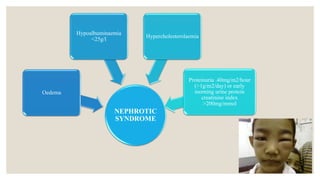
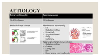
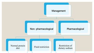
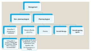
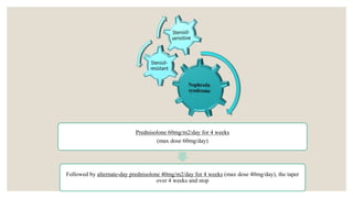
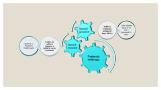
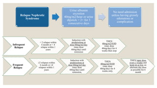
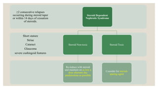
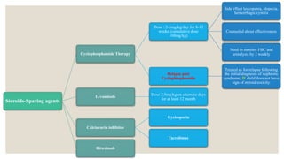
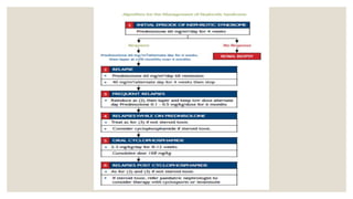
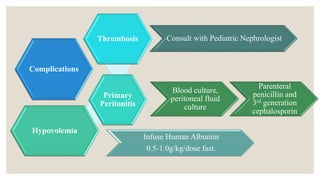
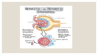
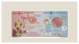
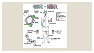
NEPHROTIC
SYNDROME
BY: NIK MOHD FAUZAN
NEPHROTIC
SYNDROME
Oedema
Hypoalbuminaemia
<25g/l
Hypercholesterolaemia
Proteinuria .40mg/m2/hour
(>1g/m2/day) or early
morning urine protein
creatinine index
>200mg/mmol
AETIOLOGY
Primary or Idiopathic Secondary causes
70-90% of cases 10% of cases
Minimal change disease Membranous nephropathy
- SLE
- Diabetes mellitus
- Hepatitis B
- Sjogren’s disease
- Drugs
- Malignany
Focal segmental glomerulosclerosis
- Diabetes mellitus
- Obesity
- Kidney loss
- HIV
- Hypertensive nephrosclerosis
History
◦ AGE : < 6 years old (peak of age)
◦ Symptoms : Tiredness, Edema, Breathless, Oliguria, Frothy urine
◦ Signs : Xantelasma, Periorbital edema, Ascites, Pedal Edema
◦ Family history : Family that having similar presentation
◦ Any clues for complications : Generalize anasarca, Peritonitis, Thrombosis
Examinations ??
◦ Height & Weight : Gross edematous can cause increase weight
◦ Blood Pressure : High in AGN & normal in NS
◦ Edema : Periorbital, sacral, genital (scrotum,labium), lower limb
◦ Oral : Injected pharynx and tonsil enlargement  post infectious or recurrent infection
◦ Neck : Cervical lymphadenopathy & JVP for fluid overload
◦ Lung : Reduce A/E ?, Crepitation
◦ Abdomen : Distended ? Soft ? Ascites ? (shiffting dullnes / Fluid trill)
Full Blood Count TWC – infection , Hb - Anemia
Renal profile : Urea, Electrolyte,
Creatinine
Renal Impairment (AKI) or Electrolyte
Imbalance
Serum Cholesterol Hyperlipidemia
Liver Function Test : Albumin Hypoalbunimia
Urinalysis, Urine Culture Why?? – Haematuria, Proteinuria
Quantitative urinary protein excretion Why?? – for diagnosis
What to expect ? More > 200mg/mmol
If urine PCI sent, still need to send 24H urine
protein collection? Yes
Issues with 24H urine collection? – Observe
proteinuria (worsening or improving)
Serum complement (C3, C4) level What to expect?
C3 level – low at onset symptom, normalizes
by 6 weeks
C4 level – usually normal limit in post
streptococcal AGN
ASOT titres Why?? ( ASOT > 200 IU/ml)
What are significant titre – Post streptococcal
infection
Antinuclear factor / anti-dsDNA What to expect? Positive to exclude SLE
Management
Non- pharmacological
Normal protein
diet
Fluid restriction
Restriction of
dietary sodium
Pharmacological
Management
Non- pharmacological Pharmacological
Penicillin V
(Prophylaxis/
treatment)
125mg BD (1-
5years), 250mg BD
(6-12years), 500mg
BD (>12years)
recommended
Human albumin
(20-25%)
0.5-1g/kg can be use in
symptomatic, grossly
oedematous case,
together with
IV Frusemide at 1-
2mg/kg
Diuretic Steroid therapy
Steroid-sparing
agents
Prednisolone 60mg/m2/day for 4 weeks
(max dose 60mg/day)
Followed by alternate-day prednisolone 40mg/m2/day for 4 weeks (max dose 40mg/day), the taper
over 4 weeks and stop
Referral to
Paediatric
Nephrologist
Failure to
achieve
response to
initial 4 weeks
treatment
Urine dipstix
trace or nil
for 3
consecutive
days
Achieve
remission
within 28
days (80%)
Infrequent
Relapse
< 2 relapse within
6 month or < 4
relapse within 1
years
Induction with
prednisolone at
dose 60mg/m2/day
(max dose
60mg/day) until
remission
THEN
40mg/m2/EOD
(max dose
40mg/day) for 4
weeks then stop
Frequent
Relapse
≥2 relapses within
6 month or ≥4
relapses within 1
years
Induction with
prednisolone at
dose 60mg/m2/day
(max dose
60mg/day) until
remission,
THEN
40mg/m2/EOD
(max dose
40mg/day) for 4
weeks only.
THEN taper dose
every 2 weeks and
keep on as low on
alternate day dose
as possible for 6
month
Relapse Nephrotic
Syndrome
Urine albumin
excretion
40mg/m2/hour or urine
dipstick > 2+ for 3
consecutive days
No need admission
unless having grossly
edematous or
complication
Short stature
Striae
Cataract
Glaucoma
severe cushingoid features
≥2 consecutive relapses
occurring during steroid taper
or within 14 days of cessation
of steroids.
Steroid Dependent
Nephrotic Syndrome
Steroid Non-toxic
Re-induce with steroid
and maintain on as low a
dose alternate day
prednisolone as possible
Steroid Toxic
Consider for steroid-
sparing agent
Steroids-Sparing agents
Cyclophosphamide Therapy
Dose : 2-3mg/kg/day for 8-12
weeks (cumulative dose
168mg/kg)
Side effect leucopenia, alopecia,
hemorrhagic cystitis
Counseled about effectiveness
Need to monitor FBC and
urinalysis by 2 weekly
Relapse post
Cyclophosphamide
Treated as for relapse following
the initial diagnosis of nephrotic
syndrome, IF child does not have
sign of steroid toxicity.
Levamisole
Dose 2.5mg/kg on alternate days
for at least 12 month
Calcineurin inhibitor
Cyclosporin
Tacrolimus
Rituximab
Complications
Thrombosis
Primary
Peritonitis
Hypovolemia
Infuse Human Albumin
0.5-1.0g/kg/dose fast.
Blood culture,
peritoneal fluid
culture
Parenteral
penicillin and
3rd generation
cephalosporin
Consult with Pediatric Nephrologist
REFERENCE
Paediatric protocols for Malaysian Hospital 4th Edition
Essential Medicine Concise Clinical Notes on Understanding & Managing Disease
Robbins and Cotran, Pathologic Basis of Disease, 8th Edition
NEPHRITIC
Characterized by inflammation in glomeruli that present with :
◦ Edema : Facial puffiness
◦ Hypertension
◦ Oliguria
◦ Azotemia : High level nitrogen-containing compound
◦ Microscopic / Macroscopic hematuria : Tea colour-urine / Cola like colour / Smoky
AETIOLOGY
POST STREPTOCOCCAL AGN ( Group A Beta Hemolytic Streptococci )
◦ Comment causes of acute nephritic syndrome, mainly due to post-streptococcal
Pharynx or skin infection
◦ Commonest at 6-10 years age
◦ Latent period :
◦ After sore throat : 1-2 weeks
◦ After skin infection : 2-3 weeks
Major Symptoms
◦ Hematuria (most frequent)
◦ Gross/microscopic >5 RBCs/HPF
◦ Urine Color : deep red-brownish, smoky, cola like color
◦ Hypertension (60-80%)
◦ Monitoring BP 4 Hourly
◦ Due to : General vasospasm, Hypervolemia or Salt water retention
◦ Oliguria (50%) <0.5mk/kg/h. Occur due to reduce GFR
◦ Edema
◦ Course : Begin with puffy eye in morning & LL edema at night
Investigation
◦ Urinalysis
◦ Hematuria : present in all patient
◦ Protenuria (trace to 2+, but mayb in nephrotic range)
◦ Red blood cell casts
◦ Pyuria may present
◦ Evidence Streptococcus infection
◦ Throat & Skin swab culture
◦ Raised ASOT (Anti Streptolysin O test) >200IU/ml
◦ Increased anti-DNAse B : better serological marker of preceding streptococcal skin infection
◦ Renal Function Test
Investigation
◦ Full Blood Count
◦ Leukocytosis may present
◦ Anaemia
◦ Complement Level
◦ C3 level : Low at onset of symptom, normalies within 6 weeks
◦ C4 level : Usually normal limits in post-streptococcal AGN
Management
◦ Strict monitoring : Fluid intake, Urine output, daily weight, BP (Nephrotic chart)
◦ Penicillin V x10/7 to eliminate B-haemolytic streptococcal infection
◦ Fluid restriction to control oedema and circulatory overload during oliguric phase until child diureses
and blood pressure controlled
◦ Day 1 : up to 400ml/m2/day. DO NOT administer IV or oral fluid if child has pulmonary edema
◦ Day 2 : till patient diureses – 400ml/m2/day
◦ When child diureses - allowed free fluid
Management
◦ Diuretics (Frusemide) should given pt that having pulmonary edema. Usually needed for treatment
hypertension
◦ Diet – NO added salt to diet. Protein restriction unnecessary
◦ Watch Out for Complication post-streptococcal AGN
Follow Up
◦ Atleast 1 year
◦ Monitor BP every visit
◦ Do urinalysis and renal function to evaluate recovery
◦ Repeat C3 levels 6 weeks later if not already normalized by time discharge
RENAL BIOPSY
Kidney biopsy is a procedure where a
doctor takes a tiny piece of kidney tissue
using a special needle. The tissue is
examined under a microscope in the
laboratory
Risk of Procedure
Kidney biopsies are usually low-risk procedures. The risk may increase depending on your child’s
condition, age and health.
◦ bleeding into the urine
◦ bleeding around or into the kidney
◦ infection
◦ needle injury to any other nearby organ
◦ urine leak from the kidney
◦ kidney damage
Procedure
1. Your child will be lying prone.
2. The interventional radiologist uses ultrasound to view the kidneys.
3. Local anaesthetic is then injected into the skin to numb the biopsy area.
4. Then, while watching the kidney using the ultrasound, the interventional radiologist passes a special
thin needle into one of the kidneys to get samples. Usually two or three samples are taken
5. The samples are about 2 to 3 centimeters (1 inch) long, and look like a piece of thread. These kidney
samples are then sent to the lab for examination.
6. You child will usually not need any stitches. A small bandage is placed over the biopsy site.
7. A kidney biopsy usually takes 45 minutes to one hour
Indication of Renal Biopsy in Nephrotic Syndrome
REFERENCE
Paediatric protocols for Malaysian Hospital 4th Edition
Essential Medicine Concise Clinical Notes on Understanding & Managing Disease
Robbins and Cotran, Pathologic Basis of Disease, 8th Edition
Nephrotic.pptx

Nephrotic.pptx

  • 1.
  • 2.
  • 3.
    AETIOLOGY Primary or IdiopathicSecondary causes 70-90% of cases 10% of cases Minimal change disease Membranous nephropathy - SLE - Diabetes mellitus - Hepatitis B - Sjogren’s disease - Drugs - Malignany Focal segmental glomerulosclerosis - Diabetes mellitus - Obesity - Kidney loss - HIV - Hypertensive nephrosclerosis
  • 4.
    History ◦ AGE :< 6 years old (peak of age) ◦ Symptoms : Tiredness, Edema, Breathless, Oliguria, Frothy urine ◦ Signs : Xantelasma, Periorbital edema, Ascites, Pedal Edema ◦ Family history : Family that having similar presentation ◦ Any clues for complications : Generalize anasarca, Peritonitis, Thrombosis
  • 5.
    Examinations ?? ◦ Height& Weight : Gross edematous can cause increase weight ◦ Blood Pressure : High in AGN & normal in NS ◦ Edema : Periorbital, sacral, genital (scrotum,labium), lower limb ◦ Oral : Injected pharynx and tonsil enlargement  post infectious or recurrent infection ◦ Neck : Cervical lymphadenopathy & JVP for fluid overload ◦ Lung : Reduce A/E ?, Crepitation ◦ Abdomen : Distended ? Soft ? Ascites ? (shiffting dullnes / Fluid trill)
  • 6.
    Full Blood CountTWC – infection , Hb - Anemia Renal profile : Urea, Electrolyte, Creatinine Renal Impairment (AKI) or Electrolyte Imbalance Serum Cholesterol Hyperlipidemia Liver Function Test : Albumin Hypoalbunimia Urinalysis, Urine Culture Why?? – Haematuria, Proteinuria Quantitative urinary protein excretion Why?? – for diagnosis What to expect ? More > 200mg/mmol If urine PCI sent, still need to send 24H urine protein collection? Yes Issues with 24H urine collection? – Observe proteinuria (worsening or improving)
  • 7.
    Serum complement (C3,C4) level What to expect? C3 level – low at onset symptom, normalizes by 6 weeks C4 level – usually normal limit in post streptococcal AGN ASOT titres Why?? ( ASOT > 200 IU/ml) What are significant titre – Post streptococcal infection Antinuclear factor / anti-dsDNA What to expect? Positive to exclude SLE
  • 8.
    Management Non- pharmacological Normal protein diet Fluidrestriction Restriction of dietary sodium Pharmacological
  • 9.
    Management Non- pharmacological Pharmacological PenicillinV (Prophylaxis/ treatment) 125mg BD (1- 5years), 250mg BD (6-12years), 500mg BD (>12years) recommended Human albumin (20-25%) 0.5-1g/kg can be use in symptomatic, grossly oedematous case, together with IV Frusemide at 1- 2mg/kg Diuretic Steroid therapy Steroid-sparing agents
  • 10.
    Prednisolone 60mg/m2/day for4 weeks (max dose 60mg/day) Followed by alternate-day prednisolone 40mg/m2/day for 4 weeks (max dose 40mg/day), the taper over 4 weeks and stop
  • 11.
    Referral to Paediatric Nephrologist Failure to achieve responseto initial 4 weeks treatment Urine dipstix trace or nil for 3 consecutive days Achieve remission within 28 days (80%)
  • 12.
    Infrequent Relapse < 2 relapsewithin 6 month or < 4 relapse within 1 years Induction with prednisolone at dose 60mg/m2/day (max dose 60mg/day) until remission THEN 40mg/m2/EOD (max dose 40mg/day) for 4 weeks then stop Frequent Relapse ≥2 relapses within 6 month or ≥4 relapses within 1 years Induction with prednisolone at dose 60mg/m2/day (max dose 60mg/day) until remission, THEN 40mg/m2/EOD (max dose 40mg/day) for 4 weeks only. THEN taper dose every 2 weeks and keep on as low on alternate day dose as possible for 6 month Relapse Nephrotic Syndrome Urine albumin excretion 40mg/m2/hour or urine dipstick > 2+ for 3 consecutive days No need admission unless having grossly edematous or complication
  • 13.
    Short stature Striae Cataract Glaucoma severe cushingoidfeatures ≥2 consecutive relapses occurring during steroid taper or within 14 days of cessation of steroids. Steroid Dependent Nephrotic Syndrome Steroid Non-toxic Re-induce with steroid and maintain on as low a dose alternate day prednisolone as possible Steroid Toxic Consider for steroid- sparing agent
  • 14.
    Steroids-Sparing agents Cyclophosphamide Therapy Dose: 2-3mg/kg/day for 8-12 weeks (cumulative dose 168mg/kg) Side effect leucopenia, alopecia, hemorrhagic cystitis Counseled about effectiveness Need to monitor FBC and urinalysis by 2 weekly Relapse post Cyclophosphamide Treated as for relapse following the initial diagnosis of nephrotic syndrome, IF child does not have sign of steroid toxicity. Levamisole Dose 2.5mg/kg on alternate days for at least 12 month Calcineurin inhibitor Cyclosporin Tacrolimus Rituximab
  • 16.
    Complications Thrombosis Primary Peritonitis Hypovolemia Infuse Human Albumin 0.5-1.0g/kg/dosefast. Blood culture, peritoneal fluid culture Parenteral penicillin and 3rd generation cephalosporin Consult with Pediatric Nephrologist
  • 17.
    REFERENCE Paediatric protocols forMalaysian Hospital 4th Edition Essential Medicine Concise Clinical Notes on Understanding & Managing Disease Robbins and Cotran, Pathologic Basis of Disease, 8th Edition
  • 23.
    NEPHRITIC Characterized by inflammationin glomeruli that present with : ◦ Edema : Facial puffiness ◦ Hypertension ◦ Oliguria ◦ Azotemia : High level nitrogen-containing compound ◦ Microscopic / Macroscopic hematuria : Tea colour-urine / Cola like colour / Smoky
  • 24.
    AETIOLOGY POST STREPTOCOCCAL AGN( Group A Beta Hemolytic Streptococci ) ◦ Comment causes of acute nephritic syndrome, mainly due to post-streptococcal Pharynx or skin infection ◦ Commonest at 6-10 years age ◦ Latent period : ◦ After sore throat : 1-2 weeks ◦ After skin infection : 2-3 weeks
  • 25.
    Major Symptoms ◦ Hematuria(most frequent) ◦ Gross/microscopic >5 RBCs/HPF ◦ Urine Color : deep red-brownish, smoky, cola like color ◦ Hypertension (60-80%) ◦ Monitoring BP 4 Hourly ◦ Due to : General vasospasm, Hypervolemia or Salt water retention ◦ Oliguria (50%) <0.5mk/kg/h. Occur due to reduce GFR ◦ Edema ◦ Course : Begin with puffy eye in morning & LL edema at night
  • 26.
    Investigation ◦ Urinalysis ◦ Hematuria: present in all patient ◦ Protenuria (trace to 2+, but mayb in nephrotic range) ◦ Red blood cell casts ◦ Pyuria may present ◦ Evidence Streptococcus infection ◦ Throat & Skin swab culture ◦ Raised ASOT (Anti Streptolysin O test) >200IU/ml ◦ Increased anti-DNAse B : better serological marker of preceding streptococcal skin infection ◦ Renal Function Test
  • 27.
    Investigation ◦ Full BloodCount ◦ Leukocytosis may present ◦ Anaemia ◦ Complement Level ◦ C3 level : Low at onset of symptom, normalies within 6 weeks ◦ C4 level : Usually normal limits in post-streptococcal AGN
  • 28.
    Management ◦ Strict monitoring: Fluid intake, Urine output, daily weight, BP (Nephrotic chart) ◦ Penicillin V x10/7 to eliminate B-haemolytic streptococcal infection ◦ Fluid restriction to control oedema and circulatory overload during oliguric phase until child diureses and blood pressure controlled ◦ Day 1 : up to 400ml/m2/day. DO NOT administer IV or oral fluid if child has pulmonary edema ◦ Day 2 : till patient diureses – 400ml/m2/day ◦ When child diureses - allowed free fluid
  • 29.
    Management ◦ Diuretics (Frusemide)should given pt that having pulmonary edema. Usually needed for treatment hypertension ◦ Diet – NO added salt to diet. Protein restriction unnecessary ◦ Watch Out for Complication post-streptococcal AGN
  • 30.
    Follow Up ◦ Atleast1 year ◦ Monitor BP every visit ◦ Do urinalysis and renal function to evaluate recovery ◦ Repeat C3 levels 6 weeks later if not already normalized by time discharge
  • 31.
    RENAL BIOPSY Kidney biopsyis a procedure where a doctor takes a tiny piece of kidney tissue using a special needle. The tissue is examined under a microscope in the laboratory
  • 32.
    Risk of Procedure Kidneybiopsies are usually low-risk procedures. The risk may increase depending on your child’s condition, age and health. ◦ bleeding into the urine ◦ bleeding around or into the kidney ◦ infection ◦ needle injury to any other nearby organ ◦ urine leak from the kidney ◦ kidney damage
  • 33.
    Procedure 1. Your childwill be lying prone. 2. The interventional radiologist uses ultrasound to view the kidneys. 3. Local anaesthetic is then injected into the skin to numb the biopsy area. 4. Then, while watching the kidney using the ultrasound, the interventional radiologist passes a special thin needle into one of the kidneys to get samples. Usually two or three samples are taken 5. The samples are about 2 to 3 centimeters (1 inch) long, and look like a piece of thread. These kidney samples are then sent to the lab for examination. 6. You child will usually not need any stitches. A small bandage is placed over the biopsy site. 7. A kidney biopsy usually takes 45 minutes to one hour
  • 34.
    Indication of RenalBiopsy in Nephrotic Syndrome
  • 38.
    REFERENCE Paediatric protocols forMalaysian Hospital 4th Edition Essential Medicine Concise Clinical Notes on Understanding & Managing Disease Robbins and Cotran, Pathologic Basis of Disease, 8th Edition