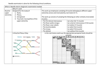
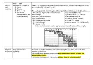
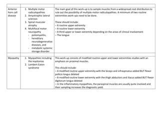
Needle examination is done for the following clinical conditions
When is Needle electro diagnostic examination needed
Why it’s used
Routine
upper
extremity
Designed for the study of
1. Roots,
2. Plexus,
3. Entrapment
4. Traumatic neuropathies of the
upper extremity
This work-up emphasizes sampling of muscles belonging to different upper
extremity nerves and innervated by root levels C5-T1.
The work-up consists of sampling the following (or other similarly innervated)
muscles:
– The first dorsal interosseous
– The flexor pollicis longus
– The flexor carpi radialis
– The brachioradialis
– The triceps
– The deltoid
(an ulnar C8, T1 muscle)
(an anterior interosseous C7,8 muscle)
(a median C7 muscle)
(a radial C5,6 muscle)
(a radial C7,8 muscle)
(an axillary C5,6 muscle)
In Brachial Plexus Palsy In the root lesions work-up, the appropriate paraspinal levels should be
sampled.
Why it’s used
Routine
lower
extremity
Designed for the study of
1. Roots,
2. Plexus,
3. Entrapment
4. Traumatic
neuropathies of the
Lower extremity
Tis work-up emphasizes sampling of muscles belonging to different lower extremity nerves
and innervated by root levels L3-S2.
The work-up consists of sampling the following (or other similarly innervated) muscles:
– The extensor digitorum brevis or extensor
hallucis longus
– The flexor digitorum longus
– The tibialis anterior
– The medial gastrocnemius
– The vastus lateralis
– The gluteus medius
a Peroneal L5-S1 muscles
a posterior tibial L5-S1,2 muscle
a peroneal L4,5 muscle
a posterior tibial S1,2 muscle
a femoral L3,4 muscle
a superior gluteal L4,5 and S1 muscle
In the root lesions work-up, the appropriate paraspinal levels should be sampled
Peripheral
neuropathy
Typical neuropathic
processes
This work-up emphasizes on distal muscles sampling because these are usually more
involved it consists of:
– A routine upper extremity examination with an extra distal muscle included, the
abductor digiti minimi
– A routine lower extremity examination with the abductor hallucis included
Anterior
horn cell
disease
1. Multiple motor
radiculopathies
2. Amyotrophic lateral
sclerosis
3. Spinal muscular
atrophy
4. Multifocal motor
neuropathy
- poliomyelitis,
- hereditary
neurodegenerative
diseases, and
- metabolic systemic
storage disorder
The main goal of this work-up is to sample muscles from a widespread root distribution to
rule out the possibility of multiple motor radiculopathies. A minimum of two routine
extremities work-ups need to be done.
These should include:
– A routine upper extremity
– A routine lower extremity
– A third upper or lower extremity depending on the areas of clinical involvement
– The tongue
Myopathy 1. Myopathies including
the myotonias
2. Lambert-Eaton
syndrome
This work-up consists of modified routine upper and lower extremities studies with an
emphasis on proximal muscles.
This should include:
– A modified routine upper extremity with the biceps and infraspinatus added BUT flexor
pollicis longus deleted
– A modified routine lower extremity with the thigh abductors and iliacus added BUT flexor
digitorum longus deleted
– In the inflammatory myopathies, the paraspinal muscles are usually quite involved and
their sampling increases the diagnostic yield.
Neuromuscular
Transmission
Defects in neuromuscular
transmission
1. Myasthenia gravis,
2. Lambert-Eaton
syndrome,
3. Botulism
4. Many drugs and
anesthetic agents
also affect
neuromuscular
junction and impulse
transmission to elicit
their effects.
Above conditions result in
muscle weakness and
paralysis
Single fiber EMG has greatly altered the traditional neuromuscular transmission
defects work-ups by needle electrodes.
Through moment to moment variation in the shape and amplitude of affected
motor unit potentials is helpful, jitter analysis by single fiber EMG is a much
more sensitive means to study defects in neuromuscular transmission.
The technique requires the use of a special needle electrode which has a 25 µm
tip on a side port to allow recording from single muscle fibers. When the tip is
positioned in the vicinity of two muscle fibers belonging to the same motor unit,
two potentials are seen firing synchronously
In neuromuscular transmission disorders, the jitter is increased early in the
course of the illness, before repetitive stimulation tests become positive. In the
later stages, impulse blocking due to total failure in neuromuscular transmission
is seen and results in the disappearance of one of the potentials on the screen.
If, by means of a delay line and a signal trigger, one of them is made to trigger the
sweep, the distance between the two potentials, or interpotential interval, is
observed to vary from discharge to discharge. The distance between the first and
second potential is measured over a certain number of tracings and the mean
interpotential interval is calculated. The standard deviation around that mean or
the mean of the consecutive differences (MCD) are used in expressing the jitter
which to a large extent represents the variability in neuromuscular transmission.
[Not needed; only if you can remember]

Needle examination [DOC 1 v2].pdf

  • 1.
    Needle examination isdone for the following clinical conditions When is Needle electro diagnostic examination needed Why it’s used Routine upper extremity Designed for the study of 1. Roots, 2. Plexus, 3. Entrapment 4. Traumatic neuropathies of the upper extremity This work-up emphasizes sampling of muscles belonging to different upper extremity nerves and innervated by root levels C5-T1. The work-up consists of sampling the following (or other similarly innervated) muscles: – The first dorsal interosseous – The flexor pollicis longus – The flexor carpi radialis – The brachioradialis – The triceps – The deltoid (an ulnar C8, T1 muscle) (an anterior interosseous C7,8 muscle) (a median C7 muscle) (a radial C5,6 muscle) (a radial C7,8 muscle) (an axillary C5,6 muscle) In Brachial Plexus Palsy In the root lesions work-up, the appropriate paraspinal levels should be sampled.
  • 2.
    Why it’s used Routine lower extremity Designedfor the study of 1. Roots, 2. Plexus, 3. Entrapment 4. Traumatic neuropathies of the Lower extremity Tis work-up emphasizes sampling of muscles belonging to different lower extremity nerves and innervated by root levels L3-S2. The work-up consists of sampling the following (or other similarly innervated) muscles: – The extensor digitorum brevis or extensor hallucis longus – The flexor digitorum longus – The tibialis anterior – The medial gastrocnemius – The vastus lateralis – The gluteus medius a Peroneal L5-S1 muscles a posterior tibial L5-S1,2 muscle a peroneal L4,5 muscle a posterior tibial S1,2 muscle a femoral L3,4 muscle a superior gluteal L4,5 and S1 muscle In the root lesions work-up, the appropriate paraspinal levels should be sampled Peripheral neuropathy Typical neuropathic processes This work-up emphasizes on distal muscles sampling because these are usually more involved it consists of: – A routine upper extremity examination with an extra distal muscle included, the abductor digiti minimi – A routine lower extremity examination with the abductor hallucis included
  • 3.
    Anterior horn cell disease 1. Multiplemotor radiculopathies 2. Amyotrophic lateral sclerosis 3. Spinal muscular atrophy 4. Multifocal motor neuropathy - poliomyelitis, - hereditary neurodegenerative diseases, and - metabolic systemic storage disorder The main goal of this work-up is to sample muscles from a widespread root distribution to rule out the possibility of multiple motor radiculopathies. A minimum of two routine extremities work-ups need to be done. These should include: – A routine upper extremity – A routine lower extremity – A third upper or lower extremity depending on the areas of clinical involvement – The tongue Myopathy 1. Myopathies including the myotonias 2. Lambert-Eaton syndrome This work-up consists of modified routine upper and lower extremities studies with an emphasis on proximal muscles. This should include: – A modified routine upper extremity with the biceps and infraspinatus added BUT flexor pollicis longus deleted – A modified routine lower extremity with the thigh abductors and iliacus added BUT flexor digitorum longus deleted – In the inflammatory myopathies, the paraspinal muscles are usually quite involved and their sampling increases the diagnostic yield.
  • 4.
    Neuromuscular Transmission Defects in neuromuscular transmission 1.Myasthenia gravis, 2. Lambert-Eaton syndrome, 3. Botulism 4. Many drugs and anesthetic agents also affect neuromuscular junction and impulse transmission to elicit their effects. Above conditions result in muscle weakness and paralysis Single fiber EMG has greatly altered the traditional neuromuscular transmission defects work-ups by needle electrodes. Through moment to moment variation in the shape and amplitude of affected motor unit potentials is helpful, jitter analysis by single fiber EMG is a much more sensitive means to study defects in neuromuscular transmission. The technique requires the use of a special needle electrode which has a 25 µm tip on a side port to allow recording from single muscle fibers. When the tip is positioned in the vicinity of two muscle fibers belonging to the same motor unit, two potentials are seen firing synchronously In neuromuscular transmission disorders, the jitter is increased early in the course of the illness, before repetitive stimulation tests become positive. In the later stages, impulse blocking due to total failure in neuromuscular transmission is seen and results in the disappearance of one of the potentials on the screen. If, by means of a delay line and a signal trigger, one of them is made to trigger the sweep, the distance between the two potentials, or interpotential interval, is observed to vary from discharge to discharge. The distance between the first and second potential is measured over a certain number of tracings and the mean interpotential interval is calculated. The standard deviation around that mean or the mean of the consecutive differences (MCD) are used in expressing the jitter which to a large extent represents the variability in neuromuscular transmission. [Not needed; only if you can remember]