
Recommended
More Related Content
What's hot
What's hot (12)
Recently uploaded
Recently uploaded (20)
msbte exam time table summer 2018
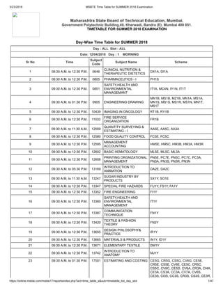
- 1. 3/23/2018 MSBTE Time Table for SUMMER 2018 Examination https://online.msbte.com/msbte17/reportsindex.php?act=time_table_s&sub=timetable_list_day_slot 1/47 Maharashtra State Board of Technical Education, Mumbai. Government Polytechnic Building,49, Kherwadi, Bandra (E), Mumbai 400 051. TIMETABLE FOR SUMMER 2018 EXAMINATION Day-Wise Time Table for SUMMER 2018 Day : ALL Slot : ALL Date: 12/04/2018 Day : 1 MORNING Sr No Time Subject Code Subject Name Scheme 1 09:30 A.M. to 12:30 P.M. 0646 CLINICAL NUTRITION & THERAPEUTIC DIETETICS DX1A, DI1A 2 09:30 A.M. to 12:30 P.M. 0805 PHARMACEUTICS - I PH1S 3 09:30 A.M. to 12:30 P.M. 0851 SAFETY,HEALTH AND ENVIRONMENTAL MANAGEMANT IT1A, MC4N, IY1N, IT1T 4 09:30 A.M. to 01:30 P.M. 0905 ENGINEERING DRAWING MN1B, MS1B, MZ1B, MN1A, MS1A, MN1S, MS1S, MS1R, MS1N, MN1T, MS1T 5 09:30 A.M. to 12:30 P.M. 10439 IMAGING IN ONCOLOGY RT1B, RY1B 6 09:30 A.M. to 12:30 P.M. 11033 FIRE SERVICE ORGANIZATION FR1B 7 09:30 A.M. to 11:30 A.M. 12559 QUANTITY SURVEYING & ESTIMATING - I AA5E, AA5C, AA3A 8 09:30 A.M. to 12:30 P.M. 12580 FOOD QUALITY CONTROL FC5E, FC5C 9 09:30 A.M. to 12:30 P.M. 12595 MANAGEMENT ACCOUNTING HM5E, HM5C, HM3B, HM3A, HM3R 10 09:30 A.M. to 12:30 P.M. 12602 BASIC HEMATOLOGY ML5E, ML5C, ML3A 11 09:30 A.M. to 12:30 P.M. 12608 PRINTING ORGNIZATIONAL MANAGEMENT PN5E, PC7E, PN5C, PC7C, PC3A, PN3A, PN3S, PN3R, PN3N 12 09:30 A.M. to 05:30 P.M. 13169 INTRODUCTION TO ANIMATION DA2E, DA2C 13 09:30 A.M. to 11:30 A.M. 13247 SUGAR INDUSTRY BY PRODUCTS SX1Y, SO1E 14 09:30 A.M. to 12:30 P.M. 13347 SPECIAL FIRE HAZARDS FU1Y, FS1Y, FA1Y 15 09:30 A.M. to 12:30 P.M. 13352 FIRE ENGINEERING FI1Y 16 09:30 A.M. to 12:30 P.M. 13360 SAFETY,HEALTH AND ENVIRONMENTAL MANAGEMENT IT1Y 17 09:30 A.M. to 12:30 P.M. 13387 COMMUNICATION TECHNIQUE FN1Y 18 09:30 A.M. to 12:30 P.M. 13420 TEXTILE & FASHION THEORY FN3Y 19 09:30 A.M. to 12:30 P.M. 13655 DESIGN PHILOSOPHY& PRACTICE IR1Y 20 09:30 A.M. to 12:30 P.M. 13665 MATERIALS & PRODUCTS IN1Y, ID1Y 21 09:30 A.M. to 12:30 P.M. 13671 ELEMENTARY TEXTILE DM1Y 22 09:30 A.M. to 12:30 P.M. 13742 INTRODUCTION TO ANATOMY MJ1Y 23 09:30 A.M. to 01:30 P.M. 17501 ESTIMATING AND COSTING CE5G, CR5G, CS5G, CV6G, CE5E, CR5E, CS5E, CV6E, CE5C, CR5C, CS5C, CV6C, CE5D, CV6A, CR3A, CI4A, CE3A, CS3A, CC3A, CV7A, CV6S, CE3S, CI3S, CC3S, CR3S, CS3S, CE3R,
- 2. 3/23/2018 MSBTE Time Table for SUMMER 2018 Examination https://online.msbte.com/msbte17/reportsindex.php?act=time_table_s&sub=timetable_list_day_slot 2/47 CR3R, CS3R, CV6M, CE3N, CR3N, CI3N, CI4N, CC4N, CC3N, CS3N, CE3O, CR3O, CS3O, CV6T 24 09:30 A.M. to 12:30 P.M. 17508 SWITCHGEAR & PROTECTION EE5G, EP5G, EU5G, EP5E, EE5E, EP5C, EE5C, EC4A, EE3A, EP3A, EG4A, EP3S, EE3S, EC4S, EG4S, EP3R, EE3R, EC4N, EP3N, EG4N, EE3N, EP3O, EE3O 25 09:30 A.M. to 12:30 P.M. 17512 OPERATING SYSTEM CM5G, CO5G, CW5G, IF5G, CD6G, IF5E, CO5E, CM5E, CD6E, CW5E, CD6C, IF5C, CO5C, CM5C, CD6A, CL4A, CM3A, CO3A, IF3A, CM3S, IF3S, CM3S, CM3R, CM3R, CO3N, CD6T 26 09:30 A.M. to 12:30 P.M. 17521 TWO WHEELER TECHNOLOGY AE5G 27 09:30 A.M. to 12:30 P.M. 17530 METROLOGY AND QUALITY CONTROL ME5G, PG5G, PT5G, MH6G, MI6G, PT5E, ME5E, MH6E, MI6E, PG5E, PT5C, ME5C, MH6C, MI6C, PG5C, MH6A, PS2A, PT3A, ME3A, MC4A, MG4A, PG3A, MH7A, PY6S, PT3S, PG3S, MC4S, ME3S, MG4S, PT3R, ME3R, PT3N, ME3N, MC3N, MG3N, ME3O, MH6T, PY6T, MH7T 28 09:30 A.M. to 11:30 A.M. 17533 COMPUTER HARDWARE & NETWORKING DE5G, EJ5G, EN5G, ET5G, EV5G, EX5G, IC5G, IE5G, IS5G, MU5G, IU6G, ED6G, EI6G, EQ5G, IE5E, IC5E, EX5E, MU5E, IU6E, IS5E, EV5E, EN5E, EJ5E, EI6E, ED6E, ET5E, DE5E, IE5C, IC5C, EX5C, MU5C, IU6C, IS5C, EN5C, DE5C, EJ5C, EI6C, ED6C, EV5C, ET5C, DE3A, EV3A 29 09:30 A.M. to 12:30 P.M. 17548 TESTING OF PLASTICS PS5G, PS5E, PS5C, PS3A 30 09:30 A.M. to 01:30 P.M. 17553 DESIGN OF FABRICATED STRUCTURAL ELEMENT FG5G, FE6G, FE6E, FE6C 31 09:30 A.M. to 12:30 P.M. 17560 HEAT TRANSFER OPERATION CH5G, CH5E, CH5C 32 09:30 A.M. to 12:30 P.M. 17563 TEXTRONICS TC5G, TX5G, TC5E, TX5E, TC5C, TX5C, TM4A 33 09:30 A.M. to 11:30 A.M. 17574 CAD-CAM IN APPAREL MANUFACTURING DC5G 34 09:30 A.M. to 12:30 P.M. 17965 GEO TECHNICAL ENGINEERING CC5G, CI5G, CI5E, CC5E, CC5C 35 09:30 A.M. to 12:30 P.M. 17967 ELECTRICAL & ELECTRONIC MEASUREMENT EC5G, EG5G, EC5E, EG5E, EC4C, EG5C 36 09:30 A.M. to 12:30 P.M. 17971 BASIC ELECTRONICS & MECHATRONICS MC5G, MG5G 37 09:30 A.M. to 11:30 A.M. 19501 QUALITY CONTROL DD5G 38 09:30 A.M. to 12:30 P.M. 19507 ADVANCED FOOD PRODUCTION HM5G 39 09:30 A.M. to 12:30 P.M. 19516 SEROLOGY ML5G 40 09:30 A.M. to 12:30 P.M. 19517 FOOD PACKAGING FC5G 41 09:30 A.M. to 12:30 P.M. 19521 MAINTENANCES OF PRINTING MACHINES PC5G, PN5G 42 09:30 A.M. to 12:30 P.M. 19529 PROCESS INSTRUMENTATION SC5G 43 09:30 A.M. to 12:30 P.M. 19533 TOUR OPERATION MANAGEMENT TR5G 44 09:30 A.M. to 12:30 P.M. 23106 SPECIAL FIRE HAZARDS FA1G, FS1G 45 09:30 A.M. to 12:30 P.M. 23112 ENVIRONMENTAL IT1G
- 3. 3/23/2018 MSBTE Time Table for SUMMER 2018 Examination https://online.msbte.com/msbte17/reportsindex.php?act=time_table_s&sub=timetable_list_day_slot 3/47 MANAGEMENT 46 09:30 A.M. to 11:30 A.M. 23125 AROMATHERAPY BY1G 47 09:30 A.M. to 11:30 A.M. 23130 ANATOMY AND PHYSIOLOGY BC1G 48 09:30 A.M. to 11:30 A.M. 23131 FUNDAMENTALS OF DESIGN DM1G 49 09:30 A.M. to 11:30 A.M. 23134 INTRODUCTION TO FASHION STUDIES FN1G 50 09:30 A.M. to 03:30 P.M. 23136 INTERIOR DESIGN ID1G, IN1G 51 09:30 A.M. to 12:30 P.M. 23141 FIRE ENGINEERING SCIENCE FR1G 52 09:30 A.M. to 12:30 P.M. 23144 FIRE ENGINEERING SCIENCE FI1G 53 09:30 A.M. to 12:30 P.M. 23149 BASICS OF BAMBOO BO1G 54 09:30 A.M. to 12:30 P.M. 6219 FIRE SERVICE EQUIPMENTS AND APPLICATIONS FL1A, FU1A, FF1A, FI1A, FS1S 55 09:30 A.M. to 12:30 P.M. 6804 MINING MACHINERY MS3B, MN3B, MZ3B Date: 12/04/2018 Day : 1 AFTERNOON Sr No Time Subject Code Subject Name Course Code 56 02:00 P.M. to 05:00 P.M. 12012 COMMUNICATION SKILLS TM2E, HC2E, VE1E, VD1E, VC1E, VB1E, VA1E, VF1E, WT2E, VW1E, VT1E, VR1E, VP1E, VN1E, VM1E, VH1E, TT2C, HC2C, VE1C, VD1C, VC1C, VB1C, VA1C, VH1C, WT2C, VW1C, VT1C, VR1C, VP1C, VN1C, VM1C, VF1C 57 02:00 P.M. to 05:00 P.M. 12877 MANAGEMENT CI8E, EG8E, CC8E, MG8E, MC8E, EC8E, CL8E, IL8E, CI6C, MG8C, MC8C, IL8C, EG7C, EC8C, CL7C, CC8C 58 02:00 P.M. to 05:00 P.M. 13059 GOOD CLINICAL PRACTICES CK2E 59 02:00 P.M. to 05:00 P.M. 13072 BASIC MEDICINES IN HEMODIALYSIS HD2E, HD2C 60 02:00 P.M. to 05:00 P.M. 13081 PARASITOLOGY, MYCOLOGY & VIROLOGY LX2E, LT2C 61 02:00 P.M. to 05:00 P.M. 13083 BASIC OF SURGERY OT2E 62 02:00 P.M. to 05:00 P.M. 13101 BASIC PRINCIPLES OF ULTRASOUND RX2E, RS2C 63 02:00 P.M. to 05:00 P.M. 13184 COMPUTER HARDWARE & TROUBLESHOOTING HN2E, HN2C 64 02:00 P.M. to 05:00 P.M. 13208 SOFTWARE ENGINEERING CP2E, CP2C 65 02:00 P.M. to 05:00 P.M. 13288 ENERGY AUDIT ER2E, EW2E 66 02:00 P.M. to 05:00 P.M. 13502 RETAIL MANAGEMENT AX2E, AM2C 67 02:00 P.M. to 05:00 P.M. 17201 COMMUNICATION SKILLS CW2G, DE2G, DC2G, ED2G, EE2G, EI2G, EJ2G, CV2G, CS2G, CR2G, CH2G, CM2G, CO2G, CE2G, CD2G, AU2G, AE2G, EN2G, EP2G, ET2G, TC2G, PT2G, PS2G, PG2G, MU2G, MI2G, MH2G, ME2G, IU2G, IS2G, IF2G, IE2G, IC2G, FG2G, EX2G, EV2G, TX2G, AA2G, EQ2G, FC2G, ML2G, PC2G, PN2G, SC2G, TR2G, EU2G, FE2E, FC2E, EX2E, EV2E, ET2E, EP2E, EN2E, IC2E, MI2E, MH2E, ME2E, IU2E, IS2E, IF2E, IE2E, EJ2E, CO2E, CM2E, CH2E, CE2E, CD2E, AE2E, AA2E, CR2E, EI2E, EE2E, ED2E, DE2E, DC2E, CW2E,
- 4. 3/23/2018 MSBTE Time Table for SUMMER 2018 Examination https://online.msbte.com/msbte17/reportsindex.php?act=time_table_s&sub=timetable_list_day_slot 4/47 CV2E, CS2E, TX2E, TC2E, SC2E, PG2E, PC2E, MU2E, ML2E, PN2E, PS2E, PT2E, ML2C, FE2C, FC2C, EX2C, ET2C, EP2C, IC2C, MI2C, MH2C, ME2C, IU2C, IS2C, IF2C, IE2C, EN2C, EJ2C, CR2C, CO2C, CM2C, CH2C, CE2C, CD2C, AE2C, CS2C, EI2C, EE2C, ED2C, DE2C, DC2C, CV2C, AA2C, TX2C, TC2C, PT2C, PS2C, PN2C, PG2C, PC2C, MU2C, SC2C, EV2C, FC1B, MU1B, IN1B, IU2A, ED2A, EI2A, FE2A, CD2A, CV2A, PY2A, MM2A, MI2A, MH2A, EC1A, EE1A, EG1A, DE1A, EV1A, EX1A, FC1A, FT1A, EJ1A, EN1A, EP1A, ET1A, CS1A, CC1A, CE1A, AA1A, CO1A, CR1A, CM1A, AE1A, CH1A, CI1A, CL1A, MU1A, ME1A, MG1A, ML1A, PS1A, PT1A, TM1A, TT1A, PC1A, PE1A, PG1A, PN1A, LO1A, IE1A, IC1A, IF1A, IL1A, MC1A, LG1A, IS1A, BC1A, IN1A, ID1A, EI2S, CD2S, CV2S, FE2S, MM2S, MH2S, IU2S, EC1S, DE1S, EE1S, CS1S, EG1S, ET1S, EX1S, FC1S, EL1S, EP1S, AE1S, CC1S, AA1S, EN1S, CM1S, CO1S, CE1S, CH1S, CI1S, CR1S, MO1S, MT1S, MC1S, ME1S, MF1S, MG1S, ML1S, MU1S, PS1S, PT1S, TM1S, TT1S, PN1S, MX1S, PC2S, PE1S, PG1S, PK1S, IS1S, IC1S, IE1S, IF1S, FT1S, KT1S, EE1R, DE1R, CS1R, EX1R, ET1R, EP1R, AE1R, CE1R, CR1R, CH1R, MO1R, MT1R, MU1R, ME1R, MF1R, ML1R, PS1R, PT1R, TM1R, PN1R, MX1R, PC2R, PE1R, PK1R, IS1R, IE1R, HM1R, FT1R, KT1R, EE1M, DE1M, CS1M, EX1M, EN1M, EP1M, ET1M, CE1M, AE1M, CM1M, CO1M, CR1M, CH1M, MU1M, ME1M, PS1M, PT1M, PE1M, IS1M, IE1M, IC1M, EI2N, FE2N, MM2N, EC1N, EE1N, EG1N, CS1N, DE1N, EL1N, ET1N, EP1N, CC1N, AA1N, AE1N, CE1N, CM1N, CO1N, CR1N, CG1N, CH1N, CI1N, FT1N, MC1N, MO1N, MT1N, MU1N, ME1N, MF1N, MG1N, ML1N, MX1N, PS1N, PT1N, TM1N, PN1N, PC2N, PE1N, PK1N, IE1N, IL1N, IS1N, HM1N, KT1N, BC1N, CV2T, ED2T, EI2T, CD2T, MH2T, IU2T, FE2T 68 02:00 P.M. to 05:00 P.M. 17916 COMMUNICATION SKILLS CC2G, CI2G, EC2G, EG2G, IL2G, MC2G, MG2G, XC2G 69 02:00 P.M. to 04:00 P.M. 19206 TRADITIONAL COSTUMES AND TEXTILES OF INDIA DD2G 70 02:00 P.M. to 05:00 P.M. 19215 HOUSEKEEPING-II HM2G 71 02:00 P.M. to 05:00 P.M. 22203 APPLIED MECHANICS CE2I, CS2I, CH2I, CR2I, AE2I, ME2I, PG2I, PT2I 72 02:00 P.M. to 05:00 P.M. 22210 APPLIED MATHEMATICS EX2I, EU2I, ET2I, EQ2I, EP2I, EN2I, EJ2I, EE2I, DE2I, IC2I, IE2I, IS2I, MU2I 73 02:00 P.M. to 05:00 P.M. 22224 APPLIED MATHEMATICS CW2I, CO2I, CM2I, IF2I 74 02:00 P.M. to 05:00 P.M. 22228 MECHANICAL ENGINEERING IN PLASTIC PRODUCTION ENGG. PS2I 75 02:00 P.M. to 05:00 P.M. 22235 BASICS OF FASHION AND APPAREL DC2I 76 02:00 P.M. to 05:00 P.M. 22240 FUNDAMENTAL OF MECHANICAL ENGINEERING TC2I, TX2I
- 5. 3/23/2018 MSBTE Time Table for SUMMER 2018 Examination https://online.msbte.com/msbte17/reportsindex.php?act=time_table_s&sub=timetable_list_day_slot 5/47 Date: 13/04/2018 Day : 2 MORNING Sr No Time Subject Code Subject Name Scheme 77 09:30 A.M. to 12:30 P.M. 0811 PHARMACEUTICS - II PH2S 78 09:30 A.M. to 12:30 P.M. 10448 RADIATION BIOLOGY RT2B, RY2B 79 09:30 A.M. to 12:30 P.M. 11037 INDUSTRIAL SAFETY FR2B 80 09:30 A.M. to 12:30 P.M. 12479 TEXTILE TESTING TC3E, TC3C, TT3C 81 09:30 A.M. to 12:30 P.M. 1272 BUSINESS ACCOUNTING ST2A 82 09:30 A.M. to 12:30 P.M. 12768 COMMUNICATION SKILLS CL3E, CI3E, CC3E, MG3E, MC3E, IL3E, EG3E, EC3E, CL3C, CI3C, CC2C, EC2C, MG3C, MC2C, IL3C, EG3C 83 09:30 A.M. to 12:30 P.M. 1311 MINE SERVEYING - I MS2A, MN2A, MS2S, MN2S, MS2R, MS2N, MS2T, MN2T 84 09:30 A.M. to 12:30 P.M. 13111 REMOVABLE PARTIAL PROSTHODONTICS DT3E, DT3C 85 09:30 A.M. to 12:30 P.M. 13200 MODELLING TECHNIQUES DA3E, DA3C 86 09:30 A.M. to 11:30 A.M. 13335 ADVANCE BEAUTY TECHNIQUE BC2Y 87 09:30 A.M. to 12:30 P.M. 13407 TEXTILE THEORY FN2Y 88 09:30 A.M. to 11:30 A.M. 13681 INTERIOR PROFESSIONAL PRACTICE ID2Y, IN3Y 89 09:30 A.M. to 12:30 P.M. 13687 INDIAN COSTUME DM2Y 90 09:30 A.M. to 11:30 A.M. 13747 RADIATION PHYSICS & RADIATION PROTECTION MJ2Y 91 09:30 A.M. to 12:30 P.M. 14091 TRANSPORT & INDUSTRIAL MANAGEMENT VA3E, VA3C 92 09:30 A.M. to 12:30 P.M. 14092 BUILDING CONSTRUCTION MANAGEMENT VB3E, VB3C 93 09:30 A.M. to 12:30 P.M. 14100 INDUSTRIAL ELECTRONICS VE3E, VE3C 94 09:30 A.M. to 12:30 P.M. 17301 APPLIED MATHEMATICS AE3G, CD3G, CE3G, CH3G, CM3G, CO3G, CR3G, CS3G, CV3G, CW3G, DE3G, ED3G, EE3G, EI3G, EJ3G, EN3G, EP3G, ET3G, EV3G, EX3G, FE3G, FG3G, IC3G, IE3G, IF3G, IS3G, IU3G, ME3G, MH3G, MI3G, MU3G, PG3G, PS3G, PT3G, EQ3G, EU3G, CD3E, CM3E, CO3E, CW3E, IF3E, EI3E, EE3E, ED3E, DE3E, PT3E, PS3E, PG3E, ME3E, EJ3E, EN3E, IU3E, IS3E, IC3E, EX3E, MU3E, EV3E, ET3E, EP3E, IE3E, MH3E, FE3E, CV3E, CS3E, CR3E, AE3E, MI3E, CH3E, CE3E, CD3C, CM3C, CO3C, IF3C, MU3C, EJ3C, EI3C, EE3C, ED3C, DE3C, PT3C, PS3C, EN3C, IE3C, IU3C, IS3C, IC3C, EX3C, EV3C, ET3C, EP3C, PG3C, MH3C, CR3C, CV3C, ME3C, FE3C, CS3C, CE3C, AE3C, MI3C, CH3C, CE3D, MU2B, MA2A, CV3A, IU3A, EI3A, ED3A, CD3A, FE3A, MH3A, MM3A, IU4A, EI4A, ED4A, IF2A, CO2A, CM2A, CL2A, CS2A, CR2A, CI2A, CE2A, CC2A, PT2A, PS2A, PG2A, MG2A, ME2A, MC2A, CH2A, AE2A, MU2A, IS2A, IL2A, IE2A, IC2A, EX2A, EV2A, ET2A, EN2A, EJ2A, DE2A, EC2A, EE2A, EG2A, EP2A, IU3S, CV3S, EI3S, FE3S, MM3S, IU4S, EI4S, IF2S, CO2S, CM2S, CS2S, CR2S, CI2S, CE2S, CC2S, PT2S, PS2S, PG2S, PE2S, MG2S, ME2S, MC2S, FT2S, CH2S, AE2S, MU2S, IS2S, IE2S, IC2S, EX2S,
- 6. 3/23/2018 MSBTE Time Table for SUMMER 2018 Examination https://online.msbte.com/msbte17/reportsindex.php?act=time_table_s&sub=timetable_list_day_slot 6/47 EV2S, ET2S, EN2S, EL2S, DE2S, EC2S, EE2S, EG2S, EP2S, CO2R, CM2R, CS2R, CR2R, CE2R, PT2R, PS2R, PE2R, ME2R, FT2R, CH2R, AE2R, MU2R, IS2R, IE2R, EX2R, EV2R, ET2R, DE2R, EE2R, EP2R, IU3N, EI3N, FE3N, MM3N, IU4N, EI4N, CO2N, CS2N, CR2N, CI2N, CE2N, CC2N, PT2N, PS2N, PE2N, MG2N, ME2N, MC2N, FT2N, EG2N, CH2N, CG2N, AE2N, MU2N, IS2N, IL2N, IE2N, EV2N, ET2N, EL2N, DE2N, CM2N, EC2N, EE2N, EP2N, CS2O, CR2O, CE2O, PT2O, PE2O, ME2O, FT2O, CH2O, AE2O, IS2O, IE2O, ET2O, DE2O, CM2O, EE2O, EP2O, IU3T, CV3T, ED3T, CD3T, EI3T, FE3T, MH3T, MM3T, IU4T, EI4T, ED4T 95 09:30 A.M. to 12:30 P.M. 17334 TEXTILE TESTING DC3G 96 09:30 A.M. to 12:30 P.M. 17343 CHEMISTRY OF DYES & PIGMENTS TC3G 97 09:30 A.M. to 12:30 P.M. 17344 YARN MANUFACTURING-II TX3G, TX3E, TX3C, TM2A 98 09:30 A.M. to 12:30 P.M. 17353 BASIC ELECTRICAL AND ELECTRONICS ENGINEERING AU3G 99 09:30 A.M. to 01:30 P.M. 19301 BUILDING CONSTRUCTION SYSTEM AA3G, AA3E, AA3C, AA2A 100 09:30 A.M. to 11:30 A.M. 19308 APPRECIATION OF WORLD COSTUMES DD3G 101 09:30 A.M. to 12:30 P.M. 19314 BASIC HOTEL ACCOUNTING HM3G 102 09:30 A.M. to 12:30 P.M. 19316 GENERAL BACTERIOLOGY ML3G, ML3E, ML3C, ML2A 103 09:30 A.M. to 12:30 P.M. 19320 PRINCIPLES OF FOOD ENGINEERING FC3G, FC3E, FC3C 104 09:30 A.M. to 12:30 P.M. 19325 PAPER & INK TECHNOLOGY PC3G, PN3G, PC3E, PN3E, PC3C, PN3C, PN1A, PC2A, PN1S, PN1R, PN1N 105 09:30 A.M. to 12:30 P.M. 19329 WESTERN WORLD TOURIST DESTINATION TR3G, TR3E, TR3C 106 09:30 A.M. to 12:30 P.M. 3508 DESIGN-II ID2N, IN3N 107 09:30 A.M. to 12:30 P.M. 6797 MINE SERVEYING - I MS2B, MN2B, MZ2B Date: 13/04/2018 Day : 2 AFTERNOON Sr No Time Subject Code Subject Name Course Code 108 02:00 P.M. to 05:00 P.M. 12250 MACHINE TOOL AND DESIGN PT6E, PG6E, PG6C, PT6C 109 02:00 P.M. to 05:00 P.M. 12270 CONTROL SYSTEMS IU7E, ET6E, EX6E, EJ6E, IC6E, IE6E, IS6E, ED7E, EI7E, DE6E, EN6E, EV6E, EJ6C, ET6C, EX6C, EN6C, EV6C, ED7C, EI7C, IC6C, IE6C, DE6C, IS6C, IU7C 110 02:00 P.M. to 05:00 P.M. 12637 ACOUSTICS & CLIMATOLOGY AA6E, AA6C, AA3A 111 02:00 P.M. to 04:00 P.M. 12647 FASHION RETAIL MANAGEMENT DD6E, DD6C 112 02:00 P.M. to 05:00 P.M. 12656 FOOD PLANT ORGANIZATION AND MANAGEMENT FC6E, FC6C 113 02:00 P.M. to 05:00 P.M. 12679 ADVANCE HEMATOLOGY ML6E, ML6C 114 02:00 P.M. to 05:00 P.M. 12683 DIGITAL PRINTING PC8E, PN6E, PC8C, PN6C, PC4A, PN3A 115 02:00 P.M. to 05:00 P.M. 12836 ELECTRICAL ESTIMATION & EG6E, EC6E, EG6C, EC6C
- 7. 3/23/2018 MSBTE Time Table for SUMMER 2018 Examination https://online.msbte.com/msbte17/reportsindex.php?act=time_table_s&sub=timetable_list_day_slot 7/47 COSTING 116 02:00 P.M. to 05:00 P.M. 17618 VEHICLE SYSTEMS MAINTENANCE AE6G, AE6E, AE6C, AE3A, AE3S, AE3R, AE3N, AE3O 117 02:00 P.M. to 05:00 P.M. 17685 KIDS WEAR FASHION DESIGNING DC6G, DC6E, DC6C 118 02:00 P.M. to 05:00 P.M. 17694 PLC AND SCADA EU6G 119 02:00 P.M. to 05:00 P.M. 17979 IRRIGATION ENGINEERING CI6G, CC6G, CC6E, CI6E, CI6C, CC6C 120 02:00 P.M. to 05:00 P.M. 17987 INDUSTRY ELECTRICAL SYSTEMS-II EC6G, EG6G 121 02:00 P.M. to 05:00 P.M. 17988 FLUID MECHANICS & MACHINARY MC6G, MG6G, MG6E, MC6E, MC6C, MG6C 122 02:00 P.M. to 06:00 P.M. 19603 ESTIMATING & COSTING AA6G 123 02:00 P.M. to 05:00 P.M. 19608 FOOD PRODUCTION MANAGEMENT HM6G 124 02:00 P.M. to 05:00 P.M. 19612 MEDICAL PARASITOLOGY ML6G 125 02:00 P.M. to 04:00 P.M. 19621 QUALITY CONTROL IN PRINTING PC6G, PN6G Date: 16/04/2018 Day : 3 MORNING Sr No Time Subject Code Subject Name Scheme 126 09:30 A.M. to 11:30 A.M. 0647 ORGANIZATION & MANAGMENT OF DIETETICS IN HOSPITAL DI1A, DX1A 127 09:30 A.M. to 12:30 P.M. 0806 PHARMACEUTICAL CHEMISTRY - I PH1S 128 09:30 A.M. to 12:30 P.M. 0952 SECRETARIAL PRACTICES ST1A 129 09:30 A.M. to 12:30 P.M. 10440 BASIC RADIOTHERAPY TECHNIQUES, RY1B, RT1B 130 09:30 A.M. to 12:30 P.M. 11034 FIRE ENGINEERING SCIENCE FR1B 131 09:30 A.M. to 12:30 P.M. 12004 ENGLISH DA1E, DA1C 132 09:30 A.M. to 12:30 P.M. 13120 ORTHODONTIC - II DT4E, DT4C 133 09:30 A.M. to 11:30 A.M. 13248 MANAGEMENT SX1Y, SO1E 134 09:30 A.M. to 11:30 A.M. 13329 HAIR DESIGNING BC1Y 135 09:30 A.M. to 12:30 P.M. 13342 FIRE ENGINEERING SCIENCE FU1Y, FS1Y, FA1Y 136 09:30 A.M. to 12:30 P.M. 13353 FIRE SERVICE ORGANISATION AND ACTS FI1Y 137 09:30 A.M. to 12:30 P.M. 13361 SAFETY ENGINEERING- I IT1Y 138 09:30 A.M. to 12:30 P.M. 13388 FUNDAMENTALS OF TEXTILE & FASHION THEORY FN1Y 139 09:30 A.M. to 12:30 P.M. 13421 MANAGEMENT STUDIES FN3Y 140 09:30 A.M. to 05:30 P.M. 13657 INTERIOR ENVIORNMENT & DESIGN IR1Y 141 09:30 A.M. to 11:30 A.M. 13666 PRIMARY SERVICES ID1Y, IN1Y 142 09:30 A.M. to 12:30 P.M. 13672 GARMENT CONSTRUCTION DM1Y 143 09:30 A.M. to 12:30 P.M. 13743 INTRODUCTION TO PHYSIOLOGY & PATHOLOGY MJ1Y 144 09:30 A.M. to 12:30 P.M. 14154 UTILIZATION OF ELECTRICAL ENERGY VE4E, VE4C 145 09:30 A.M. to 12:30 P.M. 17101 ENGLISH AE1G, CD1G, CE1G, CH1G, CM1G, CO1G, CR1G, CS1G, CV1G, CW1G,
- 8. 3/23/2018 MSBTE Time Table for SUMMER 2018 Examination https://online.msbte.com/msbte17/reportsindex.php?act=time_table_s&sub=timetable_list_day_slot 8/47 DE1G, ED1G, EE1G, EI1G, EJ1G, EN1G, EP1G, ET1G, EV1G, EX1G, FG1G, IC1G, IE1G, IF1G, IS1G, IU1G, ME1G, MH1G, MI1G, MU1G, PG1G, PS1G, PT1G, TC1G, TX1G, DC1G, AU1G, EU1G, AA1G, DD1G, EQ1G, FC1G, GT1G, ML1G, PC1G, PN1G, SC1G, TR1G, AA1E, AE1E, CD1E, CE1E, CH1E, CM1E, CO1E, CR1E, CS1E, CV1E, CW1E, DC1E, DE1E, ED1E, EE1E, EI1E, EJ1E, EN1E, EP1E, ET1E, EV1E, EX1E, FC1E, FE1E, GT1E, IC1E, IE1E, IF1E, IS1E, IU1E, ME1E, MH1E, MI1E, ML1E, MU1E, PC1E, PG1E, PN1E, PS1E, PT1E, SC1E, TC1E, TM1E, TX1E, TR2E, WT1E, AA1C, AE1C, CD1C, CE1C, CH1C, CM1C, CO1C, CR1C, CS1C, CV1C, DC1C, DE1C, ED1C, EE1C, EI1C, EJ1C, EN1C, EP1C, ET1C, EV1C, EX1C, FC1C, FE1C, GT1C, IC1C, IE1C, IF1C, IS1C, IU1C, ME1C, MH1C, MI1C, ML1C, MU1C, PC1C, PG1C, PN1C, PS1C, PT1C, SC1C, TC1C, TM1C, TT1C, TX1C, TR2C, FN1C, WT1C, MA1A, CD1A, CV1A, ED1A, EI1A, FE1A, IU1A, MH1A, MI1A, MM1A, PY1A, CD1S, CV1S, EI1S, FE1S, IU1S, MH1S, MM1S, FE1N, IU1N, MM1N, CD1T, CV1T, ED1T, FE1T, IU1T, MH1T, MM1T 146 09:30 A.M. to 12:30 P.M. 17911 ENGLISH IL1G, XC1G, GC1G, CL1G, MG1G, CI1G, EG1G, MC1G, EC1G, CC1G 147 09:30 A.M. to 12:30 P.M. 19109 FOOD PRODUCATION-I HM1G, HM1E, HM1C, HM1A, HM1R, HM1N 148 09:30 A.M. to 12:30 P.M. 23101 FIRE ENGINEERING SCIENCE FA1G, FS1G 149 09:30 A.M. to 12:30 P.M. 23113 QUALITY CONTROL IN OCCUPATIONAL SAFETY AND HEALTH IT1G 150 09:30 A.M. to 11:30 A.M. 23126 ADVANCE HAIR DRESSING BY1G 151 09:30 A.M. to 11:30 A.M. 23127 BEAUTY & SKIN CARE TECHNIQUES BC1G 152 09:30 A.M. to 11:30 A.M. 23132 INTRODUCTION TO FABRICS DM1G 153 09:30 A.M. to 11:30 A.M. 23135 INTRODUCTION TO TEXTILES FN1G 154 09:30 A.M. to 12:30 P.M. 23139 MATERIALS,PRODUCTS AND MARKET SURVEY ID1G, IN1G 155 09:30 A.M. to 12:30 P.M. 23142 SPECIAL FIRE HAZARDS FR1G 156 09:30 A.M. to 12:30 P.M. 23145 FIRE SERVICE ORGANIZATION AND ACTS FI1G 157 09:30 A.M. to 12:30 P.M. 23150 BAMBOO SEASONING & PRESERVATION BO1G 158 09:30 A.M. to 12:30 P.M. 6240 SECURITY ACTS AND LAWS FF1A 159 09:30 A.M. to 12:30 P.M. 6793 MINING TECHNOLOGY MS1B, MN1B, MZ1B 160 09:30 A.M. to 12:30 P.M. 6805 MINE MANAGEMENT LEGISLATION & SAFETY MN3B, MS3B, MZ3B Date: 16/04/2018 Day : 3 AFTERNOON Sr No Time Subject Code Subject Name Course Code 161 02:00 P.M. to 05:00 P.M. 12111 DATA STRUCTURE CM4E, CO4E, IF4E, CD4E, CW4E, CM4C, CO4C, IF4C, CD4C
- 9. 3/23/2018 MSBTE Time Table for SUMMER 2018 Examination https://online.msbte.com/msbte17/reportsindex.php?act=time_table_s&sub=timetable_list_day_slot 9/47 162 02:00 P.M. to 05:00 P.M. 12130 MECHANICAL TECHNOLOGY CH4E, CH4C 163 02:00 P.M. to 05:00 P.M. 12540 TECHNOLOGY OF SIZING & BLEACHING TC4E, TC4C 164 02:00 P.M. to 05:00 P.M. 12784 CONCRETE TECHNOLOGY CI4E, CC4E, CI4C, CC4C 165 02:00 P.M. to 05:00 P.M. 12791 APPLIED ELECTRONICS EG4E, IL4E, EC4E, EG6C, IL4C, EC5C 166 02:00 P.M. to 05:00 P.M. 12801 FUNDAMENTALS OF ELECTRONICS MG4E, MC4E, MG4C, MC4C 167 02:00 P.M. to 05:00 P.M. 12850 CONTRACT & ACCOUNTS CI7E, CC7E, CI8C, CC7C 168 02:00 P.M. to 05:00 P.M. 13020 BASIC MICROBIOLOGY & TRANSFUSION MEDICIENE HD1E 169 02:00 P.M. to 05:00 P.M. 13024 BIO CHEMISTRY (MEDICAL) LX1E, LT1E, LT1C 170 02:00 P.M. to 05:00 P.M. 13027 BASICS OF HUMAN ANATOMY AND PHYSIOLOGY OT1E, OT1C 171 02:00 P.M. to 05:00 P.M. 13046 HUMAN ANATOMY & PHYSIOLOGY RS1E, RX1E, RS1C 172 02:00 P.M. to 05:00 P.M. 13147 NETWORKING ESSENTIAL & PROTOCOLS HN1E, HN1C 173 02:00 P.M. to 05:00 P.M. 13158 DATABASE MANAGEMENT SYSTEMS CP1E, CP1C 174 02:00 P.M. to 05:00 P.M. 13226 FUNDAMENTALS OF THERMAL UTILITIES ER1E, EW1E 175 02:00 P.M. to 05:00 P.M. 13454 PURCHASING & SUPPLY CHAIN MANAGEMENT AM1E, AX1E, AM1C 176 02:00 P.M. to 05:00 P.M. 17406 HEAT ENGINEERING PG4G, PT4G 177 02:00 P.M. to 05:00 P.M. 17409 AUTOMOBILE SYSTEMS & BODY ENGINEERING AE4G, AE4E, AE4C, AE2A, AE2S, AE2O 178 02:00 P.M. to 05:00 P.M. 17410 THERMAL ENGINEERING ME4G, MH4G, MI4G, MH4E, ME4E, MI4E, MH4C, ME4C, MI4C, MH4A, AE2A, MC3A, ME2A, MG3A, AE2S, MC2S, ME2S, MG2S, PE2S, AE2R, ME2R, PE2R, AE2N, EC3N, EG3N, MC3N, ME2N, MG3N, PE2N, AE2O, ME2O, PE2O, MH4T 179 02:00 P.M. to 05:00 P.M. 17415 DC MACHINES & TRANFORMERS EE4G, EP4G, EU4G, EE4E, EP4E, EP4C, EE4C, EP2A, EG2A, EE2A, EC3A, EP2S, EG3S, EE2S, EC3S, EP2R, EE2R, EP2N, EG2N, EE2N, EC2N, EP2O, EE2O 180 02:00 P.M. to 05:00 P.M. 17418 TRANSPORTATION ENGINEERING CE4G, CR4G, CV4G, CS4G, CV4E, CS4E, CR4E, CE4E, CE4C, CR4C, CV4C, CS4C, CV4A, CC3A, CE2A, CI3A, CR2A, CS2A, CV7A, CV4S, CC3S, CE2S, CI3S, CR2S, CS2S, CV7S, CE2R, CR2R, CC3N, CE2N, CI3N, CR2N, CE2O, CR2O, CV4T, CV7T 181 02:00 P.M. to 05:00 P.M. 17423 PHYSICAL CHEMISTRY & MATERIALS OF CONSTRUCTION CH4G 182 02:00 P.M. to 05:00 P.M. 17440 ANALOG COMMUNICATION ED4G, EI4G, EJ4G, EN4G, ET4G, EX4G, EX4E, ET4E, EI4E, ED4E, EN4E, EJ4E, EN4C, EJ4C, ET4C, EI4C, ED4C, EX4C, EI4A, ED4A, EJ2A, EN2A, ET2A, EX2A, EI4N, ED4T 183 02:00 P.M. to 05:00 P.M. 17441 TV SIGNAL TRANSMISSION SYSTEM EV4G, EQ4G, EV4E, EV4C, EV2A, EV2S, EV2R, EV2N 184 02:00 P.M. to 05:00 P.M. 17442 BIO SENSOR MU4G, MU4E, MU4C 185 02:00 P.M. to 05:00 P.M. 17448 PLASTIC MATERIALS PS4G, PS4E, PS4C
- 10. 3/23/2018 MSBTE Time Table for SUMMER 2018 Examination https://online.msbte.com/msbte17/reportsindex.php?act=time_table_s&sub=timetable_list_day_slot 10/47 186 02:00 P.M. to 05:00 P.M. 17457 PROCESSES EQUIPMENTS FE4G, FG4G, FE7A, FE7S, FE7T 187 02:00 P.M. to 05:00 P.M. 17466 TEXTILE CHEMISTRY-II TX4G, TM2A 188 02:00 P.M. to 05:00 P.M. 17471 TEXTILE TESTING TC4G 189 02:00 P.M. to 05:00 P.M. 19420 PRINTERS DESIGN PC4G 190 02:00 P.M. to 06:00 P.M. 21001 DESIGN OF STEEL STRUCTURES CI7G, CC7G 191 02:00 P.M. to 05:00 P.M. 21005 MICROCONTROLLER AND APPLICATIONS EC7G, EG7G 192 02:00 P.M. to 05:00 P.M. 21011 METROLOGY & QUALITY CONTROL MC7G, MG7G Date: 17/04/2018 Day : 4 MORNING Sr No Time Subject Code Subject Name Scheme 193 09:30 A.M. to 12:30 P.M. 0812 PHARMACEUTICAL CHEMISTRY - II PH2S 194 09:30 A.M. to 12:30 P.M. 10449 CLINICAL RADIATION ONCOLOGY - I RT2B, RY2B 195 09:30 A.M. to 12:30 P.M. 11038 FIRE PREVENTION, RESCUE & PARAMEDICS FR2B 196 09:30 A.M. to 12:30 P.M. 12560 HISTORY OF ARCHITECTURE AA5E, AA5C 197 09:30 A.M. to 12:30 P.M. 12581 FERMENTATION TECHNOLOGY FC5E, FC5C 198 09:30 A.M. to 12:30 P.M. 12596 MARKETING MANAGEMENT HM5E, HM5C, HM3B, HM3A 199 09:30 A.M. to 12:30 P.M. 12603 SEROLOGY ML5E, ML5C 200 09:30 A.M. to 12:30 P.M. 12607 DIGITAL IMAGEING PN5E, PC7E, PN5C, PC7C 201 09:30 A.M. to 12:30 P.M. 1273 BUSINESS COMMUNICATION ST2A 202 09:30 A.M. to 05:30 P.M. 13170 PRINCIPLES OF ANIMATION DA2E, DA2C 203 09:30 A.M. to 11:30 A.M. 13336 HAIR DRESSING BC2Y 204 09:30 A.M. to 11:30 A.M. 13408 FASHION THEORY FN2Y 205 09:30 A.M. to 01:30 P.M. 13680 INTERIOR WORKING DRAWING IN3Y, ID2Y 206 09:30 A.M. to 12:30 P.M. 13685 TRADE MANUFACTURING THEORY DM2Y 207 09:30 A.M. to 12:30 P.M. 13748 EQUIPMENT FOR MEDICAL IMAGING-II MJ2Y 208 09:30 A.M. to 12:30 P.M. 17502 IRRIGATION ENGINEERING CE5G, CR5G, CS5G, CV6G, CE5E, CR5E, CS5E, CV6E, CE5C, CR5C, CS5C, CV6C, CE5D, CV6A, CE3A, CI3A, CR3A, CS3A, CC4A, CV7A, CV6S, CI4S, CC4S, CS3S, CE3S, CR3S, CV7S, CE3R, CR3R, CI4N, CE3N, CR3N, CC4N, CE3O, CR3O, CV6T, CV7T 209 09:30 A.M. to 12:30 P.M. 17511 A.C.MACHINES EE5G, EP5G, EU5G, EE5E, EP5E, EP5C, EE5C 210 09:30 A.M. to 12:30 P.M. 17513 SOFTWARE ENGINEERING CM5G, CO5G, CW5G, IF5G, CD6G, CM5E, CD6E, CO5E, IF5E, CW5E, CO5C, CM5C, CD6C, IF5C, IF2A, CM3A, IF3S, CM3S, CM3R 211 09:30 A.M. to 12:30 P.M. 17522 HYDRAULICS & PNEUMATICS (AE) AE5G, AE5E, AE5C, AE3A, AE3S, AE3R, AE3N, AE2O 212 09:30 A.M. to 12:30 P.M. 17527 ADVANCED MANUFACTURING PROCESSES ME5G, PG5G, PT5G, MH6G, MI6G, ME5E, MH6E, MI6E, PG5E, PT5E, FE6E, ME5C, MH6C, MI6C, PG5C, PT5C,
- 11. 3/23/2018 MSBTE Time Table for SUMMER 2018 Examination https://online.msbte.com/msbte17/reportsindex.php?act=time_table_s&sub=timetable_list_day_slot 11/47 FE6C, FE4A, MG4A, PG3A, PT3A, MC3A, ME3A, MH7A, FE4S, PY6S, MG3S, MC3S, ME3S, PG3S, PT3S, ME3R, FE2N, MG3N, MC3N, ME3N, ME3O, FE4T, PY6T, MH7T 213 09:30 A.M. to 12:30 P.M. 17534 MICROCONTROLLER DE5G, EJ5G, EN5G, ET5G, EV5G, EX5G, IC5G, IE5G, IS5G, MU5G, IU6G, ED6G, EI6G, EQ5G, EI6E, EV5E, ET5E, EN5E, EJ5E, EX5E, IU6E, IS5E, MU5E, ED6E, DE5E, IC5E, IE5E, EX5C, EJ5C, EN5C, EV5C, ET5C, IE5C, IS5C, IU6C, MU5C, ED6C, EI6C, IC5C, DE5C, IU6A, EX3A, EJ3A, IC3A, IE3A, IS3A, EN3A, ET3A, DE3A, IU7A, IU6S, IS3S, ET3S, EX3S, IE3S, EN3S, DE3S, IU7S, ET3R, EX3R, IE3R, IS3R, DE3R, IU6N, IE3N, ET3N, IS3N, DE3N, IE3O, DE3O, IU6T, IU7T 214 09:30 A.M. to 12:30 P.M. 17549 DESIGN OF MOULDS PS5G, PS5E, PS5C 215 09:30 A.M. to 12:30 P.M. 17561 CHEMICAL PROCESS INSRTUMENTATION AND CONTROL CH5G, CH5E, CH5C, CH3A, CH3S, CH3R, CH3N, CH3O 216 09:30 A.M. to 12:30 P.M. 17564 TECHNOLOGY OF DYEING- II TC5G, TC5E, TC5C 217 09:30 A.M. to 12:30 P.M. 17568 YARN MANUFACTURING-IV TX5G, TX5E, TX5C 218 09:30 A.M. to 12:30 P.M. 17966 HYDRAULICS CC5G, CI5G, CC5E, CI5E, CC5C 219 09:30 A.M. to 12:30 P.M. 17968 TRANSMISSION & DISTRIBUTION OF ELECTRICAL POWER EC5G, EG5G, EC5E, EG5E, EG4C, EC5C 220 09:30 A.M. to 12:30 P.M. 17972 THEORY OF MACHINES MC5G, MG5G, MG5E, MC5E, MG5C, MC5C 221 09:30 A.M. to 12:30 P.M. 19502 FASHION MARKETING DD5G 222 09:30 A.M. to 12:30 P.M. 19508 ADVANCED FOOD AND BEVERAGE HM5G 223 09:30 A.M. to 12:30 P.M. 19512 MEDICAL VIROLOGY AND MYCOLOGY ML5G 224 09:30 A.M. to 12:30 P.M. 19518 INDUSTRIAL DAIRY TECHNOLOGY FC5G 225 09:30 A.M. to 12:30 P.M. 19522 DIGITAL PRINTING PN5G, PC7G 226 09:30 A.M. to 12:30 P.M. 19530 TECHNOLOGY OF PAINTS-II SC5G 227 09:30 A.M. to 12:30 P.M. 19534 PLANNING IN TOURISM TR5G 228 09:30 A.M. to 12:30 P.M. 6798 ROCK ENGINEERING AND GROUND CONTROL MS2B, MN2B, MZ2B Date: 17/04/2018 Day : 4 AFTERNOON Sr No Time Subject Code Subject Name Course Code 229 02:00 P.M. to 05:00 P.M. 12389 MATHEMATICS & STATISTICS FC2E, FC2C 230 02:00 P.M. to 06:00 P.M. 12878 DESIGN OF STRUCTURES CI8E, CC8E, CI8C, CC8C 231 02:00 P.M. to 05:00 P.M. 12888 POWER ELECTRONICS AND DRIVES EC8E, EG8E, EC8C, EG8C 232 02:00 P.M. to 05:00 P.M. 13060 BIOAVAILABILITY & BIOEQUIVALENCE CK2E 233 02:00 P.M. to 05:00 P.M. 13073 HEMODIALYSIS TECHNIQUES - I HD2E, HD2C 234 02:00 P.M. to 05:00 P.M. 13077 BACTERIOLOGY, IMMUNOLOGY & SEROLOGY LX2E, LT2C
- 12. 3/23/2018 MSBTE Time Table for SUMMER 2018 Examination https://online.msbte.com/msbte17/reportsindex.php?act=time_table_s&sub=timetable_list_day_slot 12/47 235 02:00 P.M. to 05:00 P.M. 13084 SURGICAL EQUIPMENT & MACHINERY OT2E 236 02:00 P.M. to 05:00 P.M. 13102 INSTRUMENTATION OF ULTRA - SONOGRAPHY RX2E, RS2C 237 02:00 P.M. to 04:00 P.M. 13185 NETWORK OPERATING SYSTEM INFRASTRUCTURE HN2E, HN2C 238 02:00 P.M. to 05:00 P.M. 13209 WEB DESIGNING CP2E, CP2C 239 02:00 P.M. to 05:00 P.M. 13289 EVALUATION OF UTILITY SYSTEMS ER2E, EW2E 240 02:00 P.M. to 05:00 P.M. 13501 EXP. MARKETING & DOCUMENTATION AX2E, AM2C 241 02:00 P.M. to 05:00 P.M. 17216 ENGINEERING MATHEMATICS DE2G, CW2G, ED2G, EE2G, EI2G, EJ2G, EN2G, CV2G, CS2G, CO2G, CR2G, CM2G, CH2G, CE2G, CD2G, AE2G, AU2G, EP2G, EV2G, ET2G, PT2G, PS2G, MU2G, PG2G, MI2G, MH2G, EX2G, ME2G, FG2G, IE2G, IC2G, IF2G, IS2G, IU2G, EQ2G, FC2G, EU2G, FE2E, IC2E, IE2E, IF2E, IS2E, EJ2E, EN2E, EP2E, ET2E, EV2E, EX2E, PT2E, PS2E, MI2E, MU2E, TX2E, IU2E, TC2E, ME2E, MH2E, PG2E, CV2E, CO2E, CR2E, CW2E, CS2E, CM2E, AE2E, CD2E, CE2E, CH2E, EE2E, EI2E, DE2E, ED2E, IC2C, IE2C, IF2C, IS2C, IU2C, FE2C, EN2C, EP2C, ET2C, EV2C, EX2C, TC2C, PT2C, PS2C, MI2C, MU2C, PG2C, TX2C, ME2C, MH2C, CR2C, CS2C, CV2C, CO2C, AE2C, CD2C, CE2C, CH2C, CM2C, EI2C, EJ2C, EE2C, DE2C, ED2C, CV2A, CD2A, MM2A, ED2A, MI2A, PY2A, MH2A, EI2A, IU2A, FE2A, CV2S, CD2S, MH2S, MM2S, IU2S, EI2S, FE2S, MM2N, EI2N, FE2N, CV2T, CD2T, ED2T, MH2T, MM2T, IU2T, EI2T, FE2T 242 02:00 P.M. to 05:00 P.M. 17218 BASICS OF FASHION DESIGN & CLOTHING TECHNOLOGY DC2G 243 02:00 P.M. to 05:00 P.M. 17223 CHEMISTRY OF NATURAL FIBERS TC2G 244 02:00 P.M. to 05:00 P.M. 17224 YARN MANUFACTURING - I TX2G, TM1A, TM1S, TM1R, TM1N 245 02:00 P.M. to 05:00 P.M. 17917 ENGINEERING MATHEMATICS CC2G, CI2G, EC2G, EG2G, IL2G, MC2G, MG2G, XC2G, IL2E, CI2E, EG2E, EC2E, CL2E, CC2E, MG2E, MC2E, IL2C, EG2C, CC2C, EC2C, CL2C, CI2C, MG2C, MC2C 246 02:00 P.M. to 06:00 P.M. 19204 ARCHITECTURAL DRAWING-II AA2G, AA2E, AA2C 247 02:00 P.M. to 04:00 P.M. 19205 FUNDAMENTALS OF TEXTILES DD2G, DD2E, DD2C, DD1A, GT1A, DM1A 248 02:00 P.M. to 05:00 P.M. 19214 FOOD & BEVERAGE SERVICE-II HM2G, HM2E, HM2C 249 02:00 P.M. to 05:00 P.M. 19220 HUMAN BIOLOGY ML2G, ML2E, ML2C 250 02:00 P.M. to 05:00 P.M. 19222 REPRODUCTION PHOTOGRAPHY PC2G, PN2G, PC2E, PN2E, PC2C, PN2C, PC2A, PN1A, PN2A, PC3A, PN2S, PN2R, PN2N 251 02:00 P.M. to 05:00 P.M. 19227 TECHNOLOGY OF PIGMENTS - II SC2G, SC2E, SC2C 252 02:00 P.M. to 05:00 P.M. 19230 ORGANIZATION IN TOURISM INDUSTRY TR2G, TR2E, TR2C
- 13. 3/23/2018 MSBTE Time Table for SUMMER 2018 Examination https://online.msbte.com/msbte17/reportsindex.php?act=time_table_s&sub=timetable_list_day_slot 13/47 253 02:00 P.M. to 06:00 P.M. 21031 DESIGN OF R.C.C. STRUCTURES CC8G, CI8G 254 02:00 P.M. to 05:00 P.M. 21036 POWER SYSTEM OPERATION & CONTROL EC8G, EG8G 255 02:00 P.M. to 06:00 P.M. 21038 DESIGN OF MACHINE ELEMENTS MC8G, MG8G 256 02:00 P.M. to 05:00 P.M. 22212 FUNDAMENTALS OF ELECTRICAL ENGINEERING EU2I, EP2I, EE2I 257 02:00 P.M. to 05:00 P.M. 22215 ELEMENTS OF ELECTRICAL ENGINEERING ET2I, EQ2I, EN2I, EJ2I, DE2I, CW2I, CO2I, CM2I, EX2I, IC2I, IE2I, IF2I, IS2I 258 02:00 P.M. to 05:00 P.M. 22219 APPLICATIONS OF BIOMATERIALS MU2I 259 02:00 P.M. to 05:00 P.M. 22229 ORGANIC CHEMISTRY PS2I 260 02:00 P.M. to 05:00 P.M. 22231 FUNDAMENTAL OF CHEMICAL ENGINEERING CH2I 261 02:00 P.M. to 05:00 P.M. 22236 PRINCIPLES OF TEXTILE & FASHION DESIGN DC2I 262 02:00 P.M. to 05:00 P.M. 22239 ELEMENTS OF ELECTRICAL & ELECTRONICS ENGINEERING TC2I, TX2I Date: 18/04/2018 Day : 5 MORNING Sr No Time Subject Code Subject Name Scheme 263 09:30 A.M. to 12:30 P.M. 0648 FOOD PRODUCTION AND FOOD COSTING DI1A, DX1A 264 09:30 A.M. to 12:30 P.M. 0807 PHARMACOGNOSY PH1S 265 09:30 A.M. to 12:30 P.M. 0901 COMMUNICATION SKILLS MS1B, MN1B, MZ1B, MS1A, MN1A, ST1A, MS1S, MN1S, MS1R, MS1N, MS1T, MN1T 266 09:30 A.M. to 12:30 P.M. 10441 INTRODUCTION TO ANATOMY RT1B, RY1B 267 09:30 A.M. to 12:30 P.M. 11035 SAFETY MANAGEMENT FR1B 268 09:30 A.M. to 12:30 P.M. 12063 OBJECT ORIENTED PROGRAMMING IF3E, CW3E, CO3E, CM3E, CD3E, IF3C, CO3C, CM3C, CD3C 269 09:30 A.M. to 12:30 P.M. 12446 FOOD MICROBIOLOGY FC3E, FC3C 270 09:30 A.M. to 12:30 P.M. 13249 SUGAR TECHNOLOGY - I SX1Y, SO1E 271 09:30 A.M. to 12:30 P.M. 13343 ORGANISATION AND ACTS FU1Y, FA1Y, FS1Y 272 09:30 A.M. to 12:30 P.M. 13354 FIRE SAFETY IN BUILIDINGS FI1Y 273 09:30 A.M. to 12:30 P.M. 13362 SAFETY ENGINEERING - II IT1Y 274 09:30 A.M. to 12:30 P.M. 13662 BASIC DESIGN IN1Y, ID1Y 275 09:30 A.M. to 12:30 P.M. 13670 COLOUR DRAWING THEORY DM1Y 276 09:30 A.M. to 12:30 P.M. 13744 PATIENT CARE IN RADIOGRAPHY MJ1Y 277 09:30 A.M. to 12:30 P.M. 14086 CAD-CAM - II VM3E, VT3E, VM3C, VT3C 278 09:30 A.M. to 12:30 P.M. 14088 AUTO ELECTRICAL ELECTRONICS & AIR CONDITIONING SYSTEM VA3E, VA3C 279 09:30 A.M. to 12:30 P.M. 14101 ELECTRICAL MACHINE - II VE3E, VE3C 280 09:30 A.M. to 12:30 P.M. 17304 STRENGTH OF MATERIALS AE3G, FE3G, FG3G, ME3G, MH3G, MI3G, PG3G, PS3G, PT3G, AE3E, PG3E, MI3E, FE3E, ME3E, MH3E, PS3E, PT3E, ME3C, AE3C, PG3C, MI3C,
- 14. 3/23/2018 MSBTE Time Table for SUMMER 2018 Examination https://online.msbte.com/msbte17/reportsindex.php?act=time_table_s&sub=timetable_list_day_slot 14/47 MH3C, FE3C, PS3C, PT3C, MM3A, FE3A, MH3A, PT2A, PS2A, PG2A, MG2A, ME2A, MC3A, AE2A, CH2A, FE3S, MM3S, PT2S, PS2S, PG2S, PE2S, MG3S, ME2S, MC3S, FT2S, AE2S, CH2S, PT2R, PS2R, PK2R, ME2R, FT2R, AE2R, CH2R, MM3N, FE3N, PT2N, PS2N, PK2N, ME2N, FT2N, AE2N, CH2N, PT2O, ME2O, FT2O, AE2O, CH2O, FE3T, MM3T, MH3T 281 09:30 A.M. to 01:30 P.M. 17309 BUILDING DRAWING CE3G, CR3G, CS3G, CV3G, CV3E, CS3E, CR3E, CE3E, CE3C, CV3C, CR3C, CS3C, CE3D, CV4A, CS2A, CR2A, CI2A, CE2A, CC2A, CV4S, CS2S, CR2S, CE2S, CC2S, CS3R, CR2R, CE2R, CS3N, CR2N, CE2N, CC2N, CS3O, CR2O, CE2O, CV4T 282 09:30 A.M. to 12:30 P.M. 17312 INDUSTRIAL CHEMISTRY CH3G, CH3E, CH3C, CH2A, CH2S, CH2R, CH2N, CG2N 283 09:30 A.M. to 12:30 P.M. 17320 PRINCIPLES OF DIGITAL TECHNIQUES DE3G, ED3G, EI3G, EJ3G, EN3G, ET3G, EV3G, EX3G, IC3G, IE3G, IS3G, IU3G, MU3G, EQ3G, EJ3E, EN3E, ET3E, EI3E, ED3E, EV3E, MU3E, IU3E, EX3E, IC3E, IE3E, IS3E, DE3E, EJ3C, EN3C, ET3C, EI3C, ED3C, EV3C, MU3C, EX3C, IC3C, IE3C, IS3C, IU3C, DE3C, MU2B, EI3A, ED3A, IU3A, ED4A, EI4A, IU4A, DE2A, EJ2A, EN2A, ET2A, EV2A, EX2A, IC2A, IE2A, IS2A, MU2A, DE3A, IU4S, EI4S, DE2S, EL3S, EN2S, ET2S, EX2S, IC2S, IE2S, IS2S, MU2S, IE3R, ET3R, IS3R, IS2M, IE2M, EX2M, ET2M, DE2M, EI3N, IU4N, EI4N, IE3N, CM3N, EL3N, ET3N, IL3N, IS3N, CM3O, CM3O, ET3O, IS3O, IU4T, ED4T, EI4T 284 09:30 A.M. to 12:30 P.M. 17323 ELECTRICAL CIRCUITS AND NETWORKS EE3G, EP3G, EU3G, EP3E, EE3E, EP3C, EE3C, EP2A, EG2A, EE2A, EC2A 285 09:30 A.M. to 12:30 P.M. 17335 PATTERN MAKING-II DC3G, DC3E, DC3C 286 09:30 A.M. to 12:30 P.M. 17339 INDUSTRIAL CHEMISTRY TC3G, TC3E, TT3C, TC3C 287 09:30 A.M. to 12:30 P.M. 17345 FABRIC MANUFACTURING-II TX3G, TX3E, TX3C, TM2A 288 09:30 A.M. to 12:30 P.M. 17349 APPLIED MATHEMATICS (AGRICULTURE ENGINEERING) AU3G 289 09:30 A.M. to 12:30 P.M. 17929 ENGINEERING MECHANICS CC3G, CI3G, EC3G, EG3G, MC3G, MG3G, CC2E, CI2E, MG3E, EC3E, EG3E, MC3E, CC3C, CI2C, EC3C, MC3C, MG3C 290 09:30 A.M. to 12:30 P.M. 19302 STRENGTH OF MATERIALS AA3G, AA3E, AA3C, AA3A 291 09:30 A.M. to 11:30 A.M. 19306 GARMENT PRODUCTION TECHNOLOGY DD3G 292 09:30 A.M. to 12:30 P.M. 19315 HUMAN RESOURCES AND HOTEL LAW HM3G, HM3E, HM3C, HM2B, HM2A, HM2R 293 09:30 A.M. to 12:30 P.M. 19317 BIOCHEMISTRY - I ML3G, ML3E, ML3C, ML2A 294 09:30 A.M. to 12:30 P.M. 19321 FOOD BIOCHEMISTRY & HUMAN NUTRITION FC3G 295 09:30 A.M. to 12:30 P.M. 19326 SHEET FED PRESSES PC3G, PN3G, PC4E, PN3E, PC4C, PN3C, PC2A, PN2A, PN2S, PN2R, PN2N 296 09:30 A.M. to 12:30 P.M. 19330 TRAVEL AGENCY MANAGEMENT TR3G, TR3E, TR3C 297 09:30 A.M. to 12:30 P.M. 23102 FIRE SERVICE ORGANIZATION AND ACTS FA1G, FS1G
- 15. 3/23/2018 MSBTE Time Table for SUMMER 2018 Examination https://online.msbte.com/msbte17/reportsindex.php?act=time_table_s&sub=timetable_list_day_slot 15/47 298 09:30 A.M. to 12:30 P.M. 23114 SAFETY,HEALTH AND ENVIRONMENTAL LEGISLATION IT1G 299 09:30 A.M. to 12:30 P.M. 23116 FIRE SERVICE ORGANIZATION FR1G 300 09:30 A.M. to 11:30 A.M. 23119 BASIC BEAUTY BY1G 301 09:30 A.M. to 11:30 A.M. 23128 HAIR DESIGNING BC1G 302 09:30 A.M. to 11:30 A.M. 23133 BASICS OF APPAREL CONSTRUCTION DM1G 303 09:30 A.M. to 11:30 A.M. 23140 PRIMARY SERVICES ID1G, IN1G 304 09:30 A.M. to 12:30 P.M. 23146 FIRE PREVENTION AND PROTECTION SYSTEMS FI1G 305 09:30 A.M. to 12:30 P.M. 23151 BAMBOO IN HANDICRAFTS AND MANUFACTURING BO1G 306 09:30 A.M. to 12:30 P.M. 6239 SECURITY MANAGEMENT FF1A 307 09:30 A.M. to 12:30 P.M. 6806 OPENCAST MINING MS3B, MN3B, MZ3B Date: 18/04/2018 Day : 5 AFTERNOON Sr No Time Subject Code Subject Name Course Code 308 02:00 P.M. to 05:00 P.M. 12274 TELEMATICS (ELECTIVE - II) EX6E, ET6E, EN6E, EJ6E, DE6E, EI7E, ED7E, EX6C, ET6C, DE6C, EN6C, EJ6C, EI7C, ED7C 309 02:00 P.M. to 05:00 P.M. 12286 INDUSTRIAL INSTRUMENTATION (ELECTIVE - II) IS6E, IC6E, IC6C, IS6C 310 02:00 P.M. to 04:00 P.M. 12648 PRODUCT MERCHANDISING DD6E, DD6C 311 02:00 P.M. to 05:00 P.M. 12653 INDUSTRIAL DAIRY TECHNOLOGY FC6E, FC6C 312 02:00 P.M. to 05:00 P.M. 12680 IMMUNOLOGY ML6E, ML6C, ML3A 313 02:00 P.M. to 05:00 P.M. 12684 PACKAGING TECHNOLOGY PN6E, PC8E, PN6C, PC8C, PC4A, PN3A 314 02:00 P.M. to 06:00 P.M. 12849 DESIGN OF STEEL STRUCTURES CI7E, CC7E, CI6C, CC7C 315 02:00 P.M. to 05:00 P.M. 17603 CONTRACTS AND ACCOUNTS CE6G, CR6G, CS6G, CV7G, CS6E, CE6E, CR6E, CV7E, CS6C, CE6C, CR6C, CV7C, CE6D 316 02:00 P.M. to 05:00 P.M. 17609 PRODUCTION ENGINEERING & ROBOTICS FG6G, ME6G, PG6G, PT6G, FE7G, MH7G, MI7G, ME6E, PG6E, PT6E, MI7E, MH7E, FE7E, ME6C, PG6C, PT6C, MH7C, FE7C, MI7C, MH6A, FE6A, PT3A, PG3A, MG3A, ME3A, MC3A, EG4A, EE3A, EC4A, FE7A, MH7A, FE6S, FE6S, PG3S, PE3S, MG3S, ME3S, FT3S, MC3S, PT3S, EE3S, EG4S, EC4S, ME3R, FT3R, PT3R, MI6M, MG3N, FT3N, MC3N, ME3N, PT3N, ME3O, FT3O, MH6T, FE6T, FE6T, MH7T 317 02:00 P.M. to 05:00 P.M. 17617 AUTOMOTIVE ELECTRICAL AND ELECTRONIC SYSTEMS AE6G, AE6E, AE6C, AE3A 318 02:00 P.M. to 05:00 P.M. 17638 POWER ELECTRONICS EE6G, EP6G, EE6E, EP6E, EP6C, EE6C 319 02:00 P.M. to 05:00 P.M. 17649 ALCOHOL TECHNOLOGY CH6G 320 02:00 P.M. to 05:00 P.M. 17651 PETRO CHEMICAL TECHNOLOGY CH6G, CH6E, CH6C 321 02:00 P.M. to 05:00 P.M. 17652 POLYMER BLENDS AND COMPOSITES PS6G, PS6E, PS6C, PS3A 322 02:00 P.M. to 05:00 P.M. 17658 EMBEDDED SYSTEM DE6G, EJ6G, EN6G, ET6G, EV6G,
- 16. 3/23/2018 MSBTE Time Table for SUMMER 2018 Examination https://online.msbte.com/msbte17/reportsindex.php?act=time_table_s&sub=timetable_list_day_slot 16/47 EX6G, IC6G, IE6G, IS6G, MU6G, ED7G, EI7G, IU7G, EQ6G, IE6E, IC6E, EX6E, ET6E, EN6E, EJ6E, IS6E, EI7E, ED7E, IU7E, IE6C, IC6C, EX6C, ET6C, EN6C, EJ6C, IS6C, EI7C, IU7C, ED7C 323 02:00 P.M. to 05:00 P.M. 17684 HUMAN RESOURCE MANAGEMENT DC6G, DC6E, DC6C 324 02:00 P.M. to 05:00 P.M. 17689 PROCESSING OF SPECIALTY FABRICS & GARMENTS TC6G 325 02:00 P.M. to 05:00 P.M. 17692 TEXTILE MILL PLANNING TX6G, TX6E, TX6C, TM4A 326 02:00 P.M. to 05:00 P.M. 17984 INDUSTRIAL INSTRUMENTATION EC6G, EG6G, EG6E, EC6E, EG6C, EC6C 327 02:00 P.M. to 05:00 P.M. 17989 MEASUREMENT & CONTROL MC6G, MG6G, MC6E, MG6E, MG6C, MC6C 328 02:00 P.M. to 05:00 P.M. 19604 ASPECTS OF ARCHITECTURE AA6G 329 02:00 P.M. to 05:00 P.M. 19609 FOOD & BEVERAGE MANAGEMENT HM6G 330 02:00 P.M. to 05:00 P.M. 19613 APPLIED BIOCHEMISTRY ML6G 331 02:00 P.M. to 05:00 P.M. 19618 ELECTRONICS COLOUR SEPARATION & CORRECTION PN6G, PC7G Date: 19/04/2018 Day : 6 MORNING Sr No Time Subject Code Subject Name Scheme 332 09:30 A.M. to 12:30 P.M. 0813 PHARMACOLOGY & TOXICOLOGY PH2S 333 09:30 A.M. to 12:30 P.M. 10450 CLINICAL RADIATION ONCOLOGY - II RY2B, RT2B 334 09:30 A.M. to 12:30 P.M. 11039 FIRE DETECTION & FIRE FIGHTING SYSTEMS FR2B 335 09:30 A.M. to 12:30 P.M. 12323 BUILDING MATERIALS AA1E, AA1C, AA1A, AA1S, AA1N 336 09:30 A.M. to 12:30 P.M. 1274 OFFICE PROCEDURES & PRACTICES ST2A 337 09:30 A.M. to 11:30 A.M. 13121 FIXED PARTIAL PROSTHODONTICS - II DT4E, DT4C 338 09:30 A.M. to 11:30 A.M. 13337 BEAUTY THERAPY & AESTHETICS BC2Y 339 09:30 A.M. to 05:30 P.M. 13678 ADVANCE INTERIOR DESIGN ID2Y, IN2Y 340 09:30 A.M. to 12:30 P.M. 13686 ADVANCE GARMENT CONSTRUCTION DM2Y 341 09:30 A.M. to 12:30 P.M. 13749 BASIC RADIOGRAPHIC TECHNIQUES-II MJ2Y 342 09:30 A.M. to 12:30 P.M. 14153 AUTO ELECTRICIAN VE4E, VE4C 343 09:30 A.M. to 12:30 P.M. 17104 BASIC MATHEMATICS AE1G, CD1G, CE1G, CH1G, CM1G, CO1G, CR1G, CS1G, CV1G, CW1G, DE1G, ED1G, EE1G, EI1G, EJ1G, EN1G, EP1G, ET1G, EV1G, EX1G, FG1G, IC1G, IE1G, IF1G, IS1G, IU1G, ME1G, MH1G, MI1G, MU1G, PG1G, PS1G, PT1G, AU1G, EU1G, AA1G, EQ1G, FC1G, AE1E, CD1E, CE1E, CH1E, CM1E, CO1E, CR1E, CS1E, CV1E, CW1E, DE1E, ED1E, EE1E, EI1E, EJ1E, EN1E, EP1E, ET1E, EV1E, EX1E, FC1E, FE1E, IC1E, IE1E, IF1E, IS1E, IU1E, ME1E,
- 17. 3/23/2018 MSBTE Time Table for SUMMER 2018 Examination https://online.msbte.com/msbte17/reportsindex.php?act=time_table_s&sub=timetable_list_day_slot 17/47 MH1E, MI1E, MU1E, PG1E, PS1E, PT1E, TM1E, AE1C, CD1C, CE1C, CH1C, CM1C, CO1C, CR1C, CS1C, CV1C, DE1C, ED1C, EE1C, EI1C, EJ1C, EN1C, EP1C, ET1C, EV1C, EX1C, FC1C, FE1C, IC1C, IE1C, IF1C, IS1C, IU1C, ME1C, MH1C, MI1C, MU1C, PG1C, PS1C, PT1C, TM1C, FC1B, MU1B, CD1A, CV1A, ED1A, EI1A, FE1A, IU1A, MH1A, MI1A, MM1A, PY1A, MA1A, IE1A, EJ1A, EG1A, EE1A, EN1A, ET1A, EV1A, EP1A, TT1A, TM1A, IF1A, IL1A, IS1A, IC1A, EX1A, FC1A, FT1A, EC1A, CH1A, CI1A, CL1A, CM1A, AE1A, CC1A, CE1A, CS1A, CO1A, CR1A, PS1A, PT1A, DE1A, PG1A, MG1A, ME1A, PE1A, MU1A, MC1A, CD1S, CV1S, EI1S, FE1S, IU1S, MH1S, MM1S, EG1S, EE1S, EC1S, ET1S, EP1S, EN1S, TM1S, TT1S, IE1S, IF1S, IC1S, FT1S, EV1S, EX1S, CH1S, CI1S, CE1S, AE1S, CC1S, CS1S, CR1S, CM1S, CO1S, DE1S, PG1S, EL1S, PT1S, PK1S, PS1S, ME1S, MC1S, MG1S, MF1S, MO1S, IS1S, MU1S, MX1S, PE1S, MT1S, KT1S, EV1R, EE1R, ET1R, EP1R, TM1R, IE1R, EX1R, FT1R, CH1R, CE1R, AE1R, CM1R, CS1R, DE1R, CO1R, CR1R, IS1R, PT1R, PK1R, PS1R, ME1R, MF1R, MO1R, MT1R, MU1R, MX1R, PE1R, KT1R, FE1N, IU1N, MM1N, EV1N, EG1N, EE1N, EC1N, EL1N, ET1N, EP1N, TM1N, IE1N, IL1N, FT1N, IS1N, CH1N, CI1N, CG1N, AE1N, CC1N, CE1N, CM1N, DE1N, CS1N, CO1N, CR1N, PK1N, PS1N, PT1N, ME1N, MF1N, MG1N, MO1N, MU1N, MX1N, PE1N, KT1N, MC1N, MT1N, CD1T, CV1T, ED1T, EI1T, FE1T, IU1T, MH1T, MM1T 344 09:30 A.M. to 12:30 P.M. 17105 BASIC MATHEMATICS TC1G, TX1G, DC1G, TC1E, TX1E, DC1E, TC1C, TX1C, DC1C 345 09:30 A.M. to 11:30 A.M. 17912 BASIC SCIENCE (PHYSICS) IL1G, XC1G, GC1G, CL1G, MG1G, CI1G, EG1G, MC1G, EC1G, CC1G, CC1E, CI1E, CL1E, EC1E, EG1E, IL1E, MC1E, MG1E, CC1C, CI1C, CL1C, EC1C, EG1C, IL1C, MC1C, MG1C 346 09:30 A.M. to 11:30 A.M. 19104 ELEMENTS AND PRINCIPLES OF DESIGN DD1G, DD1E, DD1C 347 09:30 A.M. to 12:30 P.M. 19110 FOOD & BEVERAGE SERVICE-I HM1G, HM1E, HM1C, HM1A, HM1R, HM1N 348 09:30 A.M. to 12:30 P.M. 19113 ELEMENTARY CHEMISTRY ML1G, ML1E, ML1C, ML1A, ML1S, ML1R, ML1N 349 09:30 A.M. to 12:30 P.M. 19116 BASIC PRINTING TECHNOLOGY PC1G, PN1G, PC1E, PN1E, PC1C, PN1C, PN1A, PC1A 350 09:30 A.M. to 12:30 P.M. 19118 TECHNOLOGY OF RESINS - I SC1G 351 09:30 A.M. to 12:30 P.M. 19120 PROFILE OF TOURISM INDUSTRY TR1G, TR1E, TR1C 352 09:30 A.M. to 12:30 P.M. 6799 MINE VENTILATION MS2B, MN2B, MZ2B Date: 19/04/2018 Day : 6 AFTERNOON Sr No Time Subject Code Subject Name Course Code 353 02:00 P.M. to 05:00 P.M. 12512 BIOCHEMISTRY FC4E, FC4C
- 18. 3/23/2018 MSBTE Time Table for SUMMER 2018 Examination https://online.msbte.com/msbte17/reportsindex.php?act=time_table_s&sub=timetable_list_day_slot 18/47 354 02:00 P.M. to 05:00 P.M. 12859 TESTING & MAINTENANCE OF ELECTRICAL MACHINES EG7E, EC7E, EG7C, EC7C 355 02:00 P.M. to 05:00 P.M. 13018 ELEMENTARY HUMAN ANATOMY, PHYSIOLOGY & BIOCHEMISTRY HD1E 356 02:00 P.M. to 05:00 P.M. 13025 CLINICAL PATHOLOGY LX1E, LT1E, LT1C 357 02:00 P.M. to 05:00 P.M. 13028 ELEMENTARY PATHOLOGY AND MICROBIOLOGY OT1E, OT1C 358 02:00 P.M. to 05:00 P.M. 13047 X-RAY RADIOGRAPHIC INSTRUMENTAION RX1E, RS1E, RS1C 359 02:00 P.M. to 05:00 P.M. 13146 FUNDAMENTALS OF COMPUTER HARDWARE HN1E, HN1C 360 02:00 P.M. to 05:00 P.M. 13159 OBJECT ORIENTED PROGRAMMING WITH C++ CP1E, CP1C 361 02:00 P.M. to 05:00 P.M. 13225 RENEWABLE ENERGY SOURCES EW1E, ER1E 362 02:00 P.M. to 05:00 P.M. 14220 AUTOMOBILE ENGINES VA1E, VA1C 363 02:00 P.M. to 05:00 P.M. 17402 MANUFACTURING PROCESSES FE4G, FG4G, ME4G, MH4G, MI4G, PG4G, PT4G, PT4E, MH4E, MI4E, PG4E, FE4E, ME4E, FE4C, MH4C, MI4C, PG4C, PT4C, ME4C, FE3A, MH6A, PT2A, PG2A, FE3S, PY6S, PT2S, PG2S, AE2S, PT3R, PT2R, PE2R, ME2R, AE2R, FE3M, FE3N, PT3N, PT2N, PE2N, MG2N, ME2N, PT2O, PE2O, ME2O, AE2O, FE3T, MH6T, PY6T 364 02:00 P.M. to 05:00 P.M. 17403 AUTOMOBILE MANUFACTURING PROCESSES AE4G, AE4E, AE4C 365 02:00 P.M. to 05:00 P.M. 17416 INDUSTRY ELECTRICAL SYSTEMS-I EE4G, EP4G, EP4E, EE4E, EP4C, EE4C, EP3A, EG4A, EE3A, EC4A, EP3S, EP2S, EG4S, EG3S, EE3S, EC4S, EE3R, EG4N, EE3N, EE3O 366 02:00 P.M. to 05:00 P.M. 17419 ADVANCED SURVEYING CE4G, CR4G, CS4G, CV4G, CV4E, CS4E, CR4E, CE4E, CV4C, CS4C, CR4C, CE4C, CV4A, CV4S, CV4T 367 02:00 P.M. to 05:00 P.M. 17424 ELECTRICAL & ELECTRONICS CH4G, PS4G, CH4E, CH4C, PT2A, PS2A, PG2A, MG2A, ME2A, MC2A, CH2A, AE2A, PT2S, PS2S, PG2S, PE2S, MG3S, MG2S, ME2S, MC3S, MC2S, CH2S, PT2R, PS2R, PE2R, MT2R, ME2R, CH2R, PT2N, PS2N, PE2N, MT2N, MG2N, ME2N, MC2N, CH2N, CG2N, PT2O, PE2O, MT2O, ME2O, CH2O 368 02:00 P.M. to 05:00 P.M. 17428 COMPUTER HARDWARE & MAINTENANCE CD4G, CM4G, CO4G, CW4G, IF4G, IF4E, CO4E, CM4E, CD4E, CW4E, IF4C, CO4C, CM4C, CD4C, CM3A, CL3A, IF3A, CO3A, CD7A, CD7A, EI7A, CO3S, IF3S, CM3S, EI7S, CO3R, CM3R, CO3N, CD7T, EI7T 369 02:00 P.M. to 05:00 P.M. 17445 LINEAR INTERATED CIRCUITS DE4G, ED4G, EI4G, EJ4G, EN4G, ET4G, EV4G, EX4G, IC4G, IE4G, IS4G, IU4G, MU4G, EQ4G, MU4E, ED4E, IE4E, DE4E, EI4E, IU4E, IS4E, IC4E, EX4E, EV4E, ET4E, EN4E, EJ4E, ED4C, DE4C, EI4C, IU4C, IS4C, IE4C, IC4C, MU4C, EV4C, ET4C, EN4C, EJ4C, EX4C, MU3B, EI6A, ED6A, EV2A, IS2A, IE2A, IC2A, EX2A, ET2A, EN2A, EJ2A, DE2A, MU3A, IU7A, EI6S, IU6S, EV2S, IS3S, IE3S, IC3S, EX3S, ET3S, EN3S, EL3S, DE3S,
- 19. 3/23/2018 MSBTE Time Table for SUMMER 2018 Examination https://online.msbte.com/msbte17/reportsindex.php?act=time_table_s&sub=timetable_list_day_slot 19/47 EV2R, IS3R, IE3R, EX3R, ET3R, DE3R, EI6M, EV2N, IS3N, IE3N, ET3N, EL3N, DE3N, CO2N, CM3N, EI7N, IU7N, IS3O, IE3O, ET3O, DE3O, CM3O, ED6T, EI6T, IU6T 370 02:00 P.M. to 05:00 P.M. 17460 CLOTHING PRODUCTION MACHINERY & EQUIPMENT DC4G 371 02:00 P.M. to 05:00 P.M. 17462 YARN MANUFACTURING-III TX4G, TX4E, TX4C 372 02:00 P.M. to 05:00 P.M. 17467 TECHNOLOGY OF DYEING-I TC4G, TC4E, TC4C 373 02:00 P.M. to 05:00 P.M. 17947 TRANSPORTATION ENGINEERING CC4G, CI4G, CC4E, CI4E, CC4C 374 02:00 P.M. to 05:00 P.M. 17948 ELECTRICAL CIRCUITS & NETWORK EC4G, EG4G, EC4E, EG4E, EG4C, EC4C 375 02:00 P.M. to 05:00 P.M. 17956 STRENGTH OF MATERIALS MC4G, MG4G, MG4E, MC4E, MC4C 376 02:00 P.M. to 06:00 P.M. 19401 BUILDING TECHNOLOGY AA4G, AA4E, AA4C, AA3A 377 02:00 P.M. to 04:00 P.M. 19405 BASICS OF MERCHANDISING DD4G 378 02:00 P.M. to 05:00 P.M. 19408 MEDICAL BACTERIOLOGY ML4G, ML4E, ML4C 379 02:00 P.M. to 05:00 P.M. 19412 FLESH FOOD TECHNOLOGY FC4G 380 02:00 P.M. to 05:00 P.M. 19417 GRAVURE & FLEXOGRAPHY PRINTING PN4G, PC6G 381 02:00 P.M. to 05:00 P.M. 19421 PRINCILES OF MANAGEMENT SC4G, SC4E, SC4C 382 02:00 P.M. to 04:00 P.M. 19429 ADVENTURE TOURISM TR4G 383 02:00 P.M. to 05:00 P.M. 21002 PUBLIC HEALTH ENGINEERING CC7G, CI7G 384 02:00 P.M. to 05:00 P.M. 21006 ENERGY CONSERVATION & AUDIT EC7G, EG7G 385 02:00 P.M. to 05:00 P.M. 21012 AUTOMOBILE ENGINEERING MC7G, MG7G Date: 20/04/2018 Day : 7 MORNING Sr No Time Subject Code Subject Name Scheme 386 09:30 A.M. to 11:30 A.M. 0649 RESEARCH METHODOLOGY DX1A, DI1A 387 09:30 A.M. to 12:30 P.M. 0808 BIO-CHEM. & CLINICAL PATHOLOGY PH1S 388 09:30 A.M. to 12:30 P.M. 0902 ENGINEERING MATHEMATICS MS1B, MN1B, MZ1B, MS1A, MN1A, MN1S, MS1S, MS1R, MS1N, MN1T, MS1T 389 09:30 A.M. to 11:30 A.M. 0953 OFFICE MANAGEMENT ST1A 390 09:30 A.M. to 12:30 P.M. 10442 INTRODUCTION TO PHYSIOLOGY & PATHOLOGY RY1B, RT1B 391 09:30 A.M. to 12:30 P.M. 11036 SPECIAL HAZARDS FR1B 392 09:30 A.M. to 12:30 P.M. 12138 THEORY OF STRUCTURES CS5E, CR5E, CE5E, CV6E, CS5C, CR5C, CV6C, CE5C, CE5D 393 09:30 A.M. to 12:30 P.M. 12149 INDUSTRIAL AUTOMATION EP5E, EE5E, EP5C, EE5C 394 09:30 A.M. to 12:30 P.M. 12194 PRINCIPLES OF COMMUNICATION SYSTEMS IS5E, IC5E, IE5E, IU6E, IU6C, IS5C, IC5C, IE5C 395 09:30 A.M. to 01:30 P.M. 12556 BUILDING TECHNOLOGY AA5E, AA5C, AA3A 396 09:30 A.M. to 12:30 P.M. 12579 FRUIT & VEGETABLE TECHNOLOGY FC5E, FC5C
- 20. 3/23/2018 MSBTE Time Table for SUMMER 2018 Examination https://online.msbte.com/msbte17/reportsindex.php?act=time_table_s&sub=timetable_list_day_slot 20/47 397 09:30 A.M. to 12:30 P.M. 12592 ADVANCED FOOD PRODUCTION SKILL HM5E, HM5C 398 09:30 A.M. to 12:30 P.M. 12599 MEDICAL VIROLOGY & MYCOLOGY ML5E, ML5C, ML3A, ML2R 399 09:30 A.M. to 12:30 P.M. 12605 PRINTING MATERIAL HANDILING PC6E, PN5E, PC6C, PN5C 400 09:30 A.M. to 12:30 P.M. 13250 SUGAR TECHNOLOGY - II SX1Y, SO1E 401 09:30 A.M. to 11:30 A.M. 13331 ANATOMY & PHYSIOLOGY BC1Y 402 09:30 A.M. to 12:30 P.M. 13344 FIRE PREVENTION AND PROTECTION SYSTEMS FU1Y, FS1Y, FA1Y 403 09:30 A.M. to 12:30 P.M. 13355 EQUIPMENTS AND APPLIANCES FI1Y 404 09:30 A.M. to 12:30 P.M. 13363 QUALITY CONTROL IN OCCUPATIONAL SAFETY, HEALTH& ENV. IT1Y 405 09:30 A.M. to 03:30 P.M. 13663 INTERIOR DESIGN ID1Y, IN1Y 406 09:30 A.M. to 12:30 P.M. 13745 BASIC RADIOGRAPHIC TECHNIQUES-I MJ1Y 407 09:30 A.M. to 12:30 P.M. 17503 PUBLIC HEALTH ENGINEERING CE5G, CR5G, CS5G, CV6G, CV6A, CC4A, CE3A, CR3A, CS3A, CI4A, CV7A, CV6S, CC4S, CR3S, CS3S, CI4S, CE3S, CV7S, CR3R, CE3R, CC4N, CR3N, CE3N, CE3O, CR3O, CV6T, CV7T 408 09:30 A.M. to 12:30 P.M. 17506 ENERGY CONSERVATION & AUDIT EE5G, EP5G, EE5E, EP5E, EP5C, EE5C 409 09:30 A.M. to 12:30 P.M. 17515 JAVA PROGRAMMING CM5G, CO5G, CW5G, IF5G, CD6G, CM5E, IF5E, CD6E, CO5E, CW5E, CM5C, CO5C, IF5C, CD6C, IF3A, CM3A 410 09:30 A.M. to 12:30 P.M. 17523 ADVANCED AUTOMOBILE ENGINES AE5G, AE5E, AE5C, AE3A, AE3S, AE3R, AE3N, AE3O 411 09:30 A.M. to 12:30 P.M. 17528 MEASUREMENT & CONTROL ME5G, PG5G, PT5G, MH6G, MI6G, ME5E, MH6E, MI6E, PG5E, PS5E, PT5E, ME5C, MI6C, PG5C, PS5C, PT5C, MH6C, MM4A, FE4A, MH6A, MG3A, ME3A, MC4A, MM4S, FE4S, ME3S, MC4S, MM4N, MM4T, FE4T, MH6T 412 09:30 A.M. to 12:30 P.M. 17535 DIGITAL COMMUNICATION DE5G, EJ5G, EN5G, ET5G, EV5G, EX5G, ED6G, EI6G, EQ5G, EJ5E, EI6E, EN5E, EX5E, ET5E, ED6E, DE5E, EV5E, EN5C, EJ5C, EX5C, ET5C, EI6C, DE5C, ED6C, EV5C, EV3A, EN3A, EX3A, ET3A, EJ3A, ED7A, EX3R, ED7T 413 09:30 A.M. to 12:30 P.M. 17538 CONTROL SYSTEMS IC5G, IE5G, IS5G, IU6G, IC3A, IS3A, IU7A, IC3S, IS3S, IU7S, IS3R, IU6N, IL3N, IS3N, IS3O, IU7T 414 09:30 A.M. to 12:30 P.M. 17543 APPLICATIONS OF BIOMATERIAL MU5G 415 09:30 A.M. to 12:30 P.M. 17551 MEASUREMENT & CONTROL PS5G, PS3A 416 09:30 A.M. to 12:30 P.M. 17555 QUALITY CONTROL & INSPECTION FG5G, FE6G, FE6E, FE6C, FE7A, FE7S 417 09:30 A.M. to 12:30 P.M. 17562 CHEMICAL REACTION ENGINEERING CH5G, CH5E, CH5C, CH3A, CH3S, CH3R, CH3N, CH3O 418 09:30 A.M. to 12:30 P.M. 17565 TECHNOLOGY OF PRINTING-II TC5G 419 09:30 A.M. to 12:30 P.M. 17569 FABRIC MANUFACTURING- IV TX5G, TX5E, TX5C 420 09:30 A.M. to 12:30 P.M. 17576 FINISHING OF FABRIC AND DC5G, DC5E, DC5C
- 21. 3/23/2018 MSBTE Time Table for SUMMER 2018 Examination https://online.msbte.com/msbte17/reportsindex.php?act=time_table_s&sub=timetable_list_day_slot 21/47 GARMENTS 421 09:30 A.M. to 12:30 P.M. 17963 ADVANCED SURVEYING CC5G, CI5G, CC5E, CI5E, CC5C, CI5C 422 09:30 A.M. to 12:30 P.M. 17969 D. C. MACHINES & TRANSFORMERS EC5G, EG5G, EC5E, EG5E, EG5C, EC5C 423 09:30 A.M. to 12:30 P.M. 17973 MANUFACTURING PROCESSES MC5G, MG5G, MC5E, MG5E, MC5C, MG5C 424 09:30 A.M. to 12:30 P.M. 19503 FASHION RETAIL MANAGEMENT DD5G 425 09:30 A.M. to 12:30 P.M. 19513 CLINICAL BIOCHEMISTRY ML5G 426 09:30 A.M. to 12:30 P.M. 19523 RIGID PACKAGING TECHNOLOGY PC7G, PN5G 427 09:30 A.M. to 12:30 P.M. 19531 APPLICATION & EVALUATION OF PAINTS-II SC5G 428 09:30 A.M. to 12:30 P.M. 19535 TRAVEL DOCUMENTATION TR5G 429 09:30 A.M. to 12:30 P.M. 23103 FIRE PREVENTION AND PROTECTION SYSTEMS FA1G, FS1G 430 09:30 A.M. to 12:30 P.M. 23115 INDUSTRIAL HYGIENE AND OCCUPATIONAL HEALTH IT1G 431 09:30 A.M. to 12:30 P.M. 23117 SAFETY MANAGEMENT FR1G 432 09:30 A.M. to 11:30 A.M. 23120 GENERAL AESTHETICS BY1G 433 09:30 A.M. to 11:30 A.M. 23129 COMMUNICATION SKILLS BC1G 434 09:30 A.M. to 01:30 P.M. 23138 CONSTRUCTION TECHNIQUES-II ID1G 435 09:30 A.M. to 12:30 P.M. 23147 FIRE SERVICE EQUIPMENTS AND APPLIANCES FI1G 436 09:30 A.M. to 12:30 P.M. 23152 BAMBOO IN CONSTRUCTION BO1G 437 09:30 A.M. to 12:30 P.M. 6807 MINE ECONOMICS MS3B, MN3B, MZ3B 438 09:30 A.M. to 12:30 P.M. 9746 INDUSTRIAL SAFETY ACTS & LAWS FF1A Date: 20/04/2018 Day : 7 AFTERNOON Sr No Time Subject Code Subject Name Course Code 439 02:00 P.M. to 05:00 P.M. 12031 ELECTRONICS COMPONENTS & APPLICATIONS MU2E, IU2E, DE2E, EV2E, ED2E, EI2E, IS2E, IE2E, IC2E, EX2E, ET2E, EN2E, EJ2E, EV2C, MU2C, ED2C, DE2C, EI2C, IU2C, IS2C, IE2C, IC2C, EX2C, ET2C, EN2C, EJ2C 440 02:00 P.M. to 05:00 P.M. 12758 APPLIED SCIENCE (CIVIL) CI2E, CC2E, CI2C, CC2C 441 02:00 P.M. to 05:00 P.M. 12761 APPLIED SCIENCE ( ELECTRONICS) IL2E, EG2E, EC2E, IL2C, EG2C, EC2C 442 02:00 P.M. to 05:00 P.M. 12765 APPLIED SCIENCE (MECHANICAL) MC2E, MG2E, MC2C, MG2C 443 02:00 P.M. to 05:00 P.M. 12889 HEATING, VENTILATION & AIR CONDITIONING EC8E, EG8E, EC8C, EG8C 444 02:00 P.M. to 05:00 P.M. 12904 REFRIGERATION & AIRCONDITIONING MC8E, MG8E, MC8C, MG8C 445 02:00 P.M. to 04:00 P.M. 13061 CLINICAL DATA MANAGEMENT CK2E 446 02:00 P.M. to 05:00 P.M. 13074 HEMODIALYSIS TECHNIQUES - II HD2E, HD2C 447 02:00 P.M. to 05:00 P.M. 13078 HISTOPATHOLOGY & CYTOPATHOLOGY LX2E, LT2C 448 02:00 P.M. to 05:00 P.M. 13085 BASICS OF ANESTHESIA OT2E
- 22. 3/23/2018 MSBTE Time Table for SUMMER 2018 Examination https://online.msbte.com/msbte17/reportsindex.php?act=time_table_s&sub=timetable_list_day_slot 22/47 EQUIPMENTS & DRUGS 449 02:00 P.M. to 05:00 P.M. 13103 KNOBOLOGY OF ULTRA - SONOGRAPHY RX2E, RS2C 450 02:00 P.M. to 04:00 P.M. 13186 REMOTE NETWORK ADMINISTRATION HN2E, HN2C 451 02:00 P.M. to 05:00 P.M. 13291 PROJECT & FINANCIAL MANAGEMENT EW2E, ER3E 452 02:00 P.M. to 05:00 P.M. 13498 MERCHANDISING MANAGEMENT - II AX2E, AM2C 453 02:00 P.M. to 06:00 P.M. 17205 ENGINEERING DRAWING AU2G, PS2G, PT2G, CH2G, FG2G, ME2G, MH2G, PG2G, MI2G, AE2G, MI2E, PG2E, PS2E, MH2E, ME2E, AE2E, CH2E, FE2E, PT2E, MI2C, PG2C, PS2C, PT2C, MH2C, AE2C, CH2C, FE2C, ME2C, CV2A, FE2A, MH2A, MI2A, MM2A, PY2A, AE1A, CC1A, CE1A, CI1A, CR1A, CS1A, FT1A, MC1A, ME1A, MG1A, PE1A, PG1A, PT1A, CV1S, CV2S, FE1S, FE2S, MH1S, MH2S, MM1S, MM2S, AE1S, CC1S, CE1S, CH1S, CI1S, CM1S, CO1S, CR1S, CS1S, DE1S, EC1S, EE1S, EG1S, EL1S, EN1S, EP1S, ET1S, EX1S, FT1S, IC1S, IE1S, IF1S, IS1S, KT1S, MC1S, ME1S, MF1S, MG1S, MO1S, MT1S, MU1S, MX1S, PE1S, PG1S, PK1S, PS1S, PT1S, AE1R, CE1R, CH1R, CM1R, CO1R, CR1R, CS1R, DE1R, EE1R, EP1R, ET1R, EX1R, FT1R, IE1R, IS1R, KT1R, ME1R, MF1R, MO1R, MT1R, MU1R, MX1R, PE1R, PK1R, PS1R, PT1R, FE1N, FE2N, MM1N, MM2N, AE1N, CC1N, CE1N, CG1N, CH1N, CI1N, CM1N, CO1N, CR1N, CS1N, DE1N, EC1N, EE1N, EG1N, EL1N, EP1N, ET1N, FT1N, IE1N, IL1N, IS1N, KT1N, MC1N, ME1N, MF1N, MG1N, MO1N, MT1N, MU1N, MX1N, PE1N, PK1N, PS1N, PT1N, CV1T, CV2T, FE1T, FE2T, MH1T, MH2T, MM1T, MM2T 454 02:00 P.M. to 04:00 P.M. 17207 APPLIED SCIENCE (PHYSICS) CS2G, CV2G, CR2G, CE2G 455 02:00 P.M. to 04:00 P.M. 17210 APPLIED SCIENCE (PHYSICS) CW2G, MU2G, CO2G, CM2G, CD2G, EE2G, IS2G, IC2G, EX2G, IE2G, IF2G, IU2G, ET2G, EP2G, EN2G, EI2G, EJ2G, ED2G, EV2G, DE2G, EQ2G, EU2G 456 02:00 P.M. to 05:00 P.M. 17219 PATTERN MAKING - I DC2G, DC2E, DC2C 457 02:00 P.M. to 05:00 P.M. 17222 PHYSICAL CHEMISTRY TC2G, TC1E, TC1C 458 02:00 P.M. to 05:00 P.M. 17225 FABRIC MANUFACTURING - I TX2G, TM1A, TM1S, TM1R, TM1N 459 02:00 P.M. to 04:00 P.M. 17918 APPLIED SCIENCE (PHYSICS) CC2G, CI2G 460 02:00 P.M. to 04:00 P.M. 17921 APPLIED SCIENCE ( PHYSICS) MC2G, MG2G 461 02:00 P.M. to 04:00 P.M. 17924 APPLIED SCIENCE ( PHYSICS) EC2G, EG2G, IL2G, XC2G 462 02:00 P.M. to 05:00 P.M. 19203 APPLIED MECHANICS AA2G, AA2E, AA2C, AA1A, AA1S, AA1N 463 02:00 P.M. to 05:00 P.M. 19208 FOOD MICROBIOLOGY FC2G, FC2E, FC2C 464 02:00 P.M. to 05:00 P.M. 19213 FOOD PRODUCATION-II HM2G, HM2E, HM2C 465 02:00 P.M. to 05:00 P.M. 19219 APPLIED PHYSIC ML2G, ML2E, ML2C 466 02:00 P.M. to 05:00 P.M. 19223 PRINTERS DESIGN PN2G, PC2E, PN2E, PN2C, PC2C, PC1A, PN1A, PN1S, PN1R, PN1N
- 23. 3/23/2018 MSBTE Time Table for SUMMER 2018 Examination https://online.msbte.com/msbte17/reportsindex.php?act=time_table_s&sub=timetable_list_day_slot 23/47 467 02:00 P.M. to 05:00 P.M. 19226 TCHNOLOGY OF RESINS - II SC2G, SC2E, SC2C 468 02:00 P.M. to 05:00 P.M. 19229 INDIAN TRADITIONS & HERITAGE-II TR2G, TR2E, TR2C 469 02:00 P.M. to 05:00 P.M. 21032 SOLID WASTE MANAGEMENT CC8G, CI8G 470 02:00 P.M. to 05:00 P.M. 21035 POWER ELECTRONICS EC8G, EG8G 471 02:00 P.M. to 05:00 P.M. 21039 REFRIGERATION & AIR CONDITIONING MC8G, MG8G 472 02:00 P.M. to 05:00 P.M. 21040 RENEWABLE ENERGY SOURSES & MANAGEMENT MC8G, MG8G 473 02:00 P.M. to 05:00 P.M. 22201 APPLIED MATHEMATICS CE2I, CS2I, CR2I 474 02:00 P.M. to 05:00 P.M. 22206 APPLIED MATHEMATICS CH2I, AE2I, ME2I, PG2I, PT2I 475 02:00 P.M. to 05:00 P.M. 22213 ELEMENTS OF ELECTRONICS EU2I, EP2I, EE2I 476 02:00 P.M. to 05:00 P.M. 22216 BASIC ELECTRONICS ET2I, EQ2I, EN2I, EJ2I, DE2I, EX2I, IC2I, IE2I, IS2I 477 02:00 P.M. to 05:00 P.M. 22220 FUNDAMENTALS OF MEDICAL ELECTRONICS MU2I 478 02:00 P.M. to 05:00 P.M. 22225 BASIC ELECTRONICS CW2I, CO2I, CM2I, IF2I 479 02:00 P.M. to 05:00 P.M. 22230 POLYMER SCIENCE PS2I 480 02:00 P.M. to 05:00 P.M. 22237 PATTERN MAKING DC2I 481 02:00 P.M. to 05:00 P.M. 22241 ORGANIC CHEMISTRY TC2I 482 02:00 P.M. to 05:00 P.M. 22245 MIXING AND BLOW ROOM TX2I Date: 21/04/2018 Day : 8 MORNING Sr No Time Subject Code Subject Name Scheme 483 09:30 A.M. to 12:30 P.M. 0814 PHARMACEUTICAL JURISPRUDENCE PH2S 484 09:30 A.M. to 12:30 P.M. 10444 RADIATION INSTRUMENTATION RT2B, RY2B 485 09:30 A.M. to 12:30 P.M. 11040 FIRE SAFETY IN BUILDINGS FR2B 486 09:30 A.M. to 05:30 P.M. 1244 INTERIOR DESIGN DRAWING -II IN3B, IN3A, ID2A 487 09:30 A.M. to 12:30 P.M. 12445 BASIC FOOD CHEMISTRY FC3E, FC3C 488 09:30 A.M. to 11:30 A.M. 13682 SECONDARY SERVICES IN2Y, ID2Y 489 09:30 A.M. to 12:30 P.M. 13750 SPECIAL PROCEDURES IN MEDICAL IMAGING MJ2Y 490 09:30 A.M. to 12:30 P.M. 14089 SERVICING & OVERHAULING OF AUTOMOBILES (PETROL) VA3E, VA3C 491 09:30 A.M. to 12:30 P.M. 17303 MECHANICAL ENGINEERING MATERIALS FE3G, FG3G, ME3G, MH3G, MI3G, PG3G, PT3G, FE3E, ME3E, MH3E, MI3E, PG3E, PT3E, FE3C, ME3C, MH3C, MI3C, PT3C, PG3C, FE3A, MM3A, MM4A, FE4A, FE3S, MM3S, MM4S, FE4S, FE4N, MM3N, MM6N, CG2N, MC2N, MG2N, FE3T, MM3T, MM4T, FE4T 492 09:30 A.M. to 12:30 P.M. 17306 MATERIALS & MANUFACTURING PROCESS AE3G, AE3E, AE3C 493 09:30 A.M. to 12:30 P.M. 17310 SURVEYING CE3G, CR3G, CS3G, CV3G, CS3E, CR3E, CE3E, CV3E, CV3C, CS3C, CE3C, CR3C, CE3D, CV3A, CC2A, CE2A, CI2A, CR2A, CS2A, CV3S, CC2S, CE2S, CI2S, CR2S, CS2S, CE2R, CR2R,
- 24. 3/23/2018 MSBTE Time Table for SUMMER 2018 Examination https://online.msbte.com/msbte17/reportsindex.php?act=time_table_s&sub=timetable_list_day_slot 24/47 CS2R, CC2N, CE2N, CI2N, CR2N, CS2N, CE2O, CR2O, CS2O, CV3T 494 09:30 A.M. to 12:30 P.M. 17313 MECHANICAL OPERATION CH3G, CH3E, CH3C, CH2A, CH2S, CH2R, CH2N, CG3N, CH2O 495 09:30 A.M. to 12:30 P.M. 17318 ELECTRICAL ENGINEERING DE3G, ED3G, EI3G, EJ3G, EN3G, ET3G, EV3G, EX3G, IC3G, IE3G, IS3G, IU3G, MU3G, EQ3G, DE3E, MU3E, IU3E, IE3E, IS3E, EV3E, EN3E, ET3E, EI3E, EJ3E, EX3E, IC3E, ED3E, DE3C, ED3C, MU3C, IU3C, IE3C, IS3C, ET3C, EX3C, EV3C, IC3C, EJ3C, EN3C, EI3C, MU2B, EI2A, ED2A, CD2A, EI3A, ED3A, IU3A, IU4A, IU4A, IE2A, IC2A, EX2A, ET2A, EN2A, EJ2A, DE2A, IL2A, IS2A, MU2A, EI3S, EI3S, IU3S, IC2S, EX2S, ET2S, EN2S, EL2S, DE2S, IE2S, IS2S, EL2S, IS2S, MU2S, EX2R, ET2R, DE2R, IE2R, IS2R, DE2R, ET2R, EX2R, IS2R, EI3N, EI4N, IU3N, ET2N, EL2N, DE2N, IE2N, IL2N, IS2N, CM2N, DE2N, EL2N, ET2N, IL2N, ET2O, DE2O, CM2O, IE2O, IS2O, CM2O, DE2O, ET2O, IS2O, EI3T, ED3T, CD3T, EI3T, ED3T, IU3T 496 09:30 A.M. to 12:30 P.M. 17322 ELECTRICAL & ELECTRONIC MEASUREMENT EE3G, EP3G, EU3G, EE3E, EP3E, EE3C, EP3C, EP2A, EG2A, EE2A, EC3A, EP3S, EP2S, EG3S, EG2S, EE3S, EE2S, EC3S, EC2S, EP2R, EE2R, EP2N, EG3N, EE2N, EC4N, EC3N, EP2O, EE2O 497 09:30 A.M. to 12:30 P.M. 17326 FUNDAMENTALS OF POLYMER CHEMISTRY PS3G 498 09:30 A.M. to 12:30 P.M. 17331 ELECTRICAL TECHNOLOGY CD3G, CM3G, CO3G, CW3G, IF3G, IF2E, CW2E, CO2E, CM2E, CD2E, CD2C, IF2C, CO2C, CM2C, CL1A, CM1A, CO1A, IF1A, CM1S, CO1S, IF1S, CM1R, CO1R, CO2N 499 09:30 A.M. to 12:30 P.M. 17336 FABRIC STRUCTURE DC3G, DC3E, DC3C 500 09:30 A.M. to 12:30 P.M. 17340 ORGANIC CHEMISTRY-II TC3G, TC3E, TT3C, TC3C 501 09:30 A.M. to 12:30 P.M. 17346 TEXTILE TESTING-II TX3G, TX3E, TX3C 502 09:30 A.M. to 12:30 P.M. 17350 ENGINEERING MECHANICS(AGRICULTURE ENGINEERING) AU3G 503 09:30 A.M. to 12:30 P.M. 17931 BUILDING CONSTRUCTION CC3G, CI3G, CC3E, CI3E, CC3C, CI3C 504 09:30 A.M. to 12:30 P.M. 17933 BASIC ELECTRONICS (ELECTRICAL) EC3G, EG3G 505 09:30 A.M. to 01:30 P.M. 17940 MECHANICAL ENGINEERING DRAWING MC3G, MG3G, MC3E, MG3E, MG3C, MC3C 506 09:30 A.M. to 01:30 P.M. 19303 PERSPECTIVE & SCIOGRAPHY AA3G, AA3E, AA3C 507 09:30 A.M. to 11:30 A.M. 19307 FABRIC SCIENCE DD3G, DD3E, DD3C 508 09:30 A.M. to 11:30 A.M. 19318 BASIC LAB. INSTRUMENTS ML3G, ML3E, ML3C, ML2A 509 09:30 A.M. to 12:30 P.M. 19322 FOOD ANALYSIS FC3G 510 09:30 A.M. to 12:30 P.M. 19328 IMAGE CARRIER OFFSET PC3G, PN3G, PC3E, PN3E, PC3C, PN3C, PN2A, PC3A 511 09:30 A.M. to 12:30 P.M. 19331 BUSINESS TOURISM TR3G 512 09:30 A.M. to 12:30 P.M. 6794 METHOD OF WORKING (COAL) MS2B, MN2B, MZ2B Date: 21/04/2018 Day : 8 AFTERNOON Sr No Time Subject Subject Name Course Code
- 25. 3/23/2018 MSBTE Time Table for SUMMER 2018 Examination https://online.msbte.com/msbte17/reportsindex.php?act=time_table_s&sub=timetable_list_day_slot 25/47 Code 513 02:00 P.M. to 05:00 P.M. 12221 ENVIRONMENT ENGINEERING CS6E, CR6E, CE6E, CV7E, CS6C, CR6C, CE6C, CV7C, CE6D 514 02:00 P.M. to 05:00 P.M. 12261 ADVANCED WEB TECHNOLOGY (ELECTIVE- II) CM6E, CO6E, IF6E, CD7E, CM6C, CO6C, IF6C, CD7C 515 02:00 P.M. to 05:00 P.M. 12635 DESIGN OF R.C.C. STRUCTURES-II AA6E, AA6C, AH3N 516 02:00 P.M. to 05:00 P.M. 12654 FLESH FOOD TECHNOLOGY FC6E, FC6C 517 02:00 P.M. to 05:00 P.M. 12670 ROOMS DIVISION MANAGEMENT HM6E, HM6C 518 02:00 P.M. to 05:00 P.M. 12676 MEDICAL PARASITOLOGY ML6E, ML6C 519 02:00 P.M. to 05:00 P.M. 12682 ELECTRONICS COLOUR SEPRATION & CORRECTION PN6E, PC7E, PN6C, PC7C, PN3A, PC4A, PN3S, PN3R, PN3N 520 02:00 P.M. to 05:00 P.M. 12748 ENTERPRENEURSHIP DEVELOPMENT TR6E, TR6C 521 02:00 P.M. to 05:00 P.M. 17602 HIGHWAY ENGINEERING CE6G, CR6G, CS6G, CV7G 522 02:00 P.M. to 05:00 P.M. 17608 INDUSTRIAL FLUID POWER ME6G, PG6G, PT6G, MH7G, MI7G, PG6E, PT6E, ME6E, MH7E, MI7E, ME6C, PT6C, PG6C, MH7C, MI7C, MC3A, ME2A, MG3A, PG2A, PT2A, MG3S, PE2S, PG2S, PT2S, ME3R, PE2R, PT3R, ME2M, MG3N, PE2N, PT3N, PE2O 523 02:00 P.M. to 05:00 P.M. 17616 TRANSPORT MANAGEMENT AE6G, AE6E, AE6C, AE3A, AE3S, AE3R, AE3N, AE3O 524 02:00 P.M. to 05:00 P.M. 17630 OBJECT ORIENTED MODELLING AND DESIGN CM6G, IF6G, IF6E, CM6E, IF6C, CM6C, IF3A 525 02:00 P.M. to 05:00 P.M. 17631 ADVANCED DATABASE MANAGEMENT CM6G 526 02:00 P.M. to 05:00 P.M. 17633 FIBER OPTIC COMMUNICATION IF6G 527 02:00 P.M. to 05:00 P.M. 17634 SYSTEM PROGRAMMING CW6G, CO6E, CD7E, CW6E, CO6C, CD7C 528 02:00 P.M. to 05:00 P.M. 17637 TESTING AND MAINTENANCE OF ELECTRICAL EQUIPMENTS EE6G, EP6G, EU6G, EE6E, EP6E, EP6C, EE6C, EE3A, EC4A, EG3A, EP3A, EE3S, EC4S, EC3S, EE3R, EE3N, EC4N, EC3N, EE3O 529 02:00 P.M. to 05:00 P.M. 17648 MASS TRANSFER OPERATION CH6G, CH6E, CH6C, CH3A, CH3S, CT3S, CH3R, CH3N, CH3O 530 02:00 P.M. to 05:00 P.M. 17654 PACKAGING TECHNOLOGY PS6G, PS6E, PS6C 531 02:00 P.M. to 05:00 P.M. 17657 MOBILE COMMUNICATION DE6G, EJ6G, EN6G, ET6G, EX6G, ED7G, EI7G, EX6E, ET6E, EJ6E, EN6E, DE6E, ED7E, EI7E, EX6C, DE6C, EJ6C, EN6C, ET6C, EI7C, ED7C 532 02:00 P.M. to 05:00 P.M. 17665 PROCESS AUTOMATION SYSTEM IC6G, IS6G, PG3A, PT3A, PG3S, PT3R, PT3N 533 02:00 P.M. to 05:00 P.M. 17666 BIOMEDICAL INSTRUMENTATION IC6G, IS6G, IS3A, IC3A 534 02:00 P.M. to 05:00 P.M. 17667 INDUSTRIAL DRIVES IE6G, IU7G, IE6E, IU7E, IE6C, IU7C, IE3A, IU7A 535 02:00 P.M. to 05:00 P.M. 17668 VIDEO ENGINEERING EV6G, EQ6G, EV6E, EV6C, EV3A, EV3S, EV3R, EV3N 536 02:00 P.M. to 05:00 P.M. 17673 MEDICAL IMAGING EQUIPMENTS MU6G, MU6E, MU6C, MU3B, MU3A
- 26. 3/23/2018 MSBTE Time Table for SUMMER 2018 Examination https://online.msbte.com/msbte17/reportsindex.php?act=time_table_s&sub=timetable_list_day_slot 26/47 537 02:00 P.M. to 05:00 P.M. 17683 APPAREL MERCHANTISING DC6G, DC6E, DC6C 538 02:00 P.M. to 05:00 P.M. 17688 TEXTILE PROCESS PLANNING & MANAGEMENT TC6G 539 02:00 P.M. to 05:00 P.M. 17691 WEAVING PROCESS CONTROL TX6G, TX6E, TX6C, TM4A 540 02:00 P.M. to 05:00 P.M. 17981 CONCRETE TECHNOLOGY CC6G, CI6G 541 02:00 P.M. to 05:00 P.M. 17985 SWITCH GEAR & PROTECHTION EC6G, EG6G, EG6E, EC6E, EC6C 542 02:00 P.M. to 05:00 P.M. 17990 POWER ENGINEERING MC6G, MG6G, MG6E, MC6E, MC6C 543 02:00 P.M. to 05:00 P.M. 19610 ROOMS DIVISION MANAGEMENT HM6G 544 02:00 P.M. to 05:00 P.M. 19614 CLINICAL PATHOLOGY ML6G 545 02:00 P.M. to 05:00 P.M. 19619 PRINTING ORGANIZATIONAL MANAGEMENT PN6G, PC8G Date: 23/04/2018 Day : 9 MORNING Sr No Time Subject Code Subject Name Scheme 546 09:30 A.M. to 11:30 A.M. 0643 HUMAN PHYSIOLOGY DX1A, DI1A 547 09:30 A.M. to 12:30 P.M. 0809 HUMAN ANATOMY & PHYSIOLOGY PH1S 548 09:30 A.M. to 12:30 P.M. 0855 SAFETY, HEALTH AND ENVIRONMENTAL LEGISLATION IT1A, IY1N, IT1T 549 09:30 A.M. to 12:30 P.M. 0903 APPLIED PHYSICS MS1B, MN1B, MZ1B, MS1A, MN1A, MS1S, MN1S, MS1R, MS1N, MS1T, MN1T 550 09:30 A.M. to 12:30 P.M. 0954 HUMAN RESOURCE MANAGEMENT ST1A 551 09:30 A.M. to 12:30 P.M. 10443 INTRODUCTION TO CLINICAL ONCOLOGY RT1B, RY1B 552 09:30 A.M. to 12:30 P.M. 13122 DENTAL CERAMIC DT4E, DT4C 553 09:30 A.M. to 05:30 P.M. 13134 LIFE DRAWING DA1E, DA1C 554 09:30 A.M. to 12:30 P.M. 13251 SYSTEMS OF TECHNICAL CONTROL SX1Y, SO1E 555 09:30 A.M. to 12:30 P.M. 13345 SECURITY MANAGEMENT FU1Y 556 09:30 A.M. to 12:30 P.M. 13356 INDUSTRIAL SAFETY FS1Y 557 09:30 A.M. to 12:30 P.M. 13358 ECOLOGY AND ENVIRONMENT FA1Y 558 09:30 A.M. to 12:30 P.M. 13364 SAFETY, HEALTH & ENVIRONMENTAL LEGISLATION IT1Y 559 09:30 A.M. to 12:30 P.M. 13610 SPECIAL FIRE HAZARDS FI1Y 560 09:30 A.M. to 01:30 P.M. 13664 CONSTRUCTION TECHNIQUE IN2Y, ID1Y 561 09:30 A.M. to 12:30 P.M. 13739 PHYSICS FOR MEDICAL IMAGING MJ1Y 562 09:30 A.M. to 12:30 P.M. 14138 DESIGN OF AUTO COMPONENTS VA4E, VA4C 563 09:30 A.M. to 11:30 A.M. 17102 BASIC SCIENCE (PHYSICS) AE1G, CD1G, CE1G, CH1G, CM1G, CO1G, CR1G, CS1G, CV1G, CW1G, DE1G, ED1G, EE1G, EI1G, EJ1G, EN1G, EP1G, ET1G, EV1G, EX1G, FG1G, IC1G, IE1G, IF1G, IS1G, IU1G, ME1G, MH1G, MI1G, MU1G, PG1G, PS1G, PT1G,
- 27. 3/23/2018 MSBTE Time Table for SUMMER 2018 Examination https://online.msbte.com/msbte17/reportsindex.php?act=time_table_s&sub=timetable_list_day_slot 27/47 TC1G, TX1G, DC1G, AU1G, EU1G, EQ1G, FC1G, PC1G, PN1G, AE1E, CD1E, CE1E, CH1E, CM1E, CO1E, CR1E, CS1E, CV1E, CW1E, DE1E, ED1E, EE1E, EI1E, EJ1E, EN1E, EP1E, ET1E, EV1E, EX1E, FC1E, FE1E, IC1E, IE1E, IF1E, IS1E, IU1E, ME1E, MH1E, MI1E, MU1E, PC1E, PG1E, PN1E, PS1E, PT1E, TC1E, AE1C, CD1C, CE1C, CH1C, CM1C, CO1C, CR1C, CS1C, CV1C, DE1C, ED1C, EE1C, EI1C, EJ1C, EN1C, EP1C, ET1C, EV1C, EX1C, FC1C, FE1C, IC1C, IE1C, IF1C, IS1C, IU1C, ME1C, MH1C, MI1C, MU1C, PC1C, PG1C, PN1C, PS1C, PT1C, TC1C, MU1B, FC1B, CD1A, CV1A, ED1A, EI1A, FE1A, IU1A, MH1A, MI1A, MM1A, PY1A, TM1A, PT1A, PS1A, MU1A, PG1A, PE1A, DE1A, CS1A, EJ1A, EG1A, EE1A, EC1A, CR1A, CH1A, CC1A, AE1A, CO1A, CM1A, CL1A, CE1A, CI1A, IS1A, IL1A, IF1A, IE1A, IC1A, MC1A, MG1A, ME1A, EX1A, EN1A, ET1A, EP1A, FC1A, FT1A, CD1S, CV1S, EI1S, FE1S, IU1S, MH1S, MM1S, TM1S, TT1S, PT1S, PS1S, PK1S, MT1S, MO1S, PG1S, PE1S, MX1S, MU1S, CS1S, CR1S, DE1S, EG1S, EE1S, EC1S, CO1S, CE1S, CC1S, AE1S, CH1S, CM1S, CI1S, IS1S, EL1S, IF1S, IE1S, IC1S, KT1S, MG1S, MF1S, ME1S, MC1S, FT1S, ET1S, EN1S, EP1S, EX1S, TM1R, PT1R, PS1R, PK1R, MT1R, MO1R, PE1R, MU1R, MX1R, CS1R, CR1R, DE1R, EE1R, CE1R, AE1R, CH1R, CO1R, CM1R, IS1R, IE1R, KT1R, MF1R, ME1R, EP1R, ET1R, EX1R, FT1R, FE1N, IU1N, MM1N, TM1N, PT1N, PK1N, PS1N, MT1N, MU1N, PE1N, MX1N, CE1N, MO1N, EL1N, DE1N, CR1N, EG1N, EE1N, EC1N, CS1N, CH1N, CG1N, CC1N, AE1N, CO1N, CM1N, CI1N, KT1N, IS1N, IE1N, MG1N, MF1N, ME1N, MC1N, IL1N, ET1N, EP1N, FT1N, CD1T, CV1T, ED1T, EI1T, IU1T, MH1T, MM1T 564 09:30 A.M. to 11:30 A.M. 17913 BASIC SCIENCE (CHEMISTRY) XC1G, CL1G, GC1G, MG1G, EG1G, CI1G, MC1G, CC1G, EC1G, IL1G, CC1E, CI1E, CL1E, EC1E, EG1E, IL1E, MC1E, MG1E, CC1C, CI1C, CL1C, EC1C, EG1C, IL1C, MC1C, MG1C 565 09:30 A.M. to 01:30 P.M. 19103 ARCHITECTURAL GRAPHICS-I AA1G, AA1E, AA1C, AA1A, AA1S, AA1N 566 09:30 A.M. to 11:30 A.M. 19105 FASHION STUDIES DD1G 567 09:30 A.M. to 12:30 P.M. 19111 HOUSEKEEPING-I HM1G, HM1E, HM1C, HM1A, HM1R, HM1N 568 09:30 A.M. to 12:30 P.M. 19114 ELEMENTARY PHYSICS ML1G, ML1E, ML1C, ML1A, ML1S, ML1R, ML1N 569 09:30 A.M. to 12:30 P.M. 19119 TECHNOLOGY OF PIGMENTS - I SC1G, SC1E, SC1C 570 09:30 A.M. to 12:30 P.M. 19121 INDIA & PLACES OF TOURIST INTEREST-I TR1G, TR1E, TR1C 571 09:30 A.M. to 12:30 P.M. 23104 FIRE SERVICE EQUIPMENTS AND APPLIANCES FA1G, FS1G 572 09:30 A.M. to 12:30 P.M. 23108 INDUSTRIAL SAFETY IT1G
- 28. 3/23/2018 MSBTE Time Table for SUMMER 2018 Examination https://online.msbte.com/msbte17/reportsindex.php?act=time_table_s&sub=timetable_list_day_slot 28/47 MANAGEMENT 573 09:30 A.M. to 12:30 P.M. 23118 BUSINESS COMMUNICATION SKILLS-I FR1G 574 09:30 A.M. to 11:30 A.M. 23121 HAIR DRESSING BY1G 575 09:30 A.M. to 12:30 P.M. 23148 SPECIAL FIRE HAZARDS FI1G 576 09:30 A.M. to 12:30 P.M. 6801 METHOD OF WORKING NON-COAL MN3B, MS3B, MZ3B 577 09:30 A.M. to 12:30 P.M. 9745 INDUSTRY SAFETY MANAGEMENT FF1A Date: 23/04/2018 Day : 9 AFTERNOON Sr No Time Subject Code Subject Name Course Code 578 02:00 P.M. to 05:00 P.M. 12508 CHEMISTRY OF FOOD TECHNOLOGY FC4E, FC4C 579 02:00 P.M. to 05:00 P.M. 12541 CHEMISTRY OF SYNTHETIC FIBERS TC4E, TC4C 580 02:00 P.M. to 05:00 P.M. 12851 ENVIRONMENT ENGINEERING CC7E, CI7E, CC8C, CI7C 581 02:00 P.M. to 05:00 P.M. 12860 ILLUMINATION ENGINEERING EC7E, EG7E, EG6C, EC7C 582 02:00 P.M. to 05:00 P.M. 13006 PHARMACEUTICAL BUISNESS MANAGEMENT CK1E 583 02:00 P.M. to 05:00 P.M. 13013 HUMAN ANATOMY AND PHYSIOLOGY LX1E, LT1E, LT1C 584 02:00 P.M. to 05:00 P.M. 13019 BASIC RENAL PATHOLOGY HD1E 585 02:00 P.M. to 05:00 P.M. 13029 BASIC OBSTETRICS AND GYNAECOLOGY OT1E, OT1C 586 02:00 P.M. to 05:00 P.M. 13048 X-RAY SCANNING TECHNIQUES IN DIAGNOSTIC IMAGING RX1E, RS1E, RS1C 587 02:00 P.M. to 05:00 P.M. 13157 COMPUTER ORGANISATION AND NETWORKING CP1E, CP1C 588 02:00 P.M. to 05:00 P.M. 13227 FUNDAMENTALS OF ELECTRICAL UTILITIES EW1E, ER1E 589 02:00 P.M. to 05:00 P.M. 14015 BASIC ELECTRICITY & MEASUREMENT VE1E, VE1C 590 02:00 P.M. to 06:00 P.M. 14221 DRAWING OF AUTO- COMPONENTS VA1E, VA1C 591 02:00 P.M. to 05:00 P.M. 17407 HEAT POWER ENGINEERING AE4G, AE4E, AE4C 592 02:00 P.M. to 05:00 P.M. 17411 FLUID MECHANICS & MECHINERY FE4G, FG4G, ME4G, MH4G, MI4G, PG4G, PT4G, MH4E, ME4E, FE4E, MI4E, PG4E, PS4E, PT4E, PG4C, MI4C, MH4C, ME4C, FE4C, PS4C, PT4C, MH4A, MH4T 593 02:00 P.M. to 05:00 P.M. 17417 TRANMISSION & DISTRIBUTION OF ELECTRICAL POWER EE4G, EP4G, EU4G, EE4E, EP4E, EE4C, EP4C, EC3A, EE2A, EG3A, EP2A, EC3S, EE2S, EG3S, EP2S, EE2R, EP2R, EC3N, EE2N, EG3N, EP2N, EE2O, EP2O 594 02:00 P.M. to 05:00 P.M. 17420 GEO-TECHNICAL ENGINEERING CE4G, CR4G, CS4G, CV4G, CV4E, CE4E, CS4E, CR4E, CS4C, CR4C, CE4C, CV4C, CV3A, CS2A, CR2A, CI2A, CE2A, CC3A, CV4S, CS2S, CR2S, CI3S, CE2S, CC3S, CS2R, CR2R, CE2R, CS2N, CR2N, CI3N, CE2N, CC3N, CS2O, CR2O, CE2O, CV4T
- 29. 3/23/2018 MSBTE Time Table for SUMMER 2018 Examination https://online.msbte.com/msbte17/reportsindex.php?act=time_table_s&sub=timetable_list_day_slot 29/47 595 02:00 P.M. to 05:00 P.M. 17425 PLANT UTILITY CH4G, CH4E, CH4C, CH3A, CH3S, CH3R, CH3N, CH3O 596 02:00 P.M. to 05:00 P.M. 17431 MICROPROCESSOR & PROGRAMMING CD4G, CM4G, CO4G, CW4G, IF4G, CD4E, CM4E, CO4E, IF4E, CW4E, CD4C, CM4C, CO4C, IF4C, CD4A, CD6A, CO2A, CL2A, CO3A, CL3A, EI7A, IF2S, CO2S, CM2S, CO3S, EI7S, CO2R, CM2R, CO3R, CO2N, CM3N, CM3N, EI7N, CM3O, CM3O, CD4T, CD6T, EI7T 597 02:00 P.M. to 05:00 P.M. 17434 INDUSTRIAL MEASUREMENTS DE4G, ED4G, EI4G, EJ4G, EN4G, ET4G, EX4G, IC4G, IE4G, IS4G, IU4G, ED6A, EI6A, IS2A, IC2A, EI6S, IC3S, IS3S, IS3R, EI6N, IS3N, IS3O, EI6T 598 02:00 P.M. to 05:00 P.M. 17435 ELECTRONIC INSTRUMENTATION EV4G, EQ4G, EX4E, IU4E, EV4E, IC4E, IE4E, MU4E, IS4E, EN4E, ET4E, EJ4E, DE4E, EI4E, ED4E, EX4C, IC4C, IE4C, IU4C, IS4C, MU4C, ET4C, EN4C, EV4C, DE4C, ED4C, EJ4C, EI4C 599 02:00 P.M. to 05:00 P.M. 17436 HUMAN BIOLOGY MU4G, MU2B, MU2A 600 02:00 P.M. to 05:00 P.M. 17446 POLYMER CHEMISTRY PS4G, PS2A, PS2S, PN2S, PS2R, PN2R, PS2N, PN2N 601 02:00 P.M. to 05:00 P.M. 17451 SURVEYING & LEVELLING AU4G 602 02:00 P.M. to 05:00 P.M. 17461 INDIAN WESTERN COSTUME DC4G, DC4E, DC4C 603 02:00 P.M. to 05:00 P.M. 17463 FABRIC MANUFACTURING- III TX4G, TX4E, TX4C, TM4A 604 02:00 P.M. to 05:00 P.M. 17468 TECHNOLOGY OF PRINTING-I TC4G 605 02:00 P.M. to 05:00 P.M. 17945 MECHANICS OF STRUCTURES CC4G, CI4G, CC4E, CI4E, CI4C, CC4C 606 02:00 P.M. to 05:00 P.M. 17949 INDUSTRY ELECTRICAL SYSTEMS-I EC4G, EG4G 607 02:00 P.M. to 05:00 P.M. 17957 ELECTRICAL ENGINEERING MC4G, MG4G, MC4E, MG4E, MC4C, MG4C 608 02:00 P.M. to 05:00 P.M. 19402 DESIGN OF STEEL STRUCTURES AA4G, AA4E, AA4C, AA2A, AH5N 609 02:00 P.M. to 05:00 P.M. 19409 BIOCHEMISTRY - II ML4G, ML4E, ML4C 610 02:00 P.M. to 05:00 P.M. 19413 FERMENTATION TECHNOLOGY FC4G 611 02:00 P.M. to 05:00 P.M. 19418 WEB FED PRESSES PN4G, PC6G, PN4E, PN4C 612 02:00 P.M. to 05:00 P.M. 19422 TECHNOLOGY OF PAINTS - I SC4G, SC4E, SC4C 613 02:00 P.M. to 04:00 P.M. 19425 INTRODUCTION TO FRENCH TR4G, TR4E, TR4C 614 02:00 P.M. to 05:00 P.M. 21003 CONTRACTS & ACCOUNTS CC7G, CI7G 615 02:00 P.M. to 05:00 P.M. 21007 TESTING & MAINTENANCE OF ELECTRICAL EQUIPMENTS EC7G, EG7G 616 02:00 P.M. to 05:00 P.M. 21013 INDUSTRIAL FLUID POWER MC7G, MG7G Date: 24/04/2018 Day : 10 MORNING Sr No Time Subject Code Subject Name Scheme 617 09:30 A.M. to 12:30 P.M. 0815 DRUG STORE & BUSINESS MANAGEMENT PH2S 618 09:30 A.M. to 12:30 P.M. 10445 ADVANCED MEDICAL PHYSICS RT2B, RY2B 619 09:30 A.M. to 12:30 P.M. 12139 HIGHWAY ENGINEERING CE5E, CV6E, CS5E, CR5E, CV6C,
- 30. 3/23/2018 MSBTE Time Table for SUMMER 2018 Examination https://online.msbte.com/msbte17/reportsindex.php?act=time_table_s&sub=timetable_list_day_slot 30/47 CS5C, CR5C, CE5C, CE5D 620 09:30 A.M. to 12:30 P.M. 12158 TOOL ENGINEERING (ELECTIVE -I) MI6E, MH6E, ME5E, MI6C, MH6C, ME5C 621 09:30 A.M. to 12:30 P.M. 12160 POWER PLANT ENGINEERING (ELECTIVE-I) MH6E, ME5E, MI6E, MH6C, ME5C, MI6C 622 09:30 A.M. to 12:30 P.M. 12161 MECHATRONICS (ELECTIVE - I) PT5E, PG5E, MI6E, MH6E, ME5E, AE5E, AE5C, PT5C, PG5C, MI6C, MH6C, ME5C 623 09:30 A.M. to 12:30 P.M. 12171 VEHICLE AERODYNAMICS AND DESIGN (ELECTIVE-I) AE5E, AE5C 624 09:30 A.M. to 12:30 P.M. 12172 VEHICLE TESTING (ELECTIVE-I) AE5E, AE5C 625 09:30 A.M. to 12:30 P.M. 12173 ENVIRONMENTAL POLLUTION AND CONTROL (ELECTIVE-I) AE5E, AE5C 626 09:30 A.M. to 12:30 P.M. 12189 INDUSTRIAL ELECTRONICS EJ5E, ED6E, EX5E, EN5E, ET5E, DE5E, EI6E, EN5C, EJ5C, EI6C, EX5C, ET5C, ED6C, DE5C 627 09:30 A.M. to 12:30 P.M. 12198 BIO MEDICAL INSTRUMENTATION IS5E, IC5E, IS5C, IC5C 628 09:30 A.M. to 12:30 P.M. 12201 FUNDAMENTALS OF COMPUTER NETWORKS EV5E, EV5C 629 09:30 A.M. to 12:30 P.M. 1245 INTERIOR CONSTRUCTION - II IN3B, IN3A, ID2A 630 09:30 A.M. to 12:30 P.M. 12557 DESIGN OF R.C.C. STRUCTURES - I AA5E, AA5C 631 09:30 A.M. to 12:30 P.M. 12577 CEREAL TECHNOLOGY FC5E, FC5C 632 09:30 A.M. to 12:30 P.M. 12593 ADVANCED FOOD & BEVERAGE SKILL HM5E, HM5C, HM3B, HM3A, HM3N 633 09:30 A.M. to 12:30 P.M. 12600 CLINICAL BIOCHEMISTRY ML5E, ML5C, ML3A 634 09:30 A.M. to 12:30 P.M. 12606 MAINTENANCE OF PRINTING MACHINES PN5E, PC6E, PC6C, PN5C 635 09:30 A.M. to 12:30 P.M. 13751 DIGITAL IMAGING MJ2Y 636 09:30 A.M. to 12:30 P.M. 17504 CONCRETE TECHNOLOGY CE5G, CR5G, CS5G, CV6G, CV3A, CV3S, CV3T 637 09:30 A.M. to 12:30 P.M. 17507 INDUSTRY ELECTRICAL SYSTEMS-II EE5G, EP5G, EE5E, EP5E, EP5C, EE5C, EP3A, EE3A, EC4A, EG4A, EP3S, EG3S, EE3S, EC3S, EP3R, EE3R, EE3N, EC4N, EG4N, EP3N, EE3O, EP3O 638 09:30 A.M. to 12:30 P.M. 17514 COMPUTER SECURITY CM5G, CO5G, CD6G, CO5E, CM5E, CD6E, CW5E, CO5C, CM5C, CD6C 639 09:30 A.M. to 12:30 P.M. 17518 INFORMATION SECURITY IF5G 640 09:30 A.M. to 12:30 P.M. 17524 BASIC ELECTRICAL AND ELECTRONICS AE5G 641 09:30 A.M. to 12:30 P.M. 17526 AUTOMOBILE ENGINEERING ME5G, MH6G, MI6G, ME5E, MH6E, MI6E, ME5C, MH6C, MI6C, MC4A, ME3A, MH7A, MC4S, ME3S, MG4S 642 09:30 A.M. to 12:30 P.M. 17531 PLANT ENGINEERING PG5G, PT5G, PT5E, PG5E, PT5C, PG5C, MH4A, MH4T 643 09:30 A.M. to 12:30 P.M. 17536 CONTROL SYSTEM & PLC DE5G, EJ5G, EN5G, ET5G, EV5G, EX5G, ED6G, EI6G, EQ5G, EJ3A, EN3A, IE3A, ET3A, EX3A, DE3A, EI7A, ED7A, EI6S, EN3S, EX3S, ET3S, DE3S, EL4S, DE3R, EI5N, IL4N, DE3N, DE3O, EI6T, ED6T 644 09:30 A.M. to 12:30 P.M. 17539 ANALYTICAL INSTRUMENTATION IC5G, IS5G 645 09:30 A.M. to 12:30 P.M. 17541 INDUSTRIAL ELECTRONICS IE5G, IU6G, IE5E, IU6E, IU6C, IE5C,
- 31. 3/23/2018 MSBTE Time Table for SUMMER 2018 Examination https://online.msbte.com/msbte17/reportsindex.php?act=time_table_s&sub=timetable_list_day_slot 31/47 AND APPLICATIONS IU6A, IE3A, IU6S, IE3S, IE3R, EL3N, IE3N, IL4N, IE3O, IU6T 646 09:30 A.M. to 12:30 P.M. 17550 PLASTIC PROCESSING-II PS5G, PS5E, PS5C, PS3A 647 09:30 A.M. to 12:30 P.M. 17556 ADVANCED MANUFACTURING PROCESSES FG5G, FE6G, FE6A, FE6S, FE6N, FE6T 648 09:30 A.M. to 12:30 P.M. 17558 PLANT SAFETY & MAINTENANCE CH5G, CH5E, CH5C 649 09:30 A.M. to 12:30 P.M. 17566 TECHNOLOGY OF FINISHING-II TC5G 650 09:30 A.M. to 12:30 P.M. 17570 GARMENT TECHNOLOGY TX5G, TX5E, TX5C 651 09:30 A.M. to 12:30 P.M. 17577 MANAGEMENT OF GARMENT INDUSTRY DC5G, DC5E, DC5C 652 09:30 A.M. to 12:30 P.M. 17584 POWER ELECRTONICS EU5G 653 09:30 A.M. to 01:30 P.M. 17964 THEORY OF STRUCTURES CC5G, CI5G, CC5E, CI5E, CC5C, CI5C 654 09:30 A.M. to 12:30 P.M. 19504 ENTERPRENEURSHIP & FINANCE DD5G 655 09:30 A.M. to 12:30 P.M. 19510 ADVANCED HOTEL ACCOUNTING HM5G 656 09:30 A.M. to 12:30 P.M. 19514 HISTOPATHOLOGY & CYTOPATHOLOGY ML5G 657 09:30 A.M. to 12:30 P.M. 19532 CHEMICAL TECHNOLOGY FOR PAINT INDUSTRIES SC5G 658 09:30 A.M. to 12:30 P.M. 19537 TRAVEL RESERVATION SYSTEM TR5G 659 09:30 A.M. to 12:30 P.M. 6795 ECONOMIC AND FIELD GEOLOGY MS2B, MN2B, MZ2B Date: 24/04/2018 Day : 10 AFTERNOON Sr No Time Subject Code Subject Name Course Code 660 02:00 P.M. to 05:00 P.M. 12890 MICROPROCESSORS & MICROCONTROLLERS EC8E, EG8E, EG8C 661 02:00 P.M. to 06:00 P.M. 12903 DESIGN OF MACHINE ELEMENTS MC8E, MG8E, MC8C, MG7C 662 02:00 P.M. to 05:00 P.M. 13075 INSTRUMENTATION & OPERATION OF DIALYZER HD2E, HD2C 663 02:00 P.M. to 05:00 P.M. 13079 ADVANCED TECHNIQUES & FUTURE TRENDS IN LABORATORY SCIENCE LX2E, LT2C 664 02:00 P.M. to 05:00 P.M. 13104 REAL - TIME SOUND EQUIPMENT RX2E, RS2C 665 02:00 P.M. to 04:00 P.M. 13109 ANATOMY - II BY2E, BH2C 666 02:00 P.M. to 05:00 P.M. 13213 ADVANCED JAVA PROGRAMMING (ELECTIVE-I) CP2E, CP2C 667 02:00 P.M. to 05:00 P.M. 13290 ENERGY CONSERVATION TECHNIQUES ER3E, EW2E 668 02:00 P.M. to 05:00 P.M. 13499 GARMENT MANUFACTURING & QUALITY ASSURANCE AX2E, AM2C 669 02:00 P.M. to 05:00 P.M. 17204 ENGINEERING MECHANICS EP2G, AE2G, CH2G, CE2G, CS2G, CR2G, EE2G, CV2G, PS2G, PG2G, MH2G, ME2G, FG2G, MI2G, PT2G, EU2G, PS2E, PT2E, EE2E, CV2E, CS2E, AE2E, CR2E, CE2E, CH2E, EP2E, MI2E, MH2E, PG2E, ME2E, FE2E, PS2C, PT2C, CR2C, EP2C, EE2C, CV2C,
- 32. 3/23/2018 MSBTE Time Table for SUMMER 2018 Examination https://online.msbte.com/msbte17/reportsindex.php?act=time_table_s&sub=timetable_list_day_slot 32/47 CS2C, CE2C, CH2C, AE2C, PG2C, MH2C, MI2C, FE2C, ME2C, MU1B, CV2A, ED2A, EI2A, FE2A, IU2A, MH2A, MI2A, MM2A, PY2A, AE1A, CC2A, CE1A, CH1A, CI1A, CR1A, CS1A, DE1A, EC2A, EE1A, EG1A, EJ1A, EN1A, EP1A, ET1A, EX1A, FT1A, IC1A, IE1A, IL1A, IS1A, MC2A, ME1A, MG1A, PE1A, PG1A, PS1A, PT1A, CV1S, CV2S, FE1S, FE2S, IU1S, MH1S, MH2S, MM1S, MM2S, AE1S, CC2S, CE1S, CH1S, CI2S, CR1S, CS1S, DE1S, EC2S, EE1S, EG2S, EL2S, EN1S, EP1S, ET1S, EX1S, FT1S, IC1S, IE1S, IS1S, KT1S, MC2S, ME1S, MF1S, MG2S, MO1S, MT1S, MU1S, MX1S, PE1S, PG1S, PK1S, PS1S, PT1S, AE1R, CE1R, CH1R, CR1R, CS1R, DE1R, EE1R, EP1R, ET1R, EX1R, FT1R, IE1R, IS1R, KT1R, ME1R, MF1R, MO1R, MT1R, MU1R, MX1R, PE1R, PK1R, PS1R, PT1R, AE1M, CE1M, CH1M, CR1M, CS1M, DE1M, EE1M, EN1M, EP1M, ET1M, EX1M, IC1M, IE1M, IS1M, ME1M, MU1M, PE1M, PS1M, PT1M, FE1N, FE2N, MM1N, MM2N, AE1N, CC2N, CE1N, CG2N, CH1N, CI2N, CM1N, CR1N, CS1N, DE1N, EC2N, EE1N, EG2N, EL2N, EP1N, ET1N, FT1N, IE1N, IL2N, IS1N, KT1N, MC2N, ME1N, MF1N, MG2N, MO1N, MT1N, MU1N, MX1N, PE1N, PK1N, PS1N, PT1N, CV1T, CV2T, FE1T, FE2T, IU1T, MH1T, MH2T, MM1T, MM2T 670 02:00 P.M. to 05:00 P.M. 17212 PROGRAMMING IN C CD2G, CM2G, CO2G, CW2G, IF2G, IF2E, CD2E, CO2E, CW2E, CM2E, IF2C, CD2C, CO2C, CM2C, CD2A, MA3A, CD3T 671 02:00 P.M. to 05:00 P.M. 17215 ELEMENTS OF ELECTRONICS MU2G, IC2G, DE2G, ED2G, EI2G, EJ2G, EN2G, ET2G, IU2G, IS2G, EV2G, IE2G, EX2G, EQ2G, MU2B, EI3A, ED3A, MU2A, IS2A, IL2A, IE2A, IC2A, EX2A, ET2A, EN2A, EJ2A, DE2A, EI3S, MU2S, IS2S, IE2S, IC2S, EX2S, ET2S, EN2S, EL2S, DE2S, MU2R, IS2R, IE2R, EX2R, ET2R, DE2R, EI3N, MU2N, IS2N, IL2N, IE2N, ET2N, EL2N, DE2N, CM2N, IS2O, IE2O, ET2O, DE2O, CM2O, EI3T, ED3T 672 02:00 P.M. to 05:00 P.M. 17220 FASHION SKETCHING DC2G 673 02:00 P.M. to 05:00 P.M. 17221 ORGANIC CHEMISTRY - I TC2G, TC2E, TC2C 674 02:00 P.M. to 05:00 P.M. 17226 TEXTILE TESTING - I TX2G, TX2E, TX2C, TM2A 675 02:00 P.M. to 04:00 P.M. 17919 APPLIED SCIENCE (CHEMISTRY) CC2G, CI2G 676 02:00 P.M. to 04:00 P.M. 17922 APPLIED SCIENCE (CHEMISTRY) MC2G, MG2G 677 02:00 P.M. to 04:00 P.M. 17925 APPLIED SCIENCE (CHEMISTRY) EC2G, EG2G, IL2G, XC2G 678 02:00 P.M. to 04:00 P.M. 19202 SURVEYING AA2G, AA2E, AA2C, AA2A 679 02:00 P.M. to 05:00 P.M. 19207 BASIC FOOD CHEMISTRY FC2G 680 02:00 P.M. to 05:00 P.M. 19217 CATERING SCIENCE HM2G, HM2E, HM2C, HM1A, HM1R, HM1N 681 02:00 P.M. to 05:00 P.M. 19218 APPLIED CHEMISTRY ML2G, ML2E, ML2C, ML1A, ML1S, ML1R, ML1N 682 02:00 P.M. to 05:00 P.M. 19225 SAFETY IN COATING SC2G
- 33. 3/23/2018 MSBTE Time Table for SUMMER 2018 Examination https://online.msbte.com/msbte17/reportsindex.php?act=time_table_s&sub=timetable_list_day_slot 33/47 INDUSTRY 683 02:00 P.M. to 05:00 P.M. 19228 INDIA & PLACES OF TOURIST INTEREST-II TR2G, TR2E, TR2C 684 02:00 P.M. to 05:00 P.M. 22204 CONSTRUCTION MATERIALS CE2I, CS2I, CR2I 685 02:00 P.M. to 06:00 P.M. 22207 ENGINEERING DRAWING AE2I, ME2I, PG2I, PT2I 686 02:00 P.M. to 05:00 P.M. 22217 ELECTRONIC ENGINEERING MATERIALS ET2I, EQ2I, EN2I, EJ2I, DE2I, EX2I 687 02:00 P.M. to 05:00 P.M. 22221 BASIC ELECTRICAL ENGINEERING MU2I 688 02:00 P.M. to 05:00 P.M. 22226 PROGRAMMING IN `C` CW2I, CO2I, CM2I, IF2I 689 02:00 P.M. to 05:00 P.M. 22227 INSTRUMENTATION IN PLASTIC PROCESSES PS2I 690 02:00 P.M. to 05:00 P.M. 22232 ELECTRICAL AND ELECTRONICS TECHNOLOGY CH2I 691 02:00 P.M. to 05:00 P.M. 22234 BASICS OF TEXTILE MANUFACTURE DC2I 692 02:00 P.M. to 05:00 P.M. 22242 PHYSICAL CHEMISTRY TC2I 693 02:00 P.M. to 05:00 P.M. 22246 YARN PREPARATION FOR WEAVING TX2I Date: 25/04/2018 Day : 11 MORNING Sr No Time Subject Code Subject Name Scheme 694 09:30 A.M. to 12:30 P.M. 0644 NUTRITION AND CLINICAL BIOCHEMISTRY DX1A, DI1A 695 09:30 A.M. to 12:30 P.M. 0810 HEALTH EDU. & COMM. PHARMACY PH1S 696 09:30 A.M. to 12:30 P.M. 0904 CHEMISTRY OF ENGINEERING MATERIALS MS1B, MN1B, MZ1B, MS1A, MN1A, MS1S, MN1S, MS1R, MS1N, MN1T, MS1T 697 09:30 A.M. to 12:30 P.M. 0951 BUSINESS PROCEDURES & PRACTICES ST1A 698 09:30 A.M. to 12:30 P.M. 10437 PRINCIPLES OF MEDICAL PHYSICS RT1B, RY1B 699 09:30 A.M. to 12:30 P.M. 12038 CONCRETE TECHNOLOGY CR3E, CS3E, CV3E, CE3E, CV3C, CS3C, CR3C, CE3C, CE3D 700 09:30 A.M. to 12:30 P.M. 12073 ELCTRONIC CIRCUITS & APPLICATIONS EV3E, EV3C 701 09:30 A.M. to 12:30 P.M. 12448 FOOD PROCESSING TECHNOLOGY FC3E, FC3C 702 09:30 A.M. to 12:30 P.M. 13252 CAPACITY DESIGNING SX1Y, SO1E 703 09:30 A.M. to 12:30 P.M. 13346 SECURITY LAWS AND CRIME INVESTIGATION FU1Y 704 09:30 A.M. to 12:30 P.M. 13357 OCCUPATIONAL SAFETY & HEALTH FS1Y 705 09:30 A.M. to 12:30 P.M. 13359 ENVIRONEMENTAL POLLUTION AND CONTROL FA1Y 706 09:30 A.M. to 12:30 P.M. 13365 INDUSTRIAL HYGIENE AND OCCUPATIONAL HEALTH IT1Y 707 09:30 A.M. to 12:30 P.M. 13740 EQUIPMENT FOR MEDICAL IMAGING-I MJ1Y 708 09:30 A.M. to 11:30 A.M. 14090 TIRE REMOULDING, WHEEL BALANCING & ALIGNMENT VA3E, VA3C 709 09:30 A.M. to 12:30 P.M. 14103 SWITCH GEAR & VE3E, VE3C
- 34. 3/23/2018 MSBTE Time Table for SUMMER 2018 Examination https://online.msbte.com/msbte17/reportsindex.php?act=time_table_s&sub=timetable_list_day_slot 34/47 PROCTECTION 710 09:30 A.M. to 01:30 P.M. 17305 MECHANICAL ENGINEERING DRAWING AE3G, ME3G, MH3G, MI3G, PG3G, PT3G, PT3E, AE3E, PS3E, PG3E, ME3E, MH3E, MI3E, AE3C, PT3C, PS3C, ME3C, MH3C, MI3C, PG3C, MH3A, MM3A, MH4A, PT2A, PG2A, MG2A, ME2A, MC2A, AE2A, EI6S, MM3S, PT2S, PS2S, PG2S, PE2S, MG2S, ME2S, MC2S, AE2S, PE2R, ME2R, MM3N, PE2N, MG2N, ME2N, MC2N, CG2N, PY4N, PT2O, PE2O, ME2O, MH3T, MM3T, EI6T, ED6T, MH4T 711 09:30 A.M. to 12:30 P.M. 17314 CHEMICAL PROCESS TECHNOLOGY-I CH3G, CH3E, CH3C, CH2A, CH2S, CH2R, CH2N, CG3N, CH2O 712 09:30 A.M. to 12:30 P.M. 17319 ELECTRONICS DEVICES AND CIRCUITS DE3G, ED3G, EI3G, EJ3G, EN3G, ET3G, EV3G, EX3G, IC3G, IE3G, IS3G, IU3G, MU3G, EQ3G, MU3E, IS3E, IU3E, IE3E, EE3E, ED3E, EN3E, DE3E, EI3E, EP3E, ET3E, EX3E, IC3E, EJ3E, IS3C, IU3C, MU3C, EN3C, EI3C, EE3C, ED3C, EJ3C, ET3C, EX3C, IC3C, IE3C, EP3C, DE3C, MU2B, EI3A, ED3A, IU3A, EI4A, ED4A, EV1A, EV1A, MU2A, IS2A, IE2A, IC2A, EX2A, ET2A, EN2A, EJ2A, DE2A, EI3S, IU3S, EI4S, EV1S, EV1S, MU2S, IS2S, IE2S, IC2S, EX2S, ET2S, EN2S, EL3S, DE2S, EV1R, EV1R, MU2R, IS2R, IE2R, EX2R, ET2R, DE2R, EI3N, IU3N, EV1N, EV1N, MU2N, IS2N, IL2N, IE2N, ET2N, EL2N, DE2N, CM2N, IS2O, IE2O, ET2O, DE2O, CM2O, EI3T, ED3T, IU3T, EI4T, ED4T 713 09:30 A.M. to 12:30 P.M. 17321 BASIC ELECTRONICS (ELECTRICAL) EE3G, EP3G, EU3G, EP2A, EG2A, EE2A, EC2A, EP2S, EG2S, EE2S, EC2S, EP3R, EE3R, EP3N, EG3N, EG2N, EE3N, EC3N, EC2N, EP3O, EE3O, EE2O 714 09:30 A.M. to 01:30 P.M. 17327 BASIC MOULD DESIGN PS3G 715 09:30 A.M. to 01:30 P.M. 17328 FABRICATION ENGINEERING DRAWING FE3G, FG3G, FE3E, FE3C, FE3A, FE3S, FE3N, FE3T 716 09:30 A.M. to 12:30 P.M. 17332 RELATIONAL DATABASE MANAGEMENT SYSTEM CD3G, CM3G, CO3G, CW3G, IF3G, CW3E, IF3E, CM3E, CO3E, CD3E, CM3C, CD3C, IF3C, CO3C, CD4A, IF2A, CO2A, CM2A, CL3A, IF2S, CO2S, CM2S, CO2R, CM2R 717 09:30 A.M. to 12:30 P.M. 17337 CLOTHING CONSTRUCTION DC3G 718 09:30 A.M. to 12:30 P.M. 17341 CHEMISTRY OF SYNTHETIC FIBERS TC3G 719 09:30 A.M. to 12:30 P.M. 17347 TEXTILE CHEMISTRY-I TX3G, TX2E, TX2C 720 09:30 A.M. to 12:30 P.M. 17932 SURVEYING CC3G, CI3G, CI3E, CC3E, CI3C, CC3C 721 09:30 A.M. to 12:30 P.M. 17934 ELECTRICAL POWER GENERATION EC3G, EG3G 722 09:30 A.M. to 12:30 P.M. 17939 ELECTRONICS DEVICES AND CIRCUITS IL3G, XC3G, IL3E, EG3E, EC3E, IL3C, EG3C, EC3C 723 09:30 A.M. to 12:30 P.M. 17941 MECHANICAL ENGINEERING MATERIALS MC3G, MG3G 724 09:30 A.M. to 11:30 A.M. 19304 BUILDING SERVICES AA3G, AA3E, AA3C, AA2A 725 09:30 A.M. to 12:30 P.M. 19311 FOOD PRODUCTION OPERATIONS HM3G, HM3E, HM3C, HM2B, HM2A 726 09:30 A.M. to 12:30 P.M. 19319 COMMUNITY MEDICINE ML3G, ML3E, ML3C 727 09:30 A.M. to 12:30 P.M. 19323 BAKERY AND CONFECTIONERY FC3G
- 35. 3/23/2018 MSBTE Time Table for SUMMER 2018 Examination https://online.msbte.com/msbte17/reportsindex.php?act=time_table_s&sub=timetable_list_day_slot 35/47 TECHNOLOGY 728 09:30 A.M. to 12:30 P.M. 19327 PRINT FINISHING PN3G, PC4G, PN4E, PC4E, PN4C, PC4C, PN2A, PC3A, PN2S, PN2R, PN2N 729 09:30 A.M. to 12:30 P.M. 19332 BASIC ACCOUNTS TR3G, TR3E, TR3C 730 09:30 A.M. to 12:30 P.M. 23105 ENVIRONMENTAL MANAGEMENT FA1G 731 09:30 A.M. to 12:30 P.M. 23107 INDUSTRIAL SAFETY AND OCCUPATIONAL HEALTH FS1G 732 09:30 A.M. to 12:30 P.M. 23109 SAFETY IN ENGINEERING INDUSTRIES IT1G 733 09:30 A.M. to 11:30 A.M. 23122 ANATOMY-I BY1G 734 09:30 A.M. to 12:30 P.M. 6802 MINE ENVIRONMENT MS3B, MN3B, MZ3B Date: 25/04/2018 Day : 11 AFTERNOON Sr No Time Subject Code Subject Name Course Code 735 02:00 P.M. to 05:00 P.M. 12235 HEATING, VENTILATION & AIR CONDITIONING EE6E, EE6C 736 02:00 P.M. to 05:00 P.M. 12636 QUANTITY SURVEYING & ESTIMATING-II AA6E, AA6C, AH3N 737 02:00 P.M. to 05:00 P.M. 12655 FOOD PACKAGING FC6E, FC6C 738 02:00 P.M. to 05:00 P.M. 12671 TOURISM HM6E, HM6C, HM3B, HM3A 739 02:00 P.M. to 05:00 P.M. 12677 APPLIED BIOCHEMISTRY ML6E, ML6C 740 02:00 P.M. to 05:00 P.M. 12685 GRAVURE AND FLEXO GRAPHIC PRINTING PN6E, PC8E, PN6C, PC8C, PN3A, PC4A, PN3S, PN3R, PN3N 741 02:00 P.M. to 05:00 P.M. 12749 PLANNING IN TOURISM TR6E, TR6C 742 02:00 P.M. to 06:00 P.M. 17604 DESIGN OF R.C.C. STRUCTURES CE6G, CR6G, CS6G, CV7G, CS6E, CE6E, CR6E, CV7E, CE6C, CS6C, CR6C, CV7C, CE6D, CV6A, CV6S, CV6T 743 02:00 P.M. to 05:00 P.M. 17611 RENEWABLE ENERGY SOURCES & MANAGEMENT ME6G, MH7G, MI7G, PG6E, ME6E, AE6E, PT6E, MI7E, MH7E, PG6C, ME6C, PT6C, AE6C, MI7C, MH7C 744 02:00 P.M. to 05:00 P.M. 17612 REFRIGERATION & AIR CONDITIONING ME6G, MH7G, MI7G, ME6E, MH7E, MI7E, ME6C, MI7C, MH7C, MC4A, ME3A, MG4A, MH7A, PE3S, MI7S, PE3R, ME3R, PE3N, MG4N, MC4N, ME3N, MC4N, PE3O, ME3O, MH7T, MI7T 745 02:00 P.M. to 05:00 P.M. 17615 TOOL DESIGN PG6G, PT6G 746 02:00 P.M. to 05:00 P.M. 17622 ADVANCED FABRICATION PROCESSES FG6G, FE7G, FE6E, FE6C, FE7A, FE7S, FE7T 747 02:00 P.M. to 05:00 P.M. 17624 SOFTWARE TESTING CM6G, CO6G, CD7G, CM6E, CO6E, CD7E, CM6C, CO6C, CD7C 748 02:00 P.M. to 05:00 P.M. 17632 MOBILE COMPUTING IF6G 749 02:00 P.M. to 05:00 P.M. 17635 DISTRIBUTED OPERATING SYSTEM CW6G 750 02:00 P.M. to 05:00 P.M. 17636 DESIGN AND ANALYSIS OF ALGORITHMS CW6G, CW6E 751 02:00 P.M. to 05:00 P.M. 17639 ILLUMINATION ENGINEERING EE6G 752 02:00 P.M. to 05:00 P.M. 17643 POWER SYSTEM OPERATION AND CONTROL EP6G, EP6E, EP6C, EP2A, EG3A, EE2A, EC3A, EP2S, EG2S, EE2S, EC2S, EP2R, EE2R, EP2N, EE2N 753 02:00 P.M. to 06:00 P.M. 17647 CHEMICAL ENGINEERING DRAWING CH6G, CH3A, CH3S, CH3R, CH3N, CH3O, CH2O 754 02:00 P.M. to 05:00 P.M. 17653 ELASTOMER TECHNOLOGY PS6G, PS6E, PS6C, PS3A
- 36. 3/23/2018 MSBTE Time Table for SUMMER 2018 Examination https://online.msbte.com/msbte17/reportsindex.php?act=time_table_s&sub=timetable_list_day_slot 36/47 755 02:00 P.M. to 05:00 P.M. 17656 ADVANCED COMMUNICATION SYSTEMS DE6G, EJ6G, EN6G, ET6G, EX6G, ED7G, EI7G, DE6E, EJ6E, EN6E, ET6E, EX6E, ED7E, EI7E, DE6C, EN6C, ET6C, EX6C, EJ6C, ED7C, EI7C, EV3A, EX3A, ET3A, EN3A, EJ3A, ED7A, EV3S, EX3S, ET3S, EN3S, EL4S, EV3R, ET3R, EX3R, ET3N, EL4N, ET3O, ED7T 756 02:00 P.M. to 05:00 P.M. 17664 INDUSTRAL AUTOMATION IC6G, IE6G, IS6G, IU7G, IS6E, IE6E, IC6E, IU7E, IS6C, IC6C, IE6C, IU7C 757 02:00 P.M. to 05:00 P.M. 17669 OPTICAL FIBER & MOBILE COMMUNICATION EV6G, EQ6G, EV6E, EV6C 758 02:00 P.M. to 05:00 P.M. 17672 INTENSIVE CARE EQUIPMENT MU6G, MU6E, MU6C 759 02:00 P.M. to 05:00 P.M. 17682 FASHION COMMUNICATION DC6G, DC6E, DC6C 760 02:00 P.M. to 04:00 P.M. 17687 TESTING OF DYES AND CHEMICALS TC6G, TC6E, TC6C 761 02:00 P.M. to 05:00 P.M. 17690 SPINNING PROCESS CONTROL TX6G, TX6E, TX6C, TM4A 762 02:00 P.M. to 05:00 P.M. 17695 POWER SYSTEM STABILITY AND ANALYSIS EU6G 763 02:00 P.M. to 05:00 P.M. 17982 HIGHWAY ENGINEERING CC6G, CI6G, CI6E, CC6E, CC6C 764 02:00 P.M. to 05:00 P.M. 17986 A. C. MACHINES EC6G, EG6G, EG6E, EC6E, EG5C, EC6C 765 02:00 P.M. to 05:00 P.M. 17991 ADVANCE MANUFACTURING PROCESS MC6G, MG6G, MC6E, MG6E, MG6C, MC6C 766 02:00 P.M. to 06:00 P.M. 19602 DESIGN OF R. C. C. STRUCTURES AA6G 767 02:00 P.M. to 05:00 P.M. 19611 TRAVEL AND TOURISM HM6G 768 02:00 P.M. to 05:00 P.M. 19615 ADVANCED HAEMATOLOGY ML6G 769 02:00 P.M. to 04:00 P.M. 19620 ESTIMATING & COSTING PN6G, PC8G Date: 26/04/2018 Day : 12 MORNING Sr No Time Subject Code Subject Name Scheme 770 09:30 A.M. to 12:30 P.M. 0816 HOSPITAL & CLINICAL PHARMACY PH2S 771 09:30 A.M. to 12:30 P.M. 10446 ADVANCED RADIATION THERAPY TECHNIQUES RT2B, RY2B 772 09:30 A.M. to 05:30 P.M. 13135 IMAGE AND AUDIO-VIDEO EDITING DA1E, DA1C 773 09:30 A.M. to 12:30 P.M. 13752 MODERN IMAGING TECHNOLOGY MJ2Y 774 09:30 A.M. to 11:30 A.M. 17103 BASIC SCIENCE (CHEMISTRY) AE1G, CD1G, CE1G, CH1G, CM1G, CO1G, CR1G, CS1G, CV1G, CW1G, DE1G, ED1G, EE1G, EI1G, EJ1G, EN1G, EP1G, ET1G, EV1G, EX1G, FG1G, IC1G, IE1G, IF1G, IS1G, IU1G, ME1G, MH1G, MI1G, MU1G, PG1G, PS1G, PT1G, TC1G, TX1G, DC1G, AU1G, EU1G, EQ1G, FC1G, PC1G, PN1G, AE1E, CD1E, CE1E, CH1E, CM1E, CO1E, CR1E, CS1E, CV1E, CW1E, DE1E, ED1E, EE1E, EI1E, EJ1E, EN1E, EP1E, ET1E, EV1E, EX1E, FC1E, FE1E, IC1E, IE1E, IF1E, IS1E, IU1E, ME1E, MH1E, MI1E, MU1E, PC1E, PG1E, PN1E, PS1E, PT1E, AE1C, CD1C, CE1C, CH1C, CM1C, CO1C, CR1C, CS1C, CV1C, DE1C, ED1C, EE1C, EI1C, EJ1C, EN1C,
- 37. 3/23/2018 MSBTE Time Table for SUMMER 2018 Examination https://online.msbte.com/msbte17/reportsindex.php?act=time_table_s&sub=timetable_list_day_slot 37/47 EP1C, ET1C, EV1C, EX1C, FC1C, FE1C, IC1C, IE1C, IF1C, IS1C, IU1C, ME1C, MH1C, MI1C, MU1C, PC1C, PG1C, PN1C, PS1C, PT1C, MU1B, EI2A, ED2A, MI2A, IS1A, IL1A, IE1A, MG1A, ME1A, MC1A, ET1A, EP1A, IC1A, FT1A, EX1A, PS1A, PG1A, PT1A, PE1A, MU1A, CI1A, CR1A, CS1A, CH1A, AE1A, CE1A, CC1A, EE1A, DE1A, EG1A, EJ1A, EN1A, EC1A, TM1A, CD2S, EI2S, IE1S, ME1S, MC1S, IS1S, IC1S, EP1S, EN1S, ET1S, FT1S, EX1S, PK1S, PG1S, PE1S, PT1S, PS1S, MU1S, MT1S, MG1S, CI1S, CH1S, CR1S, CS1S, AE1S, CE1S, CC1S, EE1S, EC1S, DE1S, EL1S, EG1S, TM1S, IE1R, IS1R, ME1R, EP1R, ET1R, EX1R, PK1R, PE1R, PS1R, PT1R, MU1R, MT1R, CH1R, CR1R, CS1R, FT1R, AE1R, CE1R, EE1R, DE1R, TM1R, IS1M, IE1M, ME1M, IC1M, ET1M, EP1M, EX1M, EN1M, PS1M, PT1M, PE1M, MU1M, CR1M, CS1M, CH1M, AE1M, CE1M, EE1M, DE1M, EI2N, MG1N, IL1N, IE1N, IS1N, ME1N, ET1N, EP1N, FT1N, PK1N, PS1N, PT1N, PE1N, MT1N, MU1N, CR1N, CM1N, CI1N, CH1N, CS1N, AE1N, CG1N, CE1N, CC1N, EC1N, EE1N, DE1N, EG1N, MC1N, EL1N, TM1N, CD2T, ED2T, EI2T 775 09:30 A.M. to 12:30 P.M. 17914 BASIC MATHEMATICS XC1G, CL1G, GC1G, MG1G, CI1G, EG1G, MC1G, EC1G, CC1G, IL1G, CC1E, CI1E, CL1E, EC1E, EG1E, IL1E, MC1E, MG1E, CC1C, CI1C, CL1C, EC1C, EG1C, IL1C, MC1C, MG1C 776 09:30 A.M. to 12:30 P.M. 19102 BUILDING MATERIALS & CONSTRUCTION AA1G, AA1E, AA1C, AA1A, AA1S, AA1N 777 09:30 A.M. to 12:30 P.M. 19112 FRONT OFFICE-I HM1G, HM1E, HM1C, HM1A, HM1R 778 09:30 A.M. to 12:30 P.M. 19115 ELEMENTARY BIOLOGY ML1G, ML1E, ML1C, ML1A, ML1S, ML1R, ML1N 779 09:30 A.M. to 12:30 P.M. 19117 APPLIED CHEMISTRY SC1G, SC1E, SC1C 780 09:30 A.M. to 12:30 P.M. 19122 INDIAN TRADITIONS & HERITAGE-I TR1G, TR1E, TR1C 781 09:30 A.M. to 12:30 P.M. 22101 ENGLISH AE1I, CE1I, CH1I, CM1I, CO1I, CR1I, CS1I, CW1I, DC1I, DE1I, EE1I, EJ1I, EN1I, EP1I, EQ1I, ET1I, EX1I, FG1I, IE1I, IF1I, ME1I, MU1I, PG1I, PS1I, PT1I, TC1I, TX1I, EU1I, IC1I, IS1I 782 09:30 A.M. to 12:30 P.M. 6796 ENGINEERING MECHANICS MN2B, MS2B, MZ2B Date: 26/04/2018 Day : 12 AFTERNOON Sr No Time Subject Code Subject Name Course Code 783 02:00 P.M. to 05:00 P.M. 12112 COMPUTER GRAPHICS CO4E, CM4E, CD4E, CW4E, CO4C, CM4C, CD4C 784 02:00 P.M. to 05:00 P.M. 12126 MANAGEMENT INFORMATION SYSTEM IF4E, IF4C 785 02:00 P.M. to 05:00 P.M. 12313 PROCESS EQUIPMENT FE7E, FE7C 786 02:00 P.M. to 05:00 P.M. 12529 COLOUR SEPARATION PN4E, PC5E, PN4C, PC5C 787 02:00 P.M. to 05:00 P.M. 12544 CHEMISTRY OF DYES & PIGMENTS TC4E, TC4C 788 02:00 P.M. to 05:00 P.M. 12803 MECHANICAL ENGINEEING MATERIALS MC4E, MG4E, MC4C, MG4C
- 38. 3/23/2018 MSBTE Time Table for SUMMER 2018 Examination https://online.msbte.com/msbte17/reportsindex.php?act=time_table_s&sub=timetable_list_day_slot 38/47 789 02:00 P.M. to 05:00 P.M. 12863 INDUSTRIAL AUTOMATION (ELECTIVE - II) EG7E, EC7E, EC7C, EG7C 790 02:00 P.M. to 05:00 P.M. 13023 HEMATOLOGY AND BLOOD BANKING LT1E, LX1E, LT1C 791 02:00 P.M. to 05:00 P.M. 13228 ENERGY MANAGEMENT & INSTRUMENTATION EW1E, ER2E 792 02:00 P.M. to 05:00 P.M. 14016 BASIC ELECTRONICS VE1E, VE1C 793 02:00 P.M. to 05:00 P.M. 17412 THEORY OF MACHINES AE4G, ME4G, MH4G, MI4G, PG4G, PT4G, PS4E, AE4E, ME4E, MH4E, PG4E, MI4E, PT4E, AE4C, PT4C, ME4C, MH4C, PS4C, PG4C, MI4C, MH3A, PT2A, PG2A, MG2A, ME2A, MC3A, PS3A, ME2R, AE2R, ME2M, AE2M, MG3N, ME2N, MC3N, AE2N, ME3O, AE3O, MH3T 794 02:00 P.M. to 04:00 P.M. 17413 ELEMENTS OF MECHANICAL ENGINEERING EE4G, EP4G, FE4A, FE4S, FE4N, FE4T 795 02:00 P.M. to 05:00 P.M. 17421 HYDRAULICS CE4G, CR4G, CS4G, CV4G, CS4E, CV4E, CR4E, CE4E, CS4C, CV4C, CR4C, CE4C, CV6A, CS3A, CR3A, CI3A, CE3A, CC3A, CV4S, CS3S, CR3S, CI3S, CE3S, CC3S, CS2R, CR3R, CE3R, CV4M, CS2N, CR3N, CI3N, CE3N, CC3N, CS2O, CR3O, CE3O, CV4T 796 02:00 P.M. to 05:00 P.M. 17426 FLUID FLOW OPERATION CH4G, CH4E, CH4C 797 02:00 P.M. to 05:00 P.M. 17432 OBJECT ORIENTED PROGRAMMING CD4G, CM4G, CO4G, CW4G, IF4G, CD3A, IF2A, CO2A, CM2A, CL3A, IF3S, IF2S, CO3S, CO2S, CM2S, DE3R, CO3R, CO2R, CM2R, DE3N, CM3N, CM3O, CD4T 798 02:00 P.M. to 05:00 P.M. 17437 RADIO RECEPTION EV4G, EQ4G, EV4E, EV4C, EV2A, EV2S, EV2R, EV2N 799 02:00 P.M. to 05:00 P.M. 17438 COMMUNICATION TECHNIQUES MU4G 800 02:00 P.M. to 05:00 P.M. 17439 PRINCIPLES OF ANALOG COMMUNICATION DE4G 801 02:00 P.M. to 05:00 P.M. 17444 POWER ELECTRONICS ED4G, EI4G, EJ4G, EN4G, ET4G, EX4G, IC4G, IE4G, IS4G, IU4G, IC4E, IS4E, IU4E, IE4E, IC4C, IS4C, IU4C, IE4C, IU4A, EI6A, DE3A, ET3A, EN3A, EJ3A, EX3A, IS3A, IC3A, IE2A, IU7A, EI7A, IU4S, EN3S, EL4S, DE3S, ET3S, IS3S, IC3S, EX3S, EI7S, EX3R, DE3R, ET3R, IS3R, IU6N, EI6N, DE3N, ET3N, IS3N, IL3N, IU7N, EI7N, ET3O, IS3O, DE3O, IU4T, EI7T 802 02:00 P.M. to 05:00 P.M. 17455 WELDING TECHNOLOGY FE4G, FG4G, FE4E, FE4C, FE4A, FE4S, FE4N, FE4T 803 02:00 P.M. to 05:00 P.M. 17458 COLORATION OF TEXTILES DC4G 804 02:00 P.M. to 05:00 P.M. 17464 KNITTING TECHNOLOGY TX4G, TX4E, TX4C, TM4A 805 02:00 P.M. to 05:00 P.M. 17469 TECHNOLOGY OF FINISHING-I TC4G 806 02:00 P.M. to 06:00 P.M. 17946 BUILDING DRAWING CC4G, CI4G, CC4E, CI4E, CC4C 807 02:00 P.M. to 04:00 P.M. 17950 ELEMENTS OF MECHANICAL ENGINEERING EC4G, EG4G 808 02:00 P.M. to 05:00 P.M. 17958 THERMAL ENGINEERING MC4G, MG4G 809 02:00 P.M. to 04:00 P.M. 19403 ARCHITECTURAL PRACTICES AA4G, AA4E, AA4C, AA3A, AH5N
- 39. 3/23/2018 MSBTE Time Table for SUMMER 2018 Examination https://online.msbte.com/msbte17/reportsindex.php?act=time_table_s&sub=timetable_list_day_slot 39/47 810 02:00 P.M. to 04:00 P.M. 19410 MEDICAL LAB. INSTRUMENTS ML4G, ML4E, ML4C 811 02:00 P.M. to 05:00 P.M. 19414 FOOD PROCESS ENGINEERING FC4G, FC4E, FC4C 812 02:00 P.M. to 05:00 P.M. 19423 APPLICATION & EVALUATION OF PAINTS - I SC4G, SC4E, SC4C 813 02:00 P.M. to 05:00 P.M. 19426 BASIC OF HOTEL OPERATION TR4G 814 02:00 P.M. to 05:00 P.M. 21009 MODERN ELECTRIC TRACTION EC7G, EG7G 815 02:00 P.M. to 05:00 P.M. 21014 PRODUCTION ENGINEERING & ROBOTICS MC7G, MG7G Date: 27/04/2018 Day : 13 MORNING Sr No Time Subject Code Subject Name Scheme 816 09:30 A.M. to 12:30 P.M. 0645 COMMUNITY NUTRITION, HYGIENE & PUBLIC HEALTH DX1A, DI1A 817 09:30 A.M. to 12:30 P.M. 10438 RADIATION UNITS AND MEASUREMENTS RY1B, RT1B 818 09:30 A.M. to 01:30 P.M. 12140 DESIGN OF STEEL STRUCTURES CS5E, CV6E, CR5E, CE5E, CV6C, CS5C, CR5C, CE5C, CE5D 819 09:30 A.M. to 12:30 P.M. 12164 TOOL DESIGN PG5E, PT5E, PG5C, PT5C 820 09:30 A.M. to 12:30 P.M. 12179 DATA BASE MANAGEMENT (ELECTIVE - I) CM5E, CM5C 821 09:30 A.M. to 12:30 P.M. 12180 MULTIMEDIA AND ANIMATION TECHNIQUES (ELECTIVE-I) IF5E, CM5E, IF5C, CM5C 822 09:30 A.M. to 12:30 P.M. 12181 ADVANCE MICROPROCESSOR (ELECTIVE - I) CO5E, CO5C 823 09:30 A.M. to 12:30 P.M. 12182 WINDOWS PROGRAMMING (ELECTIVE - I) CD6E, CO5E, IF5E, CD6C, CO5C, IF5C 824 09:30 A.M. to 03:30 P.M. 12558 ARCHITECTURAL DESIGN - I AA5E, AA5C, AA3A 825 09:30 A.M. to 12:30 P.M. 12578 DAIRY PROCESSING FC5E, FC5C 826 09:30 A.M. to 12:30 P.M. 12601 HISTOPATHOLOGY ML5E, ML5C 827 09:30 A.M. to 12:30 P.M. 13246 APPLIED CHEMICAL TECHNOLOGY SX1Y, SO1E 828 09:30 A.M. to 12:30 P.M. 13366 SAFETY IN CHEMICAL INDUSTRY IT1Y 829 09:30 A.M. to 12:30 P.M. 13741 RADIOLOGICAL SCIENCE & DARK ROOM TECHNIQUES MJ1Y 830 09:30 A.M. to 01:30 P.M. 17505 DESIGN OF STEEL STRUCTURES CE5G, CR5G, CS5G, CV6G, CR3A, CS3A, CI4A, CC4A, CE3A, CV7A, CR3S, CI4S, CE3S, CC4S, CV7S, CR3R, CS3R, CE3R, CI4N, CR3N, CC4N, CE3N, CR3O, CE3O, CV7T 831 09:30 A.M. to 12:30 P.M. 17509 MICROCONTROLLER AND APPLICATION EE5G, EU5G, EG3A, EP3A, EE3A, EC3A 832 09:30 A.M. to 12:30 P.M. 17510 POWER SYSTEM ANALYSIS EP5G, EP5E, EP5C, EP3A, EP3S, EP3R, EP3N, EP3O 833 09:30 A.M. to 12:30 P.M. 17517 SYSTEM PROGRAMMING CM5G 834 09:30 A.M. to 12:30 P.M. 17520 DATA WAREHOUSING & DATA MINING CW5G, CW5E 835 09:30 A.M. to 01:30 P.M. 17525 DESIGN OF AUTOMOBILE COMPONENTS AE5G, AE5E, AE5C, AE3A, AE3S, AE3R, AE3N, AE3O