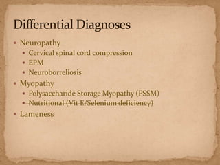
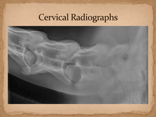
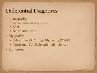
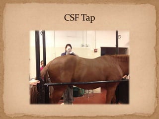
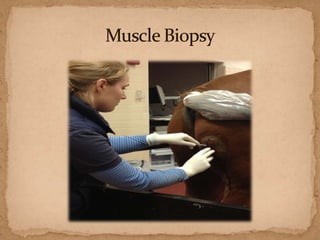
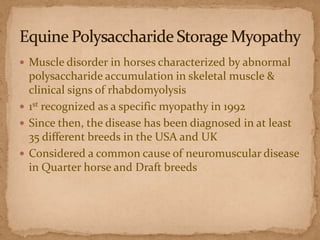
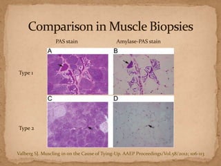
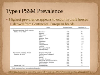
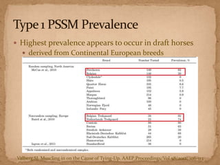
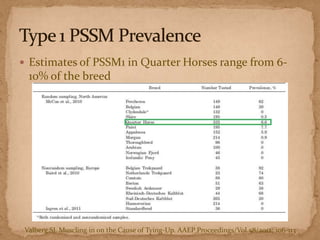
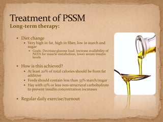
Large Animal Medicine
Ruthie Reinken, Jessica Reese, Maritza Rodriguez
 9 year old Warmblood

gelding
 Junior Equitation horse
 With current owners since
February
 4-5 month progressive onset:
 Poor performance
 Ataxia
 Personality change

 Two weeks prior to presentation, almost fell while

handwalking
 Previously treated with doxycycline and ponazuril with

no improvement.
 PE WNL
 Neuro exam:
 Grade 1 ataxia in all 4 limbs
 Weakness post-exercise

 Quickly fatigued

 Lameness exam:
 Grade 2/5 lame LF
 Grade 2/5 lame RH
 Weakness/ataxia
 History stumbling, bucking
 Lameness
 Neuropathy
 Cervical spinal cord compression
 EPM
 Neuroborreliosis

 Myopathy
 Polysaccharide Storage Myopathy (PSSM)
 Nutritional (Vit E/Selenium deficiency)

 Lameness/orthopedic
 Diagnostic analgesia
 Bilateral abaxial

 Nuclear scintigraphy
 Neuropathy
 Cervical spinal cord compression
 EPM
 Neuroborreliosis

 Myopathy
 Polysaccharide Storage Myopathy (PSSM)
 Nutritional (Vit E/Selenium deficiency)

 Lameness
 Neuropathy
 Cervical spinal cord compression
 EPM
 Neuroborreliosis

 Myopathy
 Polysaccharide Storage Myopathy (PSSM)
 Nutritional (Vit E/Selenium deficiency)

 Lameness
 Neuropathy
 Cervical spinal cord compression

 EPM
 Neuroborelliosis

 Myopathy
 Polysaccharide Storage Myopathy (PSSM)
 Nutritional (Vit E/Selenium deficiency)

 Lameness
 Neuropathy
 Cervical spinal cord compression

 EPM
 Neuroborelliosis

 Myopathy
 Polysaccharide Storage Myopathy (PSSM)
 Nutritional (Vit E/Selenium deficiency)

 Lameness
 Neuropathy
 Cervical spinal cord compression

 EPM
 Neuroborelliosis

 Myopathy
 Polysaccharide Storage Myopathy (PSSM)
 Nutritional (Vit E/Selenium deficiency)

 Lameness
 Neuropathy
 Cervical spinal cord compression

 EPM
 Neuroborelliosis

 Myopathy
 Polysaccharide Storage Myopathy (PSSM)
 Nutritional (Vit E/Selenium deficiency)

 Lameness
 Muscle disorder in horses characterized by abnormal

polysaccharide accumulation in skeletal muscle &
clinical signs of rhabdomyolysis
 1st recognized as a specific myopathy in 1992
 Since then, the disease has been diagnosed in at least
35 different breeds in the USA and UK
 Considered a common cause of neuromuscular disease
in Quarter horse and Draft breeds
 Affected horses were reported to have a 2-fold higher

muscle glycogen concentration compared to normal
horses
 Increase synthesis of glycogen, NOT an inability to

metabolize glycogen
 2 linked biochemical abnormalities associated
 1. Enhanced sensitivity to insulin in Quarter horses
 2. Abnormal regulation of glycogen synthase

 2 types of PSSM recognized
 Mutation in glycogen synthase 1 gene

(GYS1)
  resulting in excessive accumulation of

glycogen
 Inherited in an autosomal dominant

manner
 The increased activity has an association

with regulation of energy generation in
muscle fibers during exercise under
certain dietary conditions
 Muscle samples from PSSM1 horses showed less than

normal branched polysaccharide in the muscle fibers
 Related to chronic enhanced activity of GYS1 gene
 Leads to less highly branched polysaccharide

 Becomes resistant to amylase digestion when stained

 Important for differentiation between PSSM type 1 and

2 via muscle biopsy
 1st study differentiating Type 1 from Type 2 in 2009

(McCue et al)
 Current knowledge based on retrospective studies of

cases of diagnosed PSSM
 No genetic mutation associated

 Origin is yet unknown
PAS stain

Amylase-PAS stain

Type 1

Type 2

Valberg SJ. Muscling in on the Cause of Tying-Up. AAEP Proceedings/Vol.58/2012; 106-113
 Highest prevalence appears to occur in draft horses
 derived from Continental European breeds

Valberg SJ. Muscling in on the Cause of Tying-Up. AAEP Proceedings/Vol.58/2012; 106-113
 Highest prevalence appears to occur in draft horses
 derived from Continental European breeds

Valberg SJ. Muscling in on the Cause of Tying-Up. AAEP Proceedings/Vol.58/2012; 106-113
 Estimates of PSSM1 in Quarter Horses range from 6-

10% of the breed

Valberg SJ. Muscling in on the Cause of Tying-Up. AAEP Proceedings/Vol.58/2012; 106-113
 Prevalence is low to nonexistent in light horse breeds
 Thoroughbreds, Standardbreds, Arabians

Valberg SJ. Muscling in on the Cause of Tying-Up. AAEP Proceedings/Vol.58/2012; 106-113
 Warmbloods: about 80% of cases of PSSM diagnosed

by muscle biopsy (Valberg 2012)
 Quarter Horses: approximately 28% of cases of PSSM

diagnosed by muscle biopsy (Valberg 2012)
 Range significantly from one individual to another
 Pain
 Weakness  recumbency
 Muscle atrophy
 Muscle spasms/fasciculations/contractions
 Gait abnormalities
 Poor performance
 Exercise intolerance
 Exertional rhabdomyolysis
 Other problems reported by owner – back soreness, difficulty trimming/shoeing rear
hooves, behavior problems under saddle or in harness, episodic spasmodic colic
 Acute episode
 signs can last >2 hrs
 10% become recumbent
 Most affected muscles
 Gluteal, semimembranosus, semitendinosus, longissimus
 Evidence of muscle-fiber necrosis:
 Elevated CK (>350 u/L) and AST (>420 u/L) 4-6 hours

after exercise
 Other possible laboratory changes:
 Low blood selenium levels
 Low vitamin E levels
Type I:
 Gold standard – genetic testing for GYS1 mutation
 Histopathology of muscle biopsy – PAS-positive amylaseresistant inclusions in fast-twitch myofibers
 Risk of sampling error and false negatives

Type II:
 Histopathology of muscle biopsy – subsarcolemmal
aggregates of PAS-positive, amylase-sensitive (usually)
glycogen
 Risk of false positives

Histopathologic findings don’t always correlate with severity
of clinical signs or with increase in CK or AST post-exercise
Long-term therapy:
 Diet change
 Very high in fat, high in fiber, low in starch and
sugar
 Goals: Decrease glucose load, increase availability of

NEFA for muscle metabolism, lower serum insulin
levels

 How is this achieved?
 At least 20% of total calories should be from fat
additive
 Feeds should contain less than 33% starch/sugar
 Hay with 12% or less non-structural carbohydrate
to prevent insulin concentration increases
 Regular daily exercise/turnout
 Response to treatment takes up to 4 months for full fat

adaptation
 Good chance for return to acceptable performance

-Variability in prognosis is based on severity of individual’s
clinical signs
 PSSM horses will always have an increased tendency to

have muscle soreness
 CK

 Pre exercise: 125 U/L
 Post exercise: 134 U/L

 Muscle biopsy
 Results pending
 Neuropathy

 Cervical spinal cord compression
 EPM
 Neuroborelliosis
 Myopathy
 Polysaccharide Storage Myopathy (PSSM)
 Nutritional (Vit E/Selenium deficiency)

 Lameness
 Thank you
 Dr. Sweeney
 Dr. Tomlinson
 Dr. Moore
 Dr. Johnson
 Dr. Davidson
 Our rotation mates, Sam, Annie, and Alex


McCue ME, Armien AG, Lucio M, et al. Comparative skel-etal muscle histopathologic and ultrastructural features in
two forms of polysaccharide storage myopathy in horses. Vet Pathol 2009;46:1281–1291.



McCue ME, Valberg SJ. Estimated prevalence of polysac-charide storage myopathy among overtly healthy Quarter
Horses in the United States. Am J Vet Res2007;231:746–750.



McCue ME, Anderson SM, Valberg SJ, et al. Estimated prevalence of the type 1 polysaccharide storage myopathy
mutation in selected North American and European breeds. Anim Genet 2010;41 (Suppl2):145–149.



McCue ME, Ribeiro WP, Valberg SJ. Prevalence of poly-saccharide storage myopathy in horses with neuromuscular
disorders. Equine Vet J Suppl2006;36:340 –344.



Valberg SJ. Muscle disorders: Polysaccharide storage myopathy, in Proceedings. Am Assoc Equine Pract 2006;52:373380.



Valberg SJ. Muscling in on the Cause of Tying-Up. AAEP Proceedings/Vol.58/2012; 106-113



Valentine BA. Equine polysaccharide storage myopathy: tutorial article. Equine vet Educ. 2003; 15 (5): 254-262.



Valentine BA, Van Saun RJ, and Thompson KN. Role of dietary carbohydrate and fat in horses with equine
polysaccharide storage myopathy. JAVMA 2001; 219(11): 1537-1544.
Moch final

Moch final

  • 1.
    Large Animal Medicine RuthieReinken, Jessica Reese, Maritza Rodriguez
  • 2.
     9 yearold Warmblood gelding  Junior Equitation horse  With current owners since February
  • 3.
     4-5 monthprogressive onset:  Poor performance  Ataxia  Personality change  Two weeks prior to presentation, almost fell while handwalking  Previously treated with doxycycline and ponazuril with no improvement.
  • 4.
     PE WNL Neuro exam:  Grade 1 ataxia in all 4 limbs  Weakness post-exercise  Quickly fatigued  Lameness exam:  Grade 2/5 lame LF  Grade 2/5 lame RH
  • 5.
     Weakness/ataxia  Historystumbling, bucking  Lameness
  • 6.
     Neuropathy  Cervicalspinal cord compression  EPM  Neuroborreliosis  Myopathy  Polysaccharide Storage Myopathy (PSSM)  Nutritional (Vit E/Selenium deficiency)  Lameness/orthopedic
  • 7.
     Diagnostic analgesia Bilateral abaxial  Nuclear scintigraphy
  • 8.
     Neuropathy  Cervicalspinal cord compression  EPM  Neuroborreliosis  Myopathy  Polysaccharide Storage Myopathy (PSSM)  Nutritional (Vit E/Selenium deficiency)  Lameness
  • 9.
     Neuropathy  Cervicalspinal cord compression  EPM  Neuroborreliosis  Myopathy  Polysaccharide Storage Myopathy (PSSM)  Nutritional (Vit E/Selenium deficiency)  Lameness
  • 11.
     Neuropathy  Cervicalspinal cord compression  EPM  Neuroborelliosis  Myopathy  Polysaccharide Storage Myopathy (PSSM)  Nutritional (Vit E/Selenium deficiency)  Lameness
  • 13.
     Neuropathy  Cervicalspinal cord compression  EPM  Neuroborelliosis  Myopathy  Polysaccharide Storage Myopathy (PSSM)  Nutritional (Vit E/Selenium deficiency)  Lameness
  • 14.
     Neuropathy  Cervicalspinal cord compression  EPM  Neuroborelliosis  Myopathy  Polysaccharide Storage Myopathy (PSSM)  Nutritional (Vit E/Selenium deficiency)  Lameness
  • 16.
     Neuropathy  Cervicalspinal cord compression  EPM  Neuroborelliosis  Myopathy  Polysaccharide Storage Myopathy (PSSM)  Nutritional (Vit E/Selenium deficiency)  Lameness
  • 17.
     Muscle disorderin horses characterized by abnormal polysaccharide accumulation in skeletal muscle & clinical signs of rhabdomyolysis  1st recognized as a specific myopathy in 1992  Since then, the disease has been diagnosed in at least 35 different breeds in the USA and UK  Considered a common cause of neuromuscular disease in Quarter horse and Draft breeds
  • 18.
     Affected horseswere reported to have a 2-fold higher muscle glycogen concentration compared to normal horses  Increase synthesis of glycogen, NOT an inability to metabolize glycogen  2 linked biochemical abnormalities associated  1. Enhanced sensitivity to insulin in Quarter horses  2. Abnormal regulation of glycogen synthase  2 types of PSSM recognized
  • 19.
     Mutation inglycogen synthase 1 gene (GYS1)   resulting in excessive accumulation of glycogen  Inherited in an autosomal dominant manner  The increased activity has an association with regulation of energy generation in muscle fibers during exercise under certain dietary conditions
  • 20.
     Muscle samplesfrom PSSM1 horses showed less than normal branched polysaccharide in the muscle fibers  Related to chronic enhanced activity of GYS1 gene  Leads to less highly branched polysaccharide  Becomes resistant to amylase digestion when stained  Important for differentiation between PSSM type 1 and 2 via muscle biopsy
  • 21.
     1st studydifferentiating Type 1 from Type 2 in 2009 (McCue et al)  Current knowledge based on retrospective studies of cases of diagnosed PSSM  No genetic mutation associated  Origin is yet unknown
  • 22.
    PAS stain Amylase-PAS stain Type1 Type 2 Valberg SJ. Muscling in on the Cause of Tying-Up. AAEP Proceedings/Vol.58/2012; 106-113
  • 23.
     Highest prevalenceappears to occur in draft horses  derived from Continental European breeds Valberg SJ. Muscling in on the Cause of Tying-Up. AAEP Proceedings/Vol.58/2012; 106-113
  • 24.
     Highest prevalenceappears to occur in draft horses  derived from Continental European breeds Valberg SJ. Muscling in on the Cause of Tying-Up. AAEP Proceedings/Vol.58/2012; 106-113
  • 25.
     Estimates ofPSSM1 in Quarter Horses range from 6- 10% of the breed Valberg SJ. Muscling in on the Cause of Tying-Up. AAEP Proceedings/Vol.58/2012; 106-113
  • 26.
     Prevalence islow to nonexistent in light horse breeds  Thoroughbreds, Standardbreds, Arabians Valberg SJ. Muscling in on the Cause of Tying-Up. AAEP Proceedings/Vol.58/2012; 106-113
  • 27.
     Warmbloods: about80% of cases of PSSM diagnosed by muscle biopsy (Valberg 2012)  Quarter Horses: approximately 28% of cases of PSSM diagnosed by muscle biopsy (Valberg 2012)
  • 28.
     Range significantlyfrom one individual to another  Pain  Weakness  recumbency  Muscle atrophy  Muscle spasms/fasciculations/contractions  Gait abnormalities  Poor performance  Exercise intolerance  Exertional rhabdomyolysis  Other problems reported by owner – back soreness, difficulty trimming/shoeing rear hooves, behavior problems under saddle or in harness, episodic spasmodic colic  Acute episode  signs can last >2 hrs  10% become recumbent  Most affected muscles  Gluteal, semimembranosus, semitendinosus, longissimus
  • 29.
     Range significantlyfrom one individual to another  Pain  Weakness  recumbency  Muscle atrophy  Muscle spasms/fasciculations/contractions  Gait abnormalities  Poor performance  Exercise intolerance  Exertional rhabdomyolysis  Other problems reported by owner – back soreness, difficulty trimming/shoeing rear hooves, behavior problems under saddle or in harness, episodic spasmodic colic  Acute episode  signs can last >2 hrs  10% become recumbent  Most affected muscles  Gluteal, semimembranosus, semitendinosus, longissimus
  • 30.
     Evidence ofmuscle-fiber necrosis:  Elevated CK (>350 u/L) and AST (>420 u/L) 4-6 hours after exercise  Other possible laboratory changes:  Low blood selenium levels  Low vitamin E levels
  • 31.
    Type I:  Goldstandard – genetic testing for GYS1 mutation  Histopathology of muscle biopsy – PAS-positive amylaseresistant inclusions in fast-twitch myofibers  Risk of sampling error and false negatives Type II:  Histopathology of muscle biopsy – subsarcolemmal aggregates of PAS-positive, amylase-sensitive (usually) glycogen  Risk of false positives Histopathologic findings don’t always correlate with severity of clinical signs or with increase in CK or AST post-exercise
  • 32.
    Long-term therapy:  Dietchange  Very high in fat, high in fiber, low in starch and sugar  Goals: Decrease glucose load, increase availability of NEFA for muscle metabolism, lower serum insulin levels  How is this achieved?  At least 20% of total calories should be from fat additive  Feeds should contain less than 33% starch/sugar  Hay with 12% or less non-structural carbohydrate to prevent insulin concentration increases  Regular daily exercise/turnout
  • 33.
     Response totreatment takes up to 4 months for full fat adaptation  Good chance for return to acceptable performance -Variability in prognosis is based on severity of individual’s clinical signs  PSSM horses will always have an increased tendency to have muscle soreness
  • 35.
     CK  Preexercise: 125 U/L  Post exercise: 134 U/L  Muscle biopsy  Results pending
  • 36.
     Neuropathy  Cervicalspinal cord compression  EPM  Neuroborelliosis  Myopathy  Polysaccharide Storage Myopathy (PSSM)  Nutritional (Vit E/Selenium deficiency)  Lameness
  • 37.
     Thank you Dr. Sweeney  Dr. Tomlinson  Dr. Moore  Dr. Johnson  Dr. Davidson  Our rotation mates, Sam, Annie, and Alex
  • 38.
     McCue ME, ArmienAG, Lucio M, et al. Comparative skel-etal muscle histopathologic and ultrastructural features in two forms of polysaccharide storage myopathy in horses. Vet Pathol 2009;46:1281–1291.  McCue ME, Valberg SJ. Estimated prevalence of polysac-charide storage myopathy among overtly healthy Quarter Horses in the United States. Am J Vet Res2007;231:746–750.  McCue ME, Anderson SM, Valberg SJ, et al. Estimated prevalence of the type 1 polysaccharide storage myopathy mutation in selected North American and European breeds. Anim Genet 2010;41 (Suppl2):145–149.  McCue ME, Ribeiro WP, Valberg SJ. Prevalence of poly-saccharide storage myopathy in horses with neuromuscular disorders. Equine Vet J Suppl2006;36:340 –344.  Valberg SJ. Muscle disorders: Polysaccharide storage myopathy, in Proceedings. Am Assoc Equine Pract 2006;52:373380.  Valberg SJ. Muscling in on the Cause of Tying-Up. AAEP Proceedings/Vol.58/2012; 106-113  Valentine BA. Equine polysaccharide storage myopathy: tutorial article. Equine vet Educ. 2003; 15 (5): 254-262.  Valentine BA, Van Saun RJ, and Thompson KN. Role of dietary carbohydrate and fat in horses with equine polysaccharide storage myopathy. JAVMA 2001; 219(11): 1537-1544.

Editor's Notes

  • #3 Mocha is a 9 year old warmblood gelding. His owner reports that he is ½ sellefrancais, ¼ belgianwarmblood, ¼ hanoverian, and ¼ dutchwarmblood”. He works as a Junior equitation horse for a 15 year old girl. They bought him in February at which time he had a full prepurchase exam.
  • #4 Mocha was presented to NBC medicine service on October 8th for a four to five month history of progressive poor performance, characterized by bucking and reluctance to work, more recent onset of ataxia, and a gradual personality change. Mocha had been doing very well for a few months after they bought him, but over the summer, he began bucking on jump landings at shows, tripped once to his knees after a jump and even bucked his rider off in July. The owners noted that Mocha has become progressively more reactive when his girthed and is generally crankier in the barn. They also report that when turned out in recent weeks, Mocha just stands at the gate, whereas he used to be more energetic at pasture. About a month ago, Mocha was believed to begin exhibiting ataxia and his trainer reported feeling unsafe while riding. Two weeks prior to presentation, Mocha was being handwalked and his hindlegsapparantly began to buckle and he almost fell. He had been treated with doxycycline for 1 and a half months with no improvement, and after the episode two weeks prior to presentation was started on ponazuril, also with no improvement. *ASK* about doxyPreviously tx’d w ponazurilnd doxycycline to no improvement
  • #5 Mocha’s physical exam was normal, all cranial nerves were intact, and had no muscle atrophy. A subtle ataxia was noted during initial neuro exam that worsened as Mocha was exercised. At the trot, Mocha consistently scuffed his RH toe. He exhibited noticeable fatigue and had increasing difficulty with small circles, scuffing his toes and stepping on himself. Even for a horse that has been out of work for a month or two, he was abnormally fatigued. Mocha was also noticeably lame in the left front and right hind.
  • #6 To summarize, at this point our problem list consisted of weakeness/ataxia, history
  • #7 We developed three broad differentials to account for Mocha’s signs. Is his ataxia and weakness caused by a neuropathy or a myopathy? Top differentials for neuropathies that can cause ataxia in all four limbs include spinal cord compression, EPM, and lyme. Myopathies on our differential list include PSSM and nutritional causes. We also included lameness as a category, although we were unsure if lameness could account for the weakness and fatigue exhibited.(**mention lamenes/”orthopedic problem” on initial ddx**)
  • #8 To address Mocha’s lameness, diagnostic analgesia was performed. Bilateral abaxial nerve blocks in the front legs significantly improved Mocha’s front end lameness. No blocks were performed to address hind end lameness. Nerve blocks did not affect Mocha’s weakness or change his neurologic exam findings. Nuclear scintigraphy was performed, but no conclusive orthopedic problem was found to account for all of Mocha’s clinical signs. We elected to continue pursuing neuropathies and myopathies as an underlying cause of Mocha’s problems.
  • #9 Nutritional causes seem unlikely, *advance slide*as Mocha is well maintained at a show barn and gets regular turnout.
  • #10 Nutritional causes seem unlikely, *advance slide*as Mocha is well maintained at a show barn and gets regular turnout.
  • #11 Cervical radiographs were taken and although there is evidence of degenerative joint disease in some of the caudal cervical facet joints,intravertebralsaggital ratios were within normal limits, which decreases the likelihood of spinal cord compression.
  • #12 So, although we could not completely rule out spinal cord compression as a differential, it was considered it less likely.
  • #13 A CSF tap was performed to test for neuroborreliosis and EPM
  • #14 Mocha was negative for EPM on SAG2,3,4 elisa on CSF and serum.
  • #15 Mocha was also negative on the cornell multiplex Lyme test on CSF and serum.To test for PSSM, Pre- and post-exercise CK levels were measured and a muscle biopsy was performed.
  • #16 A biopsy of the semitendonosus was submitted to Minnesota for PSSM testing.
  • #17 After eliminating many of the common differentials for Mocha’s clinical signs, PSSM moved higher up on our list. So, now we’re going to discuss PSSM and its diagnosis and treatment.
  • #18 Equine polysaccharide storage myopathy is a muscle disorder in horses characterized by abnormal polysaccharide accumulation in skeletal muscle & clinical signs of rhabdomyolysisIt was first recognized as specific myopathy in 1992, however there have been individual cases of abnormal polysaccharide inclusions reported in equine muscle dating back to 1979Since then, the disease has been diagnosed in at least 35 different breeds in the US and UKIt is now considered a common cause of neuromuscular disease in quarter horse and draft breeds
  • #19 When the disease was first discovered, the initial finding was that affected horses had a 2 fold higher glycogen concentration in their skeletal muscle compared to normal horsesAdditional studies then attempted to figure out the cause of the increase in glycogen concentrationWhat was eventually discovered was that it is not an inability to metabolize glycogen because all the enzymes needed to do so are present. It is actually an increase in the synthesis of glycogen. Further studies revealed that 2 biochemical abnormalities are associated with PSSM One being an increase in insulin sensitivity in quarter horsesAnd two being an abnormal regulation of glycogen synthaseThis brings us to the 2 types of PSSM
  • #20 PSSM type 1 is a mutation in the glycogen synthase 1 gene that results in the accumulation of excessive glycogen within muscle. This is inherited in an autosomal dominant mannerThe increased activity of this gene disturbs the normal flux of muscle energy metabolism during exercise. This has been linked to certain dietary conditions and exercise regimens.So what we believe is happening, is that as these horses are exercising, they metabolize glycogen to glucose, but the genetic mutation causes the immediate addition of that glucose to a growing glycogen molecule. SO they are not effectively utilizing their glycogen storage.
  • #21 Due to the chronic enhanced activity of that glycogen synthase 1 gene, the polysaccharide becomes less branched than normalIt also becomes resistant to an amylase digestion stain on histopathologyThis Amylase digestion is an important way to differentiate Type 1 from Type 2 when looking a muscle biopsiesAnd I'll show a few pictures of that in a moment
  • #22 PSSM type 2 was first differentiated from Type 1 in 2009The authors realized that not all of the horses diagnosed with PSSM had the genetic mutation to the glycogen synthase 1 gene. So they compared skeletal muscle from horses diagnosed with PSSM that were either GYS1 negative or positive. They concluded that there were 2 forms or types of PSSM based on the differences in muscle biopsiesSince this is relatively recent, most of our current knowledge of PSSM type 2 is based on retrospective studiesAs of now, there is no genetic mutation associated with this type of PSSM so the origin is still unknown.
  • #23 These are biopsy sections of the semimembranosus muscle in horses diagnosed with PSSMThe top photos, A and B, are from a horse with PSSM type 1 and the bottom photos, C and D, are from a horse with PSSM Type 2On your left hand side, the biopsy is stained with periodic acid Schiff stainThe dark magenta stain in photo A and C is the glycogen deposition in the muscle. You can see that the glycogen concentration in higher in type 1 than type 2When the amylase-PAS stain is used on the Type 1 biopsy in photo B, you can see that the glycogen is still presentThis differs from type 2, where amylase digestion has occurred and the glycogen is no longer presentSo this is what helpdifferentiate between the two types
  • #24 In terms of the prevalance of types of PSSM in breedsA study in 2010, found that the highest glycogen-storage gene 1 mutation or type 1 PSSM was found in draft breeds originating from continental Europe
  • #25 Specifically Percherons and Belgians in North America and Belgian and Netherland Trekpaards in Europe
  • #26 Estimates of PSSM1 in quarter horses ranges from 6-10% of the breed
  • #27 Prevalence was low to non existent in light horses breeds such as throughbreds, standardbreds and arabiansThe study was done to help breed associations determine if they should screen for the GYS1 mutation and to alert veterinarians that it is a possible differential diagnosis for muscle pain, rhabdomyolysis and gait abnormalities and it should be higher on the list for certain breeds.
  • #28 As for the prevalence of PSSM type 2, about 80% percent of Warmbloods diagnosed with PSSM have type 2 and about 28% of Quarter Horses diagnosed with PSSM also have type 2.
  • #29 Clinical signs of PSSM are quite variable from one individual to another. They can include pain, weakness (which can progress to recumbency), muscle spasms/fasciculations/contractions, and muscle atrophy (which may be diffuse or confined only to the pelvic limbs). Muscle atrophy in PSSM is usually symmetrical, can mimic Equine Motor Neuron Disease, and is more common in draft breeds and warmbloods than other breeds with PSSM.Other signs include gait abnormalities, particularly in the pelvic limbs; examples of these abnormalities include stiff short-strided gait or unexplained lameness.Poor performance and exercise intolerance are also commonly reported clinical signs, and in more severe cases, episodes of exertionalrhabdomyolysis may occur (especially in QHs, WBs, Haflingers, and draft-light crossbreeds). Other problems reported by owners of PSSM horses include back soreness, difficulty trimming/shoeing rear hooves, behavior problems under saddle in or harness, and episodic spasmodic colic. In a severe acute episode of PSSM, such as onset of ER, signs may last more than 2 hours and 10% of horses become recumbent.The muscles most typically affected by PSSM include gluteal muscles, semimembranosus, semitendinosus, and longissimus, but back, abdominal, and forelimb muscles can also be affected.
  • #30 The clinical signs highlighted in red are those shared by Mocha.
  • #31 The most common laboratory changes seen in horses with PSSM are 3-fold elevations in CK and AST 4-6 hours post-light exercise. However, this is not a requirement of PSSM horses, and many with Type 2 do not have these changes. Other possible laboratory findings, which may help distinguish from other disorders, but aren’t diagnostic by any means include low blood selenium and low vitamin E levels.(*confirm*)
  • #32 The gold standard method of diagnosing Type 1 PSSM is genetic testings for the GYS1 mutation on whole blood or hair root.Additionally, diagnosis can be made by histopathology of a muscle biopsy as mentioned previously. With this method, there is a risk of sampling error because these inclusions may not be evenly distributed in all muscle. There is also a risk of false negatives in younger horses with PSSM because muscle dysfunction typically precedes the development of inclusions. Based on the criteria for diagnosing Type 2 PSSM on muscle biopsy, there is a risk of false positives in highly trained horses because they naturally have increased glycogen storage. It is important to note that histopathologic findings don’t always correlate with severity of clinical signs or with post-exercise increases in CK or AST.
  • #33 There are two main components of long-term therapy for PSSM: diet and exercise.The goals of the new diet are to decrease the glucose load, increase availability of non-esterified fatty acids for muscle metabolism, and to lower serum insulin levels. In order to achieve this, the diet should, first of all, be very high in fat; at least 20% of total calories should be from a fat additive (for comparison, most forages only contain 1.5-3% fat); fat supplements can include vegetable-based oil, rice bran fat, and animal fats. Additionally, feeds should contain less than 33% starch or sugar. Finally, it is recommended to use hay with 12% or less non-structural carbohydrate to prevent insulin concentration increases. Equally important in preventing the clinical signs of PSSM is regular daily exercise/turnout.
  • #34 While you may see some mild, transient improvement in clinical signs in the first few weeks of treatment, it takes up to 4 months for full fat adaptationWith diet change and regular exercise, there is a good chance for return to acceptable performance; however, there is variability in prognosis based on severity of clinical signs prior to diagnosis.Finally, it’s important to note that PSSM horses will always have predilection for muscle soreness.
  • #36 Mocha’s pre-exercise CK was 125 U/L and his post exercise levels were 134 U/L. Although these results are not suggestive of PSSM, they do not rule it our either. Our muscle biopsy results are still pending. Please stay tuned.
  • #37 If Mocha’s biopsy is negative for PSSM, we plan to further investigate the possibility of spinal cord compression by performing a myelogram. While we didn’t see canal narrowing on radiographs, we did have evidence of degenerative joint disease, and a myelogram will allow us to assess dynamic compression. Additionally, we can consider revisiting Mocha’s lameness as a source of his problems. Mocha is still here, boarding with us, while we all await his results.