2. OBJECTIVES
• Differentiate between the concepts of
“Innate” and “Adaptive” immunity
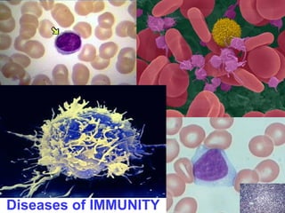
• Visually recognize and understand the basic
roles of lymphocytes, macrophages,
dendritic cells, NK cells in the immune saga
• Understand the roles of the major cytokines
in immunity
• Differentiate and give examples of the four
(4) different types of hypersensitivity
reactions
3. OBJECTIVES
• Know the common features of autoimmune
diseases, and the usual four (4) main
features (
Etiology, Pathogenesis, Morphology, and Clin
) of Systemic Lupus Erythematosus,
Rheumatoid Arthritis, Sjögrens, Systemic
Sclerosis (Scleroderma), Mixed Connective
Tissue Disease, and “Poly-” (aka, “Peri-”)
-arteritis Nodosa
• Differentiate between Primary (Genetic) and
Secondary (Acquired) Immunodeficiencies
4. OBJECTIVES
• Understand the usual four (4) main features
of AIDS, i.e., etiology, pathogenesis,
morphology, clinical expression
• Understand the usual four (4) main features
of Amyloidosis
5. IMMUNITY
• INNATE (present before
birth, “NATURAL”)
• ADAPTIVE (developed
by exposure to pathogens,
or in a broader sense,
antigens not recognized by
the MHC)
6. MHC
Major Histocompatibility Complex
• A genetic “LOCUS” on Chromosome 6,
which codes for cell surface compatibility
• Also called HLA (Human Leukocyte
Antigens) in humans and H-2 in mice
• It’s major job is to make sure all self cell
antigens are recognized and “tolerated”,
because the general rule of the immune
system is that all UN-recognized antigens
will NOT be tolerated
13. ANY ROUND CELL WITH RATHER
DENSE STAINING NUCLEUS AND
MINIMAL CYTOPLASM IN
CONNECTIVE TISSUE, A BIT
BIGGER THAN AN RBC, IS A
LYMPHOCYTE
…UNTIL PROVEN OTHERWISE
17. ANY CELL MIXED IN WITH LYMPHOCYTES BUT HAS
A LARGER MORE “OPEN”, i.e., “vesicular”, LESS
DENSE, LESS CIRCULAR NUCLEUS WITH MORE
CYTOPLASM IS A
MACROPHAGE
…UNTIL PROVEN OTHERWISE
ALMOST ALL “GRANULAR” or “PIGMENTED”
CELLS IN CONNECTIVE TISSUE ARE
MACROPHAGES. GRANULOMAS, GIANT CELLS,
ARE CHIEFLY MACROPHAGES ALSO.
18. 1) ROUND NUCLEUS
2) OVOID CYTOPLASM
3) PERIPHERAL CHROMATIN
4) “CLEAR ZONE” BETWEEN NUCLEUS AND WIDER LIP OF
CYTOPLASM
PLASMA CELLS
25. CYTOKINES/CHEMOKINES
• CYTOKINES are PROTEINS produced by
MANY cells, but usually LYMPHOCYTES
and MACROPHAGES, numerous roles in
acute and chronic inflammation, AND
immunity
–TNF, IL-1, by
macrophages
• CHEMOKINES are small proteins which are
attractants for PMNs
26. MHC
Major Histocompatibility Complex
• A genetic “LOCUS” on Chromosome 6,
which codes for cell surface compatibility
• Also called HLA (Human Leukocyte
Antigens) in humans and H-2 in mice
• It’s major job is to make sure all self cell
antigens are recognized and “tolerated”,
because the general rule of the immune
system is that all UN-recognized antigens
will NOT be tolerated
27.
28. MHC MOLECULES
(Gene Products)
• I (All nucleated cells and platelets), cell surface
glycoproteins, ANTIGENS
• II (APC’s, i.e., macs and dendritics, lymphs),
cell surface glycoproteins, ANTIGENS
• III Complement System Proteins
29. IMMUNE SYSTEM DISORDERS
WHAT CAN GO WRONG?
• HYPERSENSITIVITY REACTIONS, I-IV
• “AUTO”-IMMUNE DISEASES, aka
“COLLAGEN” DISEASES (BAD TERM)
Inflammation NOT due to external
pathogens, MHC failure.
• IMMUNE DEFICIENCY SYNDROMES,
IDS:
– PRIMARY (GENETIC)
– SECONDARY (ACQUIRED)
30. HYPERSENSITIVITY
REACTIONS (4)
• I (Immediate Hypersensitivity)
• II (Antibody Mediated
Hypersensitivity)
• III (Immune-Complex Mediated
Hypersensitivity)
• IV (Cell-Mediated Hypersensitivity)
31. Type I
IMMEDIATE HYPERSENSITIVITY
• “Immediate” means seconds to minutes
• “Immediate Allergic Reactions”, which may
lead to anaphylaxis, shock, edema, dyspnea
death
– 1) Allergen exposure
– 2) IMMEDIATE phase: MAST cell
DEgranulation, vasodilatation, vascular
leakage, smooth muscle (broncho)-spasm
– 3) LATE phase (hours, days): Eosinophils,
PMNs, T-Cells
32. TYPE II HYPERSENSITIVITY
ANTIBODY MEDIATED IMMUNITY
• ABs attach to cell surfaces
– OPSONIZATION (basting the turkey)
– PHAGOCYTOSIS
– COMPLEMENT FIXATION (cascade of
C1q, C1r, C1s, C2,
C3, C4, C5….. )
– LYSIS (destruction of cells by
rupturing or breaking of the cell
membrane)
33. TYPE II DISEASES
• Autoimmune Hemolytic Anemia, AHA
• Idiopathic Thrombocytopenic Purpura,
ITP
• Goodpasture Syndrome (Nephritis and
Lung hemorrhage)
• Rheumatic Fever
• Myasthenia Gravis
• Graves Disease
• Pernicious Anemia, PA
34. TYPE III HYPERSENSITIVITY
IMMUNE COMPLEX MEDIATED
• Antigen/Antibody “Complexes”
• Where do they go?
– Kidney (Glomerular Basement Membrane)
– Blood Vessels
– Skin
– Joints (synovium)
• Common Type III Diseases- SLE (Lupus),
Poly(Peri)arteritis Nodosa,
Poststreptococcal Glomerulonephritis,
Arthus reaction (hrs), Serum sickness
(days)
35. TYPE IV HYPERSENSITIVITY
CELL-MEDIATED (T-CELL)
DELAYED HYPERSENSITIVITY
• Tuberculin Skin Reaction
• DIRECT ANTIGENCELL CONTACT
– GRANULOMA FORMATION
– CONTACT DERMATITIS
36.
37. SUMMARY
• I
Acute allergic reaction
• II Antibodies directed against cell
surfaces
• III Immune complexes
• IV Delayed Hypersensitivity, e.g., Tb
skin test
40. AUTO-IMMUNE DISEASES
• Failure of SELF RECOGNITION
• Failure of SELF TOLERANCE
• TOLERANCE
– CENTRAL (Death of self reactive lymphocytes)
– PERIPHERAL (anergy, suppression by T-cells,
deletion by apoptosis, sequestration (Ag masking))
• STRONG GENETIC PREDISPOSITION
• OFTEN RELATED TO OTHER AUTOIMMUNE
DISEASES
• OFTEN TRIGGERED BY INFECTIONS
69. REVERSE TRANSCRIPTASE
• The enzyme reverse transcriptase
(RT) is used by retroviruses to
transcribe their single-stranded
RNA genome into single-stranded
DNA and to subsequently construct
a complementary strand of DNA,
providing a DNA double helix
capable of integration into host cell
chromosomes.
Just like we said “EVERY disease is a genetic disease”, we can also say “EVERY disease is an ‘immune’ disease.”
Unlearned vs. Learned
Toll-like receptors (TLRs) are a class of single membrane-spanning non-catalytic receptors on macrophages and other APCs that recognize structurally conserved molecules derived from microbes once they have breached physical barriers such as the skin or intestinal tract mucosa, and activate immune cell responses. They are very WELCOME evolutionary leftovers. You might say they are “learned” only in the evolutionary sense.
Adaptive immunity is “learned”. It relies on PREVIOUS EXPOSURE to the pathogen or foreign antigen, or even native antigen at times.
The classic types of adaptive immunity are:
Humoral, largely learned
Cellular, direct contact, no need for circulating antibodies
If you wanted to make this simple you can say there are 3 types of cells, T-lymphocytes, B-lymphocytes, and Macrophages or APC’s.
If you wanted to make it incredibly simple then just say lymphocytes and macrophages, i.e., the “monos” we saw in chronic inflammation, because dendritic cells have macrophage roots and functions, and plasma cells and NK cells have lymphocyte roots.
The many faces of a lymphocyte, NONE of which we will be seeing in histopathology lab.
Note even a normal lymph has “cartwheeling” best seen on TEM, like a plasma cell.
About ½ trillion lymphocytes in the human body, or 1% of all cells.
Even though some classify “dendritic” cells as being separate from “macrophages” you can imagine that the function of all APC’s (Antigen Presenting Cells” is to maximize surface area.
Even though we call a macropahge a “mono”-cyte, conventionally, it’s nucleus can be as convoluted or “cerebrated” as a neutrophil.
Are almost all “pigmented” cells in the body, intrinsic or extrinsic, macrophages? Yes!
Pathologists love to call macrophages “histiocytes”, historically.
It is very important to understand the “misnomer”.
We called neutrophils “polys” because of “poly” or “multi” lobes in its nucleus.
And we called lymphs and macrophages “monos” because we said the nuclei were “mono”nucleaded.
But in reality, the nucleus of a macrophage (aka, tissue monocyte) can be VERY convoluted, or “cerebrated”.
It might also be allowed to call a macrophage a “APC”, i.e., an Antigen Presenting Cell, of cellular immunity.
Plasma are B-lymphocytes that have dedicated themselves to be antibody factories, stuffing their golgi apparatus with immunoglobulins.
If you see a cell with these 4 features, it can only be a plasma cell, even if it has, say 2-3 features, it still may very well be a plasma cell!
A Dendridic cell is a type of macrophage with many spiny cytoplasmic processes, found in many places especially skin (Langhans cells) and brain (microglia), and many other less known places like liver. They are also APC’s. They are found in many many places however.
Can you call dendritic cells “max” macs? I think so, because they maximize their surface area.
NK cells are types of lymphocytes which specialize in direct killing of cells which the come in contact with, hence the term NK, Natural Killer.
Natural killer cells (or NK cells) are a type of cytotoxic lymphocyte that constitute a major component of the innate immune system. NK cells play a major role in the rejection of tumors and cells infected by viruses. The cells kill by releasing small cytoplasmic granules of proteins called perforin and granzyme that cause the target cell to die by apoptosis. The cell reminds me of the Rodney Dangerfield’s joke where he says his football team was so tough, after they sacked the quarterback, they then went after his family.
NK cells do not “phagocytize”, they just Kill, Naturally.
The NK cell is a crucial gatekeeper or sentry of the MHC. Halt, who goes there? Friend or foe?
An NK cell is activated or not, whether it recognizes a MHC I cell or not.
So what kills the virus infected cell, the virus or the NK cell? They BOTH may!
General roles of B and T cells. Which cell is most akin to a NK cell? ANS: CD8, aka, cytotoxic T-lymphocyte.
Just as EPO is to red cells, CSF is to granulocytes and macrophages
This is the same EXACT slide from our discussion of acute inflammation.
Why these 3?
This is the outline of our entire chapter 6.
Why were autoimmune diseases called “collagen” diseases? Because fibrosis often follows chronic inflammation, and the MAIN pattern of autoimmune diseases is CHRONIC inflammation, NOT usually acute, i.e., many systemic autoimmune diseases eventually evoke fibrosis.
A good understanding of the 4 different types of classical “hypersensitivity” reactions, should be obtained. These are always taught as the general FOUR types of hypersensitivity, but are by no means complete or mutually exclusive. These 4 items have ALWAYS been taught this way, even if more recent knowledge implies it is ridiculous to classify them this way.
The MAST cell is the key cell if Type I Hypersensitivity.
What are the granules in mast cells composed of?
Attacking cell or microbial membranes is the key feature of Type II Hypersensitivity.
Complement cascades are needed for LYSIS of cells, NOT just antibody attachment.
Understandably, these are all “AUTO”-immune diseases, or FAILURES of the MHC. Note most are organ-specific (i.e., “local”) rather than systemic.
But beware, many autoimmune diseases which have an organ’s name in them may very well be SYSTEMIC, like RA, SjS, DMS
An Arthus reaction is a local vasculitis associated with deposition of immune complexes and activation of complement. Immune complexes form in the setting of high local concentration of vaccine antigens and high circulating antibody concentration. Arthus reactions are characterized by severe pain, swelling, induration, edema, hemorrhage, and occasionally by necrosis. These symptoms and signs usually occur 4–12 hours after vaccination.
Serum sickness symptoms can take as long as fourteen days after exposure to appear, and may include signs and symptoms commonly associated with allergic reactions or infections, such as rashes, itching, joint pain (arthralgia), fever, and swollen lymph nodes (lymphadenopathy), and malaise. Historically, it was a result of animal serum injections.
Think of GBM, Blood Vessels, SKIN, and SYNOVIUM as all being MAGNETS for Ag/Ab complexes.
No antibodies involved.
Schematic for granuloma formation in Type IV Hypersensitivity. The key association between interferon-γ and granulomas is that the cytokine interferon-γ activates macrophages so that they become more powerful in killing intracellular organisms.
Clinical vs. pathological, acute and chronic.
As expected, immediate endothelial responses are hyperacute, cellular infiltrates are acute to chronic, and fibrosis is chronic. We have seen this time and time again.
Central tolerance occurs DURING lymphocyte development and operates in the thymus and bone marrow. Here, T and B lymphocytes that recognize self antigens are deleted before they develop into fully immunocompetent cells, preventing autoimmunity.
Peripheral tolerance is immunological tolerance developed AFTER T and B cells mature and enter the periphery.
Acquired or induced tolerance refers to the immune system's adaptation to external antigens characterized by a specific non-reactivity of the lymphoid tissues to a given antigen that in other circumstances would likely induce cell-mediated or humoral immunity. One of the most important natural kinds of acquired tolerance is immune tolerance in pregnancy, where the fetus and the placenta must be tolerated by the maternal immune system.
Anergy is a term in immunobiology that describes a lack of reaction by the body's defense mechanisms to foreign substances, and consists of a direct induction of peripheral lymphocyte tolerance.
Please do not think that because the names of some of these SYSTEMIC auto-immune diseases seem to localize to certain areas, like joints, salivary glands, or skin, that they are NOT SYSTEMIC diseases.
Many/most of these may have some kind of ANA positivity.
It is always dangerous to call a disease “local” because the more we study it, the more we realize it isn’t.
Nevertheless, this is the classic list of “local” autoimmune diseases.
Would it be fair to say EVERY disease is autoimmune? Probably NOT!
Would it be fair to say almost every disease can result as a failure of some immune process? Probably!
Would it fair to say inflammatory diseases can be divided into pathogen-caused and autoimmune? Perhaps.
Please note that there are 4 classical patterns of ANA, and there is a clinical difference between these patterns, but there is so much overlap, it is probably not worth going into this deeper.
Homo: MANY autoimmune diseases, anti-DNA antibody
Speckled: Sjogrens, Scleroderma, MCD
RIM: anti-ds(double stranded)DNA
Nucleolar: Scleroderma, Polymyositis
Does this remind you of type III hypersensitivity?
Libman-Sacks vegetations, also called Libman-Sacks endocarditis, are on BOTH sides of the leaflet.
BIG TIME board question?
Renal failure in a young woman is always highly suspect of lupus.
Does it look like there is any major body system which lupus ignores? Answer: NO
Rheumatoid factor (RF or RhF) is an autoantibody (antibody directed against an organism's own tissues) most relevant in rheumatoid arthritis. It is an antibody against the Fc portion of IgG, which is itself also an antibody.
In rheumatoid arthritis the primary areas of the body ravaged by autoimmune destruction are synovium and blood vessels.
Keratoconjunctivitis “sicca” (i.e., “dry”) is another name for Sjögren Syndrome, another SYSTEMIC auto-immune disease. If this involves salivary glands why is it systemic? (kidneys and lungs, etc., too!)
Normal salivary gland for comparison?
Which salivary gland is primarily serous acini?
Which salivary gland is primarily mucus (mucinous) acini?
Which salivary gland is a good mixture of both?
In this massively inflamed salivary gland you only see a few remnants of epithelial structures, i.e., ducts and acini. Would you also diagnose this as “severe chronic sialadenitis”? Of course!
You might mistake this salivary gland for a lymph node, or at least lymphoid tissue.
Scleroderma is progressive small vessel vasculitis and fibrosis. Auto-antibodies have always been difficult to find, but as with most systemic autoimmune diseases, OTHER markers may be present, e.g., ANA, RF.
This is a classical hand appearance of scleroderma, i.e., systemic sclerosis.
The main reason why the name scleroderma was changed to SYSTEMIC sclerosis was due to the fact that it needed to be emphasized that there was progressive of INTERNAL ORGANS also, especially GI, NOT just skin!
I think you should, as a knee jerk reflex, ALWAYS know, if you hearese diseases mentioned, that they are ALL autoimmune diseases, but rather localized rather than systemic.
Don’t you find it interesting that the most common causes of adult hypo- and hyper- thyroidism are BOTH autoimmune diseases?
22q11.2 deletion syndrome, also known as Velocardiofacial Syndrome, DiGeorge Syndrome and Strong Syndrome is a disorder caused by the deletion of a small piece of chromosome 22. The deletion occurs near the middle of the chromosome at a location designated q11.2. It has a prevalence estimated at 1:4000. Do you remember what CATCH is the mnemonic for? Hint: the “T” stands for “T”hymic aplasia.
SCID: Chronic diarrhea, ear infections, recurrent Pneumocystis jirovecii pneumonia, and profuse oral candidiasis commonly occur. These babies, if untreated, usually die within 1 year due to severe, recurrent infections. However, treatment options are much improved since David Vetter, “the Boy in the Bubble”.
Wiskott-Aldrich syndrome (WAS) is a rare X-linked recessive disease characterized by eczema, thrombocytopenia (low platelet count), immune deficiency, and bloody diarrhea (secondary to the thrombocytopenia). It is also sometimes called the eczema-thrombocytopenia-immunodeficiency syndrome in keeping with Aldrich's original description in 1954.
You should probably know the clinical presentation “profiles” of these patients with PIDS (PRIMARY Immune Deficiency Syndromes)
Bruton’s: Males (of course) with infections, especially enteroviral, after a few months of life, after maternal antibodies are gone.
COMMON VARIABLE: Most patients in 20’s, UTIs, LTIs
IgA Deficiency: Usually NO symptoms
Hyper IgM: Recurrent pyogenic infections, pneumonia, PCP, neutropenia, thrombocytopenia.
DiGeorge: Birth defects, learning disabilities, infections, thymus problems. C-A-T-C-H:
Cardiac Abnormality (especially Fallot's Tetralogy)Abnormal faciesThymic aplasiaCleft palateHypocalcemia
SCID: Candidiasis, diaper rash, failure to thrive, “The Boy in the Bubble”
The PIDS are genetic defects in which there is innefective or absent maturation and differentiation of lymphocytes.
This is a MOST important slide for understanding the UNIFYING concepts between ALL the PRIMARY immunodeficiencies.
Bruton’s x-linked agammaglobulemia: NO tyrosine kinase (BTK gene)
COMMON VARIABLE: Various genetic defects, both B and T cells involved.
IgA deficiency: Unknown
Hyper IgM: CD40-L gene defect
DiGeorge: 22q11 deletion, failure of development of 3rd and 4th pharyngeal pouch.
SCID: Early T-Cell failure. Would you think the “C” in combined stands for T, B, or T4,T8?
Would you imagine an ADA deficiency would be the worst of all possible defects? Ans: YES
If there were no red arrows in this diagram, would it be a good diagram to explain lymphocyte “differentiation”? Ans: YES
This is a useful slide for understanding, VERY GENERALLY, which kinds of infections result from which kinds of deficiencies. As you can see, there is considerable overlap. Bear with me on this one, please.
Note that the RED pathogens are fairly typical for defects in the class of defects ABOVE them
Would it make sense that as the “viral load” (i.e., HIV-RNA) goes UP, the the CD4 count goes down? Ans: YES
Three levels of therpeutic rationale:
Block surface attachment
Anti-RT
Anti-Protease
Would it also make sense that an antibody to an EXTERNAL antigen become positive BEFORE an antibody to an INTERNAL (i.e., “core”) antigen?
Hoping to understand the process of “reverse” transcription RNADNA.
Reverse transcriptase creates single stranded DNA from a RNA template.
This is NOT intended for memorization. Just understand the gene/gene-product scheme of things.
How many genes does HIV have? Only 9
AIDS , rather than HIV infection, is characterized by multiple opportunistic infections.
Where in this spectrum would the “HIV” (i.e. antibody) test be positive?
When would the viral load (RNA) be the highest?
On the left is plotted CD4 cells and viremia (viral load), on the right are various antibodies.
In general. SURFACE antibodies appear earlier than deeper core antibodies. This is true of most viral illnesses, especially hepatitis.
Cryptosporidium is a protozoan pathogen of the Phylum Apicomplexa and causes a diarrheal illness called cryptosporidiosis
Note BOTH the radiologists AND the pathologists use the word “WOOLY”. Why?
“Wooly” infiltrates on the chest x-ray, radiologically
Cotton “wooly” exudates in the alveoli, microscopically
We are all exposed to the protozoan, and can tolerate it. AIDS patients have a much harder time.
What is the mother of all caseating granulomatous diseases? Ans: TB
Why is amyloid called amyloid? Because in the early days, it took up STARCH stains applied to GROSS specimens, e.g., IODINE stains.
But of course now we know it’s a PROTEIN, chiefly immunoglobulin protein chronic buildup.
It is therefore not surprising that diseases which have chronic immunoglobulin buildup over many years are associated with amyloidosis, i.e., multiple myeloma (also called plasma cell “dyscrasias”), granulomatous diseases, classically.
Apple green birefringence under polarized light of congo red stained amyloid is DIAGNOSTIC of amyloidosis.
Apple green birefringence under polarized light of congo red stained amyloid is DIAGNOSTIC of amyloidosis.
Apple green birefringence under polarized light of congo red stained amyloid is DIAGNOSTIC of amyloidosis.
Apple green birefringence under polarized light of congo red stained amyloid is DIAGNOSTIC of amyloidosis.
Apple green birefringence under polarized light of congo red stained amyloid is DIAGNOSTIC of amyloidosis.
Apple green birefringence under polarized light of congo red stained amyloid is DIAGNOSTIC of amyloidosis.
Apple green birefringence under polarized light of congo red stained amyloid is DIAGNOSTIC of amyloidosis.
Diseases in which there is a cumulative buildup of immunoglobulin would be a setting for amyloidosis, so myelomas and chronic granulomatous diseases are at the top of the list.