
Recommended
More Related Content
Similar to method of analysis and protein binding.pptx
Similar to method of analysis and protein binding.pptx (20)
Recently uploaded
Recently uploaded (20)
method of analysis and protein binding.pptx
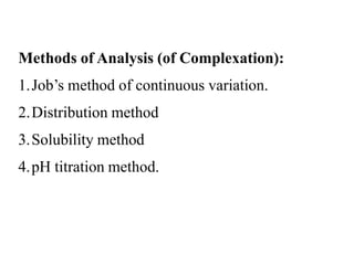
- 1. Methods of Analysis (of Complexation): 1.Job’s method of continuous variation. 2.Distribution method 3.Solubility method 4.pH titration method.
- 3. 1. Method of continuous variation. 1. Dielectric constant 2. Refractive index 3. Spectrophotometric extinction coefficient Physical properties Characteristics of species. A+B No complexation Complexation Physical properties are additive values Physical properties values different
- 5. 2. Distribution method: Partition coefficient / Distribution changes due to complexation. By conducting 2 experiments stability constant is estimated.
- 6. 3. Solubility method: • When mixture form complexes solubility of one component may increase/ decrease. • Experiments are conducted to estimate parameters Experiment: 1. Caffeine (Complexing agent) taken in different concentrations into a series of flask. 2. Add PABA, Agitate, Filter & analyze drug content.
- 7. 4. pH titration method. This method is suitable if complexation produces change in pH. Experiment: 1.Glycine solution (75 ml) titrated with NaoH, pH is recorded. 2.Glycine solution (75 ml) + Cu+2 Complex titrated with NaoH, pH is recorded. (Complexation releases Protons and pH decreases) Quantity of alkali = Concentration of ligand bound.
- 8. PROTEIN BINDING The interacting molecules are generally the macromolecules such as protein, DNA or adipose. The proteins are particularly responsible for such an interaction. The phenomenon of complex formation of drug with protein is called as protein binding of drug.
- 9. • As a protein bound drug is neither metabolized nor excreted hence it is pharmacologically inactive due to its pharmacokinetic and Pharmacodynamics inertness. • Protein + drug ⇌ Protein-drug complex. • Protein binding may be divided into – 1. Intracellular binding. 2. Extracellular binding.
- 10. Protein Binding may thus: • Facilitate the distribution of drugs throughout the body • Inactivate a drug by binding so firmly that sufficient concentration is not available at the receptor site. • Retard the excretion of a drug which may accumulate in the body. • Alter the duration of action of a drug. • Display body hormones or a co-administered agent.
- 11. Mechanisms of protein drug binding: Binding of drugs to proteins is generally of reversible and irreversible. • Reversible generally involves weak chemical bond such as: 1. Hydrogen bonds 2.Hydrophobic bonds 3. Ionic bonds 4. Van der Waal’s forces. • Irreversible drug binding, though rare, arises as a result of covalent binding and is often a reason for the carcinogenicity or tissue toxicity of the drug.
- 12. • Absorption - As we know the conventional dosage form follow first order kinetics. So when there is more protein binding then it disturbs the absorption equilibrium. • Distribution - A protein bound drug in particular does not cross the BBB, the placental barrier, the glomerulus. Thus protein binding decreases the distribution of drugs. • Metabolism - Protein binding decreases the metabolism of drugs and enhances the biological half life. Only unbound fractions get metabolized. e.g. Phenylbutazone and Sulfonamide. • Elimination – Only the unbound drug is capable of being eliminated. Protein binding prevent the entry of drug to the metabolizing organ (liver) and to glomerulus filtration. e.g. Tetracycline is eliminated mainly by glomerular filtration.
- 13. Systemic solubility of drug – Lipoprotein act as vehicle for hydrophobic drugs like steroids, heparin, oil soluble vitamin. Drug action - Protein binding inactivates the drugs because sufficient concentration of drug cannot be build up in the receptor site for action. e.g. Naphthoquinone. Sustain release – The complex of drug protein in the blood act as a reservoir and continuously supply the free drug. e.g. Suramin sodium-protein binding for antitrypanosomal action. Diagnosis – The chlorine atom of chloroquine replaced with radiolabeled I- 131 can be used to visualize-melanomas of eye and disorders of thyroid gland.
- 14. Binding of drug to blood plasma proteins – • The binding of drugs to plasma proteins is reversible. • The extent or order of binding of drug to plasma proteins is: • Albumin › ὰ1-Acid glycoprotein › Lipoproteins › Globulins. • Binding of drug to human serum Albumin – • It is the most abundant plasma protein (59 %). • Having M.W. of 65,000 with large drug binding capacity. • Both endogenous compounds such as fatty acid, bilirubin as well as drug bind to • HSA.
- 15. • Four different sites on HSA for drug binding. • Site I: warfarin and azapropazone binding site. • Site II: diazepam binding site. • Site III: digitoxin binding site. • Site IV: tamoxifen binding site.
- 16. Binding of drug to α1-Acid glycoprotein – • It is called as orosomucoid. It has a M.W. 44,000. • Its plasma conc. range of 0.04 to 0.1 g %. • It binds to no. of basic drugs like imipramine, lidocaine, propranolol, and quinidine. • Binding of drug to Lipoproteins – • Binding by Hydrophobic Bonds, Non-competative. • Mol wt: 2-34 Lacks dalton. • Lipid core composed of: Inside: triglyceride & cholesteryl esters. Outside: • Apoprotein. e.g. Acidic: Diclofenac. Neutral: Cyclosporin A. Basic: Chlorpromazine. • Its types are LDL, HDL, VLDLand Chylomicrons.
- 17. Binding of drug to Globulins – • α1 Globulin (Transcortine /Corticosteroid Binding globulin) - Steroidal drugs, • Thyroxin & Cyanocobalamine (Vit B12). • α2 Globulin (Ceruloplasmine) - Vitamin A, D, E, K. • β1 Globulin (Transferin) - Ferrous ions. • β2 Globulin – Carotinoids. • γ Globulin – Antigens.
- 18. Patient related factors: Age – Neonates have low albumin content, thus less drug binding. Disease state – Disease sate alter the drug binding.
- 23. Patient related factors: Age – Neonates have low albumin content, thus less drug binding. Disease state – Disease state alter the drug binding. Drug Interaction: • Displacement reaction • Allosteric changes in protein molecules.