Bio Inspired Self-Curing Composite: A Leap into Augmented Enactment
MEM395 ManisagarN 36x48 unmounted
1. 3D Biopolymer Micro/Nano-meter Scaled Fibers
Noreshvarman Manisagar | Mentor : Mingkun Wang | Advisor : Dr. Li-Hsin(Leo) Han
Hand-spun, Mass Produced, Micro/Nano-meter Scaled Fibers as a Novel Biomaterial for Tissue Engineering
Biopolymer
Microfabrication Lab
Introduction Method & Technology Results Discussion/Future Work
In the recent decade, methods for culturing cells in 3D
environments have received growing attention in the field of
tissue engineering. These 3D approaches are closely mimicking
natural, in vivo bioactivities of cells. To engineer desired cellular
processes and tissue formation, 3D biomimetic scaffolds that
incorporates different biochemical, mechanical or architectural
cues have been developed with extensive efforts.
Hydrogel-based scaffolds are widely used for 3D tissue
engineering.
Advantage: Tissue-like water content, tunable biochemical
properties, and ease for cell encapsulation.
Disadvantage: Lack macroporosity, non-ideal for cell
bioactivities, weak mechanical strength.
Microfibers are constantly being researched on for these
applications as well.
Advantage: High mechanical strength, highly porous scaffolds,
ease of modification for biochemical cues.
Disadvantage: Method of production (electro-spinning) produces
a structure that may result in poor cell infiltration and distribution.
To tackle these, a microribbon-like scaffold made from gelatin is
produced that combines the advantages of both scaffold
productions stated above.
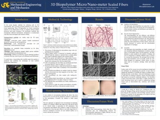
Figure 2: Schematics of fabrication process of microribbon-based scaffolds.
(retrieved from Advanced Functional Material Journal by Li-Hsin(Leo) Han,
Stephanie Yu, Tianyi Wang, Anthony W. Behn,and Fan Yang)
Uniform Cell Distribution
The hand-spun micro/nanofibers are paste-like in water,
and thus enable direct cell mixing and cell encapsulation,
which promotes uniform cell distribution and enables the
control of fiber density.
Ease for Zonal Organization
The micro/nanofibers are non diffusive, and different
types of micro/nanofibers made of different biopolymers
can be dispensed in desired location in 3D. Such character
facilitates the formation of multi-zonal scaffolds that
mimic the zonal organization of native tissues, such as the
layered structures of arteries and the osteochondral
organization of cartilages.
Versatility
The hand-spun micro/nanofibers are highly versatile and
can incorporate different biopolymers to achieve various
types of extracellular matrix (ECM) properties. The fiber
diameter is easily tunable by the number of folding, and
can be customized to replicate different types of tissues
by varying fiber dimensions. We anticipate that the hand-
spun micro/nanofibers will be broadly useful for cell-
based therapies for repairing a broad variety of tissue
types such as of muscles, cartilage, bones and blood
vessels.
Figure 4: Scanning Electron Microscope (SEM) images of microribbon
revealing the macroporous structure. (SEM session by Mingkun Wang)
50 µm
50 µm
50 µm
50 µm
I. The process of fabricating the microribbons starts with wet
spinning process of gelatin.
II. Type-A gelatin (GelA) is dissolved in dimethyl sulfoxide
(DMSO) and the solution is injected from a syringe pump
into a bath of anhydrous ethanol with constant stirring.
III.Pulled by gravity and stabilized by a high surface tension,
the ejected GelA solution drips unbrokenly and forms a fine
thread with continuous flow into the ethanol bath.
IV.After wet-spinning, the microfibers are transferred and dried
in acetone bath for 3 hours at either 25 °C or 60 °C.
V. Drying by acetone causes a rapid and asymmetrical collapse
of microfibers which leads to formation of the microribbon
structure.
VI.The as-formed microribbons are washed 3-times by ethanol
and then dissociated into short segments of less than 1 mm
using a homogenizer.
VII.The microribbons are then treated with methacrylic
anhydride.
VIII.The methacrylated microribbons are further treated with
glutaraldehyde at 40 °C for either 3 hours or 12 hours.
IX.The aldehyde-fixed microribbons are finally neutralized by
lysine solution.
X. Upon exposure to light, the gelatin microribbons crosslinks
like hydrogels and forms a macroporous gelatin network.
Hand-spinning Technology
A new method of wet-spinning replacing the above has been
discovered by our lab and the production method is undisclosed
in this presentation. Currently, the method is under patent
approval.
This new approach, as opposed to the conventional wet-spinning,
enables us to produce ribbons that range from 500 micron down
to 0.1 micron which is equivalent to 100 nanometers. The
conventional microribbons only produces a range of between 20
to 200 micron. Since the native fibers in our body range from 0.1
micron to hundreds of micron, our new method enables us to
closely replicate the structures.
Future Work
Further study will be carried out on the mechanical strength and
the techniques on manipulating the growth of stem cells by
shaping the nanofibers to resemble the shape of collagen fibers.
Moving forward, in near future, with such technology we hope
to open doors for healing of diseases, injuries and birth defects
that cannot be reversed by traditional medicine. The flexibility
of these micro/nano-meter scaled fibers is highly desirable for
shock absorbing tissues and is useful in engineering cartilage
tissues such as intervertebral disc, meniscus, and articular
cartilage.
Figure 1: Cell spreading and proliferation in microribbon scaffolds with
tunable biochemical, mechanical, and topographical cues.(retrieved from
Advanced Material Journal by Li-Hsin(Leo) Han, Xinming Tong, and Fan
Yang)
Figure 3: Structures of collagen and elastin fibers which typically range
from 0.1 micron to few hundred nanometer. (retrieved from boundless.com)
Figure 5: Image of stem cells in culture medium. (Shot by Noreshvarman
Manisagar)
References
1) Han, Li-Hsin, Stephanie Yu, Tianyi Wang, Anthony W. Behn, and Fan Yang.
"Microribbon-Like Elastomers for Fabricating Macroporous and Highly Flexible
Scaffolds That Support Cell Proliferation in 3D." Adv. Funct. Mater. Advanced
Functional Materials 23.3 (2012): 346-58. Web.
2) Han, Li-Hsin, Xinming Tong, and Fan Yang. "Photo-crosslinkable PEG-Based
Microribbons for Forming 3D Macroporous Scaffolds with Decoupled Niche
Properties." Adv. Mater. Advanced Materials 26.11 (2013): 1757-762. Web.
3) Loose Connective Tissue. Digital image. Boundless.com. N.p., n.d. Web.
<https://www.boundless.com/biology/textbooks/boundless-biology-textbook/the-
animal-body-basic-form-and-function-33/animal-primary-tissues-193/connective-
tissues-loose-fibrous-and-cartilage-738-11968/>.
4) SEM images by Mingkun Wang.
5) Stem cell image by Noreshvarman Manisagar.
Discussion/Future Work
Advantages
Mass production
Different from electrospinning, which often has a slow
production rate, the folding/pulling protocol for
micro/nanofibers fabrication is highly efficient, low cost,
and suitable for mass production.
Acknowledgement
I would like to thank my advisor Dr. Li-Hsin(Leo) Han, my
mentor Mr. Mingkun Wang, and the Biopolymer Microfabrication
Lab Group for the experience and knowledge acquired, and Dr.
Nicholas P. Cernansky for the opportunity to be a part of the Hess
Undergraduate Research Community.
(cont’d)