Reese_Masters Plan II Report Supplement_Spring 2015
1. 1
Directing cell migration and organization via nanocrater-patterned cell-repellent interfaces
Master’s Plan II Project Report Supplement
By: Willie Mae Reese
Abstract
Although cell adhesion to nanostructured interfaces has been extensively studied, few studies have focused on tuning nano-
topographical surfaces to direct cell migration for cell patterning. Using multi-photon ablation lithography, my collaborators fabricated
arrays of nanoscale craters in quartz substrates with a variety of geometries and spacing (i.e. pitch). Changing the nanocrater diameter
(600-1000 nm), depth (110-350 nm), and/or pitch (1-10 um) alters the planar surface area available for cells to establish stable focal
adhesions (FAs) and induces migration away from regions of high nanocrater density. This persistent migration can be used to dictate
cell patterning (e.g., lines, circles) according to the nanocrater parameters. By using immunofluorescence to visualize focal adhesion
size, I was able to conclude that nanocrater features significantly dictated focal adhesion formation, which I concluded leads to high
turnover of focal adhesions and increased migration. To further investigate interactions of these patterned surfaces and cellular
adhesion mechanisms, I also probed the effects of intracellular contractile protein (e.g., Talin) activation on patterning. I found that cells
that overexpress the N-terminus of Talin-1, which is one of the major proteins responsible for stable focal adhesion formation, lack the
ability to quickly spread and migrate from low pitch to high pitch regions due to integrin over activation. To continue this study, I am
currently beginning a study to pattern similar nanoscale craters using nanoimprint technology in materials that are more relevant to
biomaterial studies such as tissue culture polystyrene and other novel biomaterials. These nanoscale surfaces serve as tools for
mechanobiology studies and understanding the attributes of surfaces necessary to physically pattern cells.
Introduction
Surfaces that manipulate cell adhesion, motility, and differentiation are a large focus of many areas of biomaterials research including
tissue regeneration, cell patterning, and biocompatibility. Various methods have been extensively employed to alter the cell-substrate
interaction through modification of surface chemistry, geometry, topography, and mechanical properties. Here, we explored the cellular
response to nanoscale surface features, specifically their influence on focal adhesions (FAs) and cell motility. This research has
recently been accepted for publication in Nature Materials.
Arrays of nano-sized craters with diameters of 500-1000 nm and depths of 45-350 nm were
patterned in quartz using direct write multiphoton lithography. Thereby, we were able to vary not
only the geometry of the craters, but also the distance (i.e. pitch) between the pits in the pattern (1-
10 µm). By systematically changing these variables we are able to use these surfaces to decipher
the complex interaction of cells and topography. Our ability to pattern regular structures on the
order of the FA size (~1-10 µm) distinguishes our surfaces from other topographical work by more
closely mimicking the natural dimensions of the extracellular matrix topography. Our nano-patterned
surfaces go beyond solely altering cell adhesion, the prominent approach for cell patterning, and
instead guide cell migration by disrupting mature FA formation. Certain combinations of diameter,
depth, and pitch promote cell migration, which can be exploited to pattern cells into lines, circles,
or other structures.
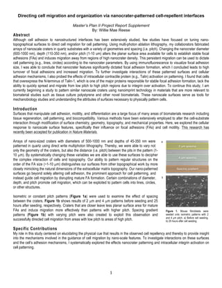
Isometric or constant pitch patterns (Figure 1a) were used to examine the effect of spacing
between the craters. Figure 1b shows results of 2 µm and 4 µm patterns before seeding and 25
hours after seeding, respectively. Craters that are closer leave less planar surface area for mature
FAs and induce migration more effectively than patterns with higher pitch. Spacing gradient
patterns (Figure 1b) with varying pitch were also created to exploit this observation and
successfully directed cell migration from areas with low pitch to areas of high pitch.
Specific Contributions
My role in this study centered on elucidating the physical cue that results in the observed cell repellency and thereby to provide insight
into the mechanisms involved in the guidance of cell migration by nano-scale features. To investigate interactions on these surfaces
and the cell’s adhesion mechanisms, I systematically explored the effects nanocrater patterning and intracellular integrin activation on
cell patterning.
2 μm spacing
4 μm spacing
Spacing gradient
a
.
b
.
Figure 1. Mouse fibroblasts were
seeded onto isometric patterns with 2
and 4 µm pitch. a) Before cell seeding.
b) 25 hours after cell seeding.
2. 2
Nanoscale craters reduce focal adhesion size resulting in nascent adhesions.
Previous studies showed that focal adhesion size (i.e. area) is closely correlated
(r=0.93) with cell migration speed,1 which dictates the ability of the surface to
effectively pattern cells. To investigate if focal adhesion distribution could provide
insight into the mechanism of cell patterning, cells were plated on the patterned
surfaces in serum-containing medium for 1 hour and fixed. Focal adhesions were
visualized using immunofluorescent staining of vinculin, an intercellular adhesion
protein, to locate and measure FA area (Figure 2). The size of the focal adhesions
was quantified using ImageJ software and a standardized image processing
protocol and statistically analyzed using ANOVA in Graphpad Prism.
We found that nanocraters impair the cells’ ability to form mature focal adhesions.
Depending on pattern pitch, cells revealed different morphology and focal adhesion
distribution, where cells on surfaces with lower pitch have smaller and less
pronounced focal adhesions that are primarily distributed at the leading and trailing
edges of cells. Focal adhesions located on low pitch regions or near nanocraters
are smaller and therefore more nascent while larger and mature focal adhesions
were located in on the planar spaces between the nanocraters. In contrast,
lamellipodia in the cells on flat surfaces (no patterns) were symmetrically
distributed. In this study, the 4µm pattern most significantly (p≤0.0001) reduced
focal adhesion size (Figure 3).
Integrin activation reduces migration speed.
To investigate intracellular effects, I transfected wild type NIH-3T3 fibroblasts with
DNA plasmids containing the sequence of a GFP-tagged Talin-1 globular N-
terminus using the method describe by Coyer and others4. Talin-1, an intracellular
contractile protein, increases integrin activation and stabilizes focal adhesion
formation, which has been shown to enable cells to overcome focal adhesion area
limitations4. Talin-1+ fibroblasts were seeded after fibronectin was physio-adsorbed
onto quartz nano-patterned surfaces. These cells lack the necessary balance
between integrin activation and turnover to quickly spread and migrate from low
pitch to high pitch regions. It should be noted that the cells do not lose their ability
to adhere to the surface, but cannot turnover formed focal adhesions and form new
ones, a necessary step of migration. These results support other studies in the
conclusion Talin1+ activation enables cells to overcome area limitations to form
focal adhesions.
Future Investigations
Nano-imprinting is an alternative to multi-photon lithography.
In quartz, the unwanted perturbations produced by laser ablation lithography are very small and the
increase in surface roughness is negligible compared to the size of the pits (see Figure 4). In
materials with significantly lower melting points and in polymers with glass transition temperatures
near 100 °C, however, these perturbations approach the size of the craters themselves.
Consequently, patterning and roughness effects are superimposed and the ability to make arrays of
nano-craters with dimensions that have influential effects on FA formation is limited. Polymer
surfaces (e.g. tissue culture treated poly(styrene) or poly(urethane)) that direct cell migration for cell
patterning or cell adhesion resistance would be beneficial for many biological studies applications. In
order to fabricate structured polymer surfaces similar to the nano-patterned quartz substrates, new
techniques are needed. Nano-imprinting shows great promise to develop surfaces at even smaller
sizes (10 nm) with regular, reproducible structures using photo-crosslinkable polymers.
Microimprinting allows the exploration with soft materials and also greatly reduces fabrication time.
Currently our patterns are 750 µm squares and take two hours to fabricate. In the same time with nano-imprinting, 150 mm square
patterns could be fabricated. In the next phase of this project, I have begun to develop methods for processing, characterizing, and
C
ontrol
8µm
6µm
4µm
2µmC
ontrol
4µm
2µm
0.0
0.5
1.0
1.5
Isometric Spacing
FAArea
(µm2)
*
****
****
Wild Type Talin-1 +
Figure 2. Immunofluorescence images of fibroblast cultured on
patterned quartz surfaces with craters (1 µm in diameter, 350 nm in
depth) with inverted DIC. Scale bars =10 µm.
Figure 3. Focal Adhesion area of wild type NIH 3T3’s on unpatterned
(control) and isometrically patterned nanocrater surfaces. 4 µm
spacing showed the most significant reduction in focal adhesion area.
Talin-1+ transfected cells had significantly larger focal adhesions on
all surfaces, but did not exhibit significantly different focal adhesion
distributions between isometric spacings. *p≤ 0.05,**p≤ 0.01,
***p≤0.001, ****p≤0.0001 (Kruskal-Wallis ANOVA).
Figure 4. Tapping AFM cross sectional
profile of nano-craters in quartz.
3. 3
testing different polymer-based nano-topographical substrates for biological studies. These surfaces will provide tools for studying
mechanobiological studies with more relevant materials for biomedical and biological applications.
References:
1. D.-H. Kim and D. Wirtz, FASEB J., 2013, 27, 1351–61.
2. E. Ruoslahti and M. D. Piershbacher, Science (80)., 1987, 238, 491–497.
3. S. Aota, M. Nomizu, and K. M. Yamada, J. Biol. Chem., 1994, 269, 24756–61.
4. S. R. Coyer, A. Singh, D. W. Dumbauld, D. a Calderwood, S. W. Craig, E. Delamarche, and A. J. García, J. Cell Sci., 2012, 125, 5110–23.