

Recommended
More Related Content
What's hot
What's hot (20)
Similar to Yang2015
Similar to Yang2015 (20)
More from Dr. Carlos Joel Sequeira.
More from Dr. Carlos Joel Sequeira. (20)
Recently uploaded
Recently uploaded (20)
Yang2015
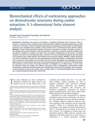
- 1. Biomechanical effects of corticotomy approaches on dentoalveolar structures during canine retraction: A 3-dimensional finite element analysis Chongshi Yang,a Chao Wang,b Feng Deng,c and Yubo Fand Chongqing and Beijing, China Introduction: Corticotomy has proven to be effective in facilitating orthodontic tooth movement. There is, however, no relevant study to compare the biomechanical effects of different corticotomy approaches on tooth movement. In this study, a series of corticotomy approaches was designed, and their impacts on dentoalveolar structures were evaluated during maxillary canine retraction with a 3-dimensional finite element method. Methods: A basic 3-dimensional finite element model was constructed to simulate orthodontic retraction of the maxillary canines after extraction of the first premolars. Twenty-four corticotomy approach designs were simulated for variations of position and width of the corticotomy. Displacement of the canine, von Mises stresses in the canine root and trabecular bone, and strain in the canine periodontal ligament were calculated and compared under a distal retraction force directed to the miniscrew implants. Results: A distal corticotomy cut and its combinations showed the most approximated biomechanical effects on dentoalveolar structures with a continuous circumscribing cut around the root of the canine. Mesiolabial and distopalatal cuts had a slight influence on dentoalveolar structures. Also, the effects decreased with the increase of distance between the corticotomy and the canine. No obvious alteration of displacement, von Mises stress, or strain could be observed among the models with different corticotomy widths. Conclusions: Corticotomies enable orthodontists to affect biomechanical responses of dentoalveolar structures during maxillary canine retraction. A distal corticotomy closer to the canine may be a better option in corticotomy-facilitated canine retraction. (Am J Orthod Dentofacial Orthop 2015;148:457-65) T wo main challenges are often confronted in orthodontic treatment for adult patients: long duration and risk of side effects such as root resorption and marginal bone loss.1,2 To cope with these problems, surgical techniques are used to facilitate orthodontic tooth movement, including alveolar osteotomy, corticotomy, and distraction osteogenesis.3-5 Compared with osteotomy and distraction osteogenesis, corticotomy has become popular gradually because of the minor surgical injury, flexible approach, and operational simplicity. Corticotomy has been demonstrated to facilitate orthodontic tooth movement effectively in the clinic. It has many advantages over more traditional orthodon- tics, including reduced orthodontic treatment time, more significant tooth movement, and less anchorage required.5,6 Wilkco et al5 attributed these effects of corticotomy to the regional acceleration phenomenon, which is characterized by an increase in the rate of bone turnover. The remodeling of alveolar bone and periodontal ligament (PDL) would be activated and aggravated after being exposed to an insult such as a corticotomy or a cortical perforation. En-block a Graduate student, Chongqing Key Laboratory of Oral Diseases and Biomedical Sciences, Stomatological Hospital of Chongqing Medical University, Chongqing, China. b Assistant professor, Chongqing Key Laboratory of Oral Diseases and Biomedical Sciences, Stomatological Hospital of Chongqing Medical University, Chongqing, China. c Professor, Chongqing Key Laboratory of Oral Diseases and Biomedical Sciences, Stomatological Hospital of Chongqing Medical University, Chongqing, China. d Professor, Chongqing Key Laboratory of Oral Diseases and Biomedical Sciences, Chongqing Medical University, Chongqing; professor, Key Laboratory for Biome- chanics and Mechanobiology of Chinese Education Ministry, International Research Center for Implantable and Interventional Medical Devices, Beihang University, Beijing, China. Funded by the National Natural Science Foundation of China (numbers 11421202 and 11120101001). Address correspondence to: Yubo Fan, Stomatological Hospital of Chongqing Medical University, No. 426, Yubei District, Chongqing, China; e-mail, yubofan@buaa.edu.cn. Submitted, September 2014; revised and accepted, March 2015. 0889-5406/$36.00 Copyright Ó 2015 by the American Association of Orthodontists. http://dx.doi.org/10.1016/j.ajodo.2015.03.032 457 ORIGINAL ARTICLE
- 2. movement of the bone segment was another explanation for this technique.7,8 Corticotomy can reduce the resistance of alveolar bone to orthodontic tooth movement by breaking the continuity of the bone layer, leading to a bone-bending effect, and possibly even a greenstick fracture of the alveolar bone.9 Orthodontic tooth movement is the result of remodeling of the alveolar bone and the PDL around the tooth.10 Two stages are included in this procedure: the delivery of orthodontic force and the biologic responses to the force.11 The force generated by the orthodontic appliance is transmitted to the PDL by the teeth and then to surrounding alveolar bone, contributing to the change of the mechanical environment of the dentoalveolar structure. The mechanical stimulation can trigger a series of biologic responses to regulate the deposition or resorption of the alveolar bone temporarily and spatially to facilitate tooth movement. The internal strength and distribution of the mechanical stimulation can be denoted accurately with stress and strain, rather than with orthodontic force, in consideration of the shape and size of the den- toalveolar units.12 The stress and strain in dentoalveolar structure can be altered by breaking the continuity of the cortical bone after a corticotomy.13 The change of stress or strain can be calculated using 3-dimensional (3D) finite element methods. Three-dimensional finite element analysis is a numeric technique for simulating mechanical processes of a real physical system; it is considered to be a valid and reliable approach for calculating stress, strain, and displacement of dentoalveolar structures.14,15 This technique can be used to simulate orthodontic processes with different treatment plans and to compare their biomechanical effects without increasing the numbers of patients or animals in the sample, unlike clinical or animal investigations. The routine treatment plan for patients with maxillary or bimaxillary protrusion is retraction of the anterior teeth after extraction of the premolars. With regard to these patients, a 2-step treatment can be used to make the canine move first during space closure.16 To shorten the treatment time and save anchorage, corticotomies have been used to facilitate canine movement in the clinic.17,18 Although these studies have provided useful findings, the corticotomy approach was limited. More importantly, little research regarding the biomechanical effects of corticotomy approaches on dentoalveolar structures has been done. Therefore, the aims of this study were to design different corticotomy approaches and compare their biomechanical impacts on dentoalveolar structures during retraction of the maxillary canine and to select an optimal approach for clinical adoption. MATERIAL AND METHODS Construction of the basic finite element model was based on computed tomography scans (120 kV; 175 mA; slice thickness, 0.625 mm) (LightSpeed VCT; GE Healthcare, Wauwatosa, Wis) of the skull of an adult with maxillary protrusion. A surface 3D model of the maxillary alveolar bone and the dentition with the first premolars extracted was constructed from the scanned images through thresholding, region growing, and calculating 3D operations with Mimics software (version 9.0; Materialise, Leuven, Belgium). Scaling and Boolean operations were performed on the surface model of the alveolar bone and dentition to generate the cortical bone with an average thickness of 2.0 mm and the PDL around the tooth with an average thickness of 0.2 mm based on the studies of Wang et al14 and Lim et al19 with Rapidform software (version 6.5; INUS, Seoul, Korea). The orthodontic appliances, including edgewise brackets for the anterior teeth and premolars, buccal tubes for the molars, square wires, and miniscrew implants, were designed and modeled according to clinical applications in computer-assisted design software (version 2011; Solidworks, Velizy-Villacoublay, France). The brackets and buccal tubes were placed on the center of the labial or buccal surface of the clinical crown of the teeth. The implants were placed in the interradicular space between the first molar and the second premolar. All components were saved in initial graphics exchange specification format and imported into Abaqus (version 6.11; Solidworks). The bases of the brackets and buccal tubes were subtracted from the corresponding teeth to create a matching geometry. Then the alveolar bone, teeth, PDL and orthodontic appliances were assembled to form the geometric model. Volumetric meshes of these components in the model were created separately. The types and numbers of elements and nodes are listed in Table I The first step of simulation of the corticotomy approach was to design and build a shear band Table I. Types of elements and numbers of nodes and elements Types of element Nodes (n) Elements (n) Teeth C3D4 34037 166707 PDL C3D20R 589521 140402 Trabecular bone C3D4 29405 139600 Cortical bone C3D10 79619 40356 Bracket C3D4 28097 116846 Square wire C3D8R 1233 540 458 Yang et al September 2015 Vol 148 Issue 3 American Journal of Orthodontics and Dentofacial Orthopedics
- 3. corresponding to the approach in Solidworks. Then cortical bone was subtracted by the shear band to generate the model in Abaqus. Twenty-four models were built in this study. These models can be classified as follows. For the corticotomy approaches with the position of the cuts varied, the corticotomy cuts were in cortical bone around the maxillary canine. There were 4 basic cuts: mesiolabial, mesiopalatal, distolabial, and distopalatal. Other approaches were the various combinations of these cuts and combinations with labial and palatal cuts. There were 20 corticotomy approaches designed and modeled (Fig 1). For the corticotomy approaches with the distance to the canine varied, 2 other cuts parallel to the distal corticotomy were designed at the extraction side. They were 2 and 4 mm from the distal corticotomy cut, respectively (Fig 1). For the corticotomy approaches with the width of the corticotomy cut varied, the width of the corticotomy cuts above was set at 0.4 mm according to the diameter of the bur used for corticotomies in the clinic. Two other cuts with widths of 0.6 and 0.8 mm were designed on the base of the distal corticotomy cut (Fig 1). Five materials were used in this study. All materials were assumed to be homogeneous, isotropic, and linearly elastic. The material properties were determined from the values documented in the literature (Table II). Then the material properties were assigned to cortical bone, trabecular bone, teeth, PDL, and orthodontic appliances. Canine distalization is the first step of space closure in a 2-step treatment. To eliminate the complex constraints of square wire on the canine and to facilitate its movement, the segmented arch technique was used in this study. The maxillary incisors, ipsilateral molars, and second premolars were connected with 3 pieces of square wire. The bilateral canines were not tied to the Fig 1. Designs of corticotomy approaches for the maxillary canine: A, B, and C, continuous circumscribing cut around the canine from the labial, lingual, and occlusal views; D, definition of canine position for the designs of corticotomy approaches below; E, corticotomy approaches with the positions of the cuts varied; F, corticotomy approaches with the distance to the canine varied; G, corticotomy approaches with the width of the corticotomy cut varied. Table II. Material properties Young's modulus (MPa) Poisson's ratio Reference Cortical bone 10.7E3 0.3 Aversa et al20 Trabecular bone 9.7E2 0.3 Aversa et al20 Teeth 20.7E3 0.3 Lee and Baek21 PDL 50 0.45 Lin et al22 Brackets and stainless steel wire 200E3 0.3 Canales et al23 American Journal of Orthodontics and Dentofacial Orthopedics September 2015 Vol 148 Issue 3 Yang et al 459
- 4. square wire to let them move freely. To prevent separation and slippage, binding constraints were used to define these interfaces: square wire-bracket, tooth-bracket, PDL-tooth, trabecular bone-PDL, and cortical bone-trabecular bone. The miniscrew implants were not involved in calculations of finite element models, and they were used to indicate the direction of the orthodontic load (Fig 2). All these models were restrained at the nodes on the maxillary bone at the base of nasal cavity in all degrees of freedom. To simulate retraction of the maxillary canine, a 100-cN concentrated force was applied to the middle points of the bracket slot toward the crown notch of the miniscrew implant according to the clinical condition (Fig 2). The displacement of the canine, stress distributions in the canine root, and strain distributions in the PDL and the trabecular bone were analyzed with Abaqus software. RESULTS The displacements of the canines were observed in a newly defined coordinate system: origin-anterior nasal spine; x-axis, the line through posterior nasal spine; and the x-y plane–middle sagittal plane. The initial displacement of the canine showed distal movement combined with clockwise rotation, and the largest displacement of the canine was highly concentrated on the cusp in all models (Fig 3). The position of the corticotomy had an obvious effect on the displacement of the canine. The maxillary canine in the model with the continuous circumscribing cut showed the greatest displacement, and larger displacements were found in models with distal corticotomy cuts and their combinations. However, no notable difference of canine displacement was observed in models with distopalatal and mesiolabial cuts and in the model with no corticotomy. As the distance between the canine and the corticotomy increased, the displacement of the canine decreased gradually. The width of the corticotomy cut had no obvious influence on the canine displacement (Fig 3). The von Mises stress distribution in the canine root is illustrated in Figure 4. The maximum von Mises stress was centered on the distolabial side of the cervix in all models. The position of the corticotomy cuts had similar variations on the distribution of the von Mises stresses to the distribution of displacements of the canine. When the distance and the width of the corticotomy cut were increased, the maximum stress value was reduced, especially for a corticotomy with the distance increased. In general, the differences of the maximum stress in the canine roots were not evident among these models. The maximum strain was concentrated mainly in the cervical region of the PDL (Fig 5). There were significant differences in the strain values in the PDL with the changes of corticotomy position. The maximum strain values decreased in the models with the continuous circumscribing cut and the distal cut and its combinations, but the values were enhanced in the models with the mesial cut and its combinations compared with the model with no corticotomy. The maximum strain in the PDL increased gradually when the distance to the canine increased. The influence of the farthest corticotomy, which was about 5 mm from the canine, got close to the effect with no corticotomy. The width of the corticotomy had no remarkable influence on the strain distribution in the PDL of the canine. von Mises stress in the trabecular bone was mainly concentrated on the alveolar crest at the distal side of the canine in the model without a corticotomy. But in models with a corticotomy, the area of stress concentration transferred to the alveolar crest near the corticotomy. Thus, the position of the corticotomy can affect the distribution of stress in trabecular bone remarkably. The trabecular bone showed the maximum stress value in the model with a continuous circumscribing corticotomy cut, a lower stress value in the model with the distal corticotomy cut and its combi- nations, and still a lower stress value in the models with mesial and minor cuts. When the distance between the Fig 2. Basic geometric model and illustration of the boundary and load: A, frontal view; B, lateral view. Diamonds, Boundary; arrow, orthodontic load. September 2015 Vol 148 Issue 3 American Journal of Orthodontics and Dentofacial Orthopedics 460 Yang et al
- 5. corticotomy and the canine increased, the maximum stress value was reduced. No obvious alteration of the stress distributions was observed among the models with different corticotomy widths (Fig 6). DISCUSSION The continuous circumscribing cut around the root of the tooth proposed by Wilkco et al5 was reported in many studies, and its effect on facilitating tooth movement has been confirmed.18,24 In this approach, 4 vertical and 2 horizontal cuts around the root need to be made after elevation of the full-thickness mucoperiosteal flap, as for the canine. Although this technique proved to be effective in shortening treatment time, it was difficult for most orthodontic patients to accept because of its postoperative discomfort and risk of complications. To reduce the surgical injury and the difficulty of the operation, new corticotomy approaches with minor cuts were designed by changing the continuous circumscribing cut in this study. The biomechanical effects of these approaches on dentoalveolar structures were studied and compared with the models with the continuous circumscribing corticotomy and with no corticotomy. At the same time, we also considered the distance between the corticotomy and the canine, and considered the width of the corticotomy cut in this study. Because of the deviations between the finite element models and the actual situation in geometric and mechanical similarities, and owing to material property and boundary conditions, the results of this study should be interpreted with caution. For instance, the PDL was assumed to be isotropic, homogeneous, and linearly elastic in this study, but the PDL is a nonlinear, viscoelastic, and anisotropic material.25 Meanwhile, the thickness of the PDL was set at 0.2 mm, and the thickness of cortical bone was set at 2.0 mm. However, the PDL and the cortical bone are not uniform in thickness.26,27 In addition, it is difficult to simulate the real situation of alveolar sockets after the extraction of teeth. Because the healing of an alveolar socket is a dynamic process, the morphology and mechanical characteristics of alveolar bone in this area change markedly. Therefore, the alveolar sockets were excluded in these finite element models, and the height and width of the alveolar process at the Fig 3. Comparison of the maximum initial displacements of the canine in the models: A, with the position of corticotomy cut varied; B, with the distance between the corticotomy cut and the canine varied; C, with the widths of corticotomy varied; D, distribution of the initial displacements of the canine. American Journal of Orthodontics and Dentofacial Orthopedics September 2015 Vol 148 Issue 3 Yang et al 461
- 6. extraction site were decreased slightly. Canine movement occurred after the socket was well healed in this study. Nevertheless, for comparisons, the relative results have more value than the absolute results. In this study, the different biomechanical responses of the dentoalveolar structures with corticotomy approaches were observed, and influences of these approaches on orthodontic canine distalization were seen. Several studies have been published about the impact of acceleration of canine movement and the histologic findings. Aboul-Ela et al17 evaluated the clinical effects of corticotomy-facilitated orthodontics during maxillary canine retraction. Their results showed that the rate of canine retraction with corticotomy was about twice that of the control side in the first 2 months. Kim et al28 investigated the biologic effects of corticotomy on alveolar remodeling during canine retraction in an animal study. Their histomorphometric analysis showed an extensive direct resorption of bundle bone with less hyalinization. Many researchers have attributed these effects to the regional acceleration phenomenon, where the acceleration of bone turnover was observed through animal research.29,30 These studies confirmed the effects of corticotomy as a stimulating factor that can increase the rate of bone turnover. The change of the mechanical situation in alveolar bone may be another stimulating factor that can strengthen the remodeling of alveolar bone, leading to rapid canine movement, which has been proven in previous studies.31,32 In this study, we found that the mechanical responses of dentoalveolar structures can be affected by a corticotomy; the responses were mainly reflected in the stress in the trabecular bone. The maximum stress value in the model with the continuous circumscribing cut increased several times in the alveolar bone near the corticotomy compared with the model with no corticotomy. The maximum displacement of the canine was concentrated on the cusp of its crown under an Fig 4. Comparison of the maximum stresses in the canine roots in the models: A, with the position of the corticotomy cut varied; B, with the distance between the corticotomy cut and the canine varied; C, with the widths of the corticotomy varied; D, distribution of the von Mises stress in the canine root from the labial and distal views. September 2015 Vol 148 Issue 3 American Journal of Orthodontics and Dentofacial Orthopedics 462 Yang et al
- 7. orthodontic load directed to the miniscrew implant. This result demonstrated that the canine had clockwise rotation, since the orthodontic force did not pass the resistance center of the canine. This result is consistent with the results reported by Xue et al.13 Alveolar bone on the distolabial side of the canine was the main source of resistance to its movement when the canine showed clockwise rotation. Therefore, the maximum stress in the canine root was focused on its cervix at the distolabial side. The effect of accelerated tooth movement and the safety of the periodontal tissues are primary concerns when performing a corticotomy. Changes of strain in the PDL may have a significant influence on orthodontic tooth movement. For one thing, the PDL is thought to be the medium of orthodontic force transfer. For another, the PDL itself takes part in the reactions promoting orthodontic tooth movement.33 In this study, the distal corticotomy approach and its combinations can reduce the strain value of the PDL of the canine, but a mesial corticotomy and its combinations can increase the strain value. This result may contribute to the lower the rate of orthodontic external root resorption, according to Viecilli et al.12 In these models with different corticotomy approaches, the model with the continuous circumscrib- ing cut around the root of the canine affected the mechanical responses of dentoalveolar structures the most, and the distal corticotomy and its combinations had similar impacts with the continuous circumscribing cut compared with the model with no corticotomy. These results can be generalized as to how corticotomy cuts can greatly reduce the resistance to tooth move- ment and how corticotomy cuts can affect the mechan- ical responses of dentoalveolar structures greatly. Therefore, a distal corticotomy approach may be a better alternative to continuous circumscribing, and it can be accepted readily by patients because of its minor injury to dentoalveolar structures. A corticotomy cut closer to the canine may benefit its distalization more because the impact of the corticotomy decreased with the increased distance between the cut and the canine. Fig 5. Comparison of the maximum strains in the PDL of canine in the models: A, with the position of the corticotomy cut varied; B, with the distance between the corticotomy cut and the canine varied; C, with the widths of the corticotomy varied; D, distribution of strains in the PDL of the canine from the labial and distal views. American Journal of Orthodontics and Dentofacial Orthopedics September 2015 Vol 148 Issue 3 Yang et al 463
- 8. CONCLUSIONS In this study, 24 corticotomy approaches used for facilitating canine distalization were designed and simulated, taking into consideration position, distance, and width of the corticotomy. The biomechanical responses of dentoalveolar structures were analyzed and compared with 3D finite element analysis. From the results of this study, the following conclusions can be drawn. 1. Corticotomy surgery can influence the mechanical responses of dentoalveolar structures during maxillary canine retraction. 2. The position of the corticotomy can affect the mechanical responses of dentoalveolar structures. A distal corticotomy has similar biomechanical effects as a continuous circumscribing cut around the root of the canine. 3. When the distance between the corticotomy cut and the canine increased, the biomechanical effects of the corticotomy decreased gradually. 4. A distal corticotomy closer to the canine may be a better option for rapid canine retraction, taking into account of its biomechanical effects and surgical injury. SUPPLEMENTARY DATA Supplementary data related to this article can be found online at http://dx.doi.org/10.1016/j.ajodo. 2015.03.032 REFERENCES 1. Fisher MA, Wenger RM, Hans MG. Pretreatment characteristics associated with orthodontic treatment duration. Am J Orthod Dentofacial Orthop 2010;137:178-86. 2. Killiany DM. Root resorption caused by orthodontic treatment: an evidence-based review of literature. Semin Orthod 1999;5:128-33. 3. Ren A, Lv T, Kang N, Zhao B, Chen Y, Bai D. Rapid orthodontic tooth movement aided by alveolar surgery in beagles. Am J Orthod Dentofacial Orthop 2007;131:160.e1-10. 4. Liou EJ, Huang CS. Rapid canine retraction through distraction of the periodontal ligament. Am J Orthod Dentofacial Orthop 1998; 114:372-82. Fig 6. Comparison of the maximum stresses in the maxillary trabecular bones in the models: A, with the position of corticotomy cut varied; B, with the distance between the corticotomy cut and the canine varied; C, with the widths of corticotomy varied; D, distribution of stresses in trabecular bone in the models with the continuous circumscribing cut and with no corticotomy. September 2015 Vol 148 Issue 3 American Journal of Orthodontics and Dentofacial Orthopedics 464 Yang et al
- 9. 5. Wilcko WM, Wilcko T, Bouquot JE, Ferguson DJ. Rapid orthodontics with alveolar reshaping: two case reports of decrowding. Int J Periodontics Restorative Dent 2001;21:9-19. 6. Mostafa YA, Fayed MM, Mehanni S, ElBokle NN, Heider AM. Comparison of corticotomy-faciliated vs standard tooth- movement techniques in dogs with miniscrews as anchor units. Am J Orthod Dentofacial Orthop 2009;136:570-7. 7. K€ole H. Surgical operations on the alveolar ridge to correct occlusal abnormalities. Oral Surg Oral Med Oral Pathol 1959;12: 515-29. 8. Chung KR, Oh MY, Ko SJ. Corticotomy-assisted orthodontics. J Clin Orthod 2001;35:331-9. 9. Chung KR, Kim SH, Lee BS. Speedy surgical orthodontic treatment using temporary anchorage devices as an alternative to orthognathic surgery. Am J Orthod Dentofacial Orthop 2009; 135:787-98. 10. Henneman S, Von den Hoff JW, Maltha JC. Mechanobiology of tooth movement. Eur J Orthod 2008;30:299-306. 11. Krishnan V, Davidovitch Z. Cellular, molecular, and tissue-level reactions to orthodontic force. Am J Orthod Dentofacial Orthop 2006;129:469.e1-32. 12. Viecilli RF, Kar-Kuri MH, Varriale J, Budiman A, Janal M. Effects of initial stresses and time on orthodontic external root resorption. J Dent Res 2013;92:346-51. 13. Xue J, Ye N, Yang X, Wang S, Wang J, Wang Y, et al. Finite element analysis of rapid canine retraction through reducing resistance and distraction. J Appl Oral Sci 2014;22:52-60. 14. Wang C, Han JY, Li Q, Wang LZ, Fan YB. Simulation of bone remodelling in orthodontic treatment. Comput Methods Biomech Biomed Engin 2014;17:1042-50. 15. Ammar HH, Ngan P, Crout RJ, Mucino VH, Mukdadi OM. Three-dimensional modeling and finite element analysis in treatment planning for orthodontic tooth movement. Am J Orthod Dentofacial Orthop 2011;139:e59-71. 16. Vig PS, Weintraub JA, Brown C, Kowalski CJ. The duration of orthodontic treatment with and without extraction: a pilot study of five selected practices. Am J Orthod Dentofacial Orthop 1990; 97:45-51. 17. Aboul-Ela SM, El-Beialy AR, El-Sayed KM, Selim EM, El-Mangoury NH, Mostafa YA. Miniscrew implant-supported maxillary canine retraction with and without corticotomy- facilitated orthodontics. Am J Orthod Dentofacial Orthop 2011; 139:252-9. 18. Iino S, Sakoda S, Miyawaki S. An adult bimaxillary protrusion treated with corticotomy-facilitated orthodontics and titanium miniplates. Angle Orthod 2006;76:1074-82. 19. Lim JE, Lee SJ, Kim YJ, Lim WH, Chun YS. Comparison of cortical bone thickness and root proximity at maxillary and mandibular interradicular sites for orthodontic mini-implant placement. Orthod Craniofac Res 2009;12:299-304. 20. Aversa R, Apicella D, Perillo L, Sorrentino R, Zarone F, Ferrari M, et al. Non-linear elastic three-dimensional finite element analysis on the effect of endocrown material rigidity on alveolar bone remodeling process. Dent Mater 2009;25:678-90. 21. Lee NK, Baek SH. Stress and displacement between maxillary protraction with miniplates placed at the infrazygomatic crest and the lateral nasal wall: a 3-dimensional finite element analysis. Am J Orthod Dentofacial Orthop 2012;141:345-51. 22. Lin TS, Tsai FD, Chen CY, Lin LW. Factorial analysis of variables affecting bone stress adjacent to the orthodontic anchorage mini-implant with finite element analysis. Am J Orthod Dentofacial Orthop 2013;143:182-9. 23. Canales C, Larson M, Grauer D, Sheats R, Stevens C, Ko CC. A novel biomechanical model assessing continuous orthodontic archwire activation. Am J Orthod Dentofacial Orthop 2013;143: 281-90. 24. Kim SH, Kim I, Jeong DM, Chung KR, Zadeh H. Corticotomy- assisted decompensation for augmentation of the mandibular anterior ridge. Am J Orthod Dentofacial Orthop 2011;140:720-31. 25. Fill TS, Toogood RW, Major PW, Carey JP. Analytically determined mechanical properties of, and models for the periodontal ligament: critical review of literature. J Biomech 2012;45:9-16. 26. Toms SR, Eberhardt AW. A nonlinear finite element analysis of the periodontal ligament under orthodontic tooth loading. Am J Orthod Dentofacial Orthop 2003;123:657-65. 27. Ozdemir F, Tozlu M, Germec-Cakan D. Cortical bone thickness of the alveolar process measured with cone-beam computed tomography in patients with different facial types. Am J Orthod Dentofacial Orthop 2013;143:190-6. 28. Kim SJ, Park YG, Kang SG. Effects of corticision on paradental remodeling in orthodontic tooth movement. Angle Orthod 2009; 79:284-91. 29. Baloul SS, Gerstenfeld LC, Morgan EF, Carvalho RS, Van Dyke TE, Kantarci A. Mechanism of action and morphologic changes in the alveolar bone in response to selective alveolar decortication-facilitated tooth movement. Am J Orthod Dentofacial Orthop 2011;139(4 Suppl):S83-101. 30. Wang L, Lee W, Lei DL, Liu YP, Yamashita DD, Yen SL. Tissue responses in corticotomy- and osteotomy-assisted tooth movements in rats: histology and immunostaining. Am J Orthod Dentofacial Orthop 2009;136:770.e1-11:discussion, 770-1. 31. Melsen B. Tissue reaction to orthodontic tooth movement—a new paradigm. Eur J Orthod 2001;23:671-81. 32. Kawarizadeh A, Bourauel C, Zhang D, G€otz W, J€ager A. Correlation of stress and strain profiles and the distribution of osteoclastic cells induced by orthodontic loading in rat. Eur J Oral Sci 2004;112: 140-7. 33. McCormack SW, Witzel U, Watson PJ, Fagan MJ, Gr€oning F. The biomechanical function of periodontal ligament fibres in orthodontic tooth movement. PLoS One 2014;9:e102387. American Journal of Orthodontics and Dentofacial Orthopedics September 2015 Vol 148 Issue 3 Yang et al 465