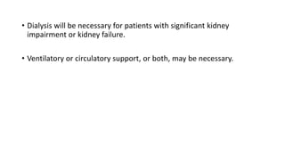
2. Introduction
• Second abundant intracellular cation.
• Serve as a cofactor >300 enzyme reaction i.e. involve ATP.
• Proper functioning of the Na+-K+ exchange pump that generates the
electrical gradient across cell membranes.
• Regulates the movement of calcium into smooth muscle cells
( maintenance of cardiac contractile strength and peripheral vascular
tone).
3. Magnesium Distribution in Adults
• The average-sized adult contains approximately 24 g (1 mole, or 2,000
mEq) of magnesium (over half is located in bone, <1% is located in
plasma).
4. Serum Magnesium
• Serum is favored over plasma for magnesium assays
Ionized Magnesium
• About 67% of the magnesium in plasma is in the ionized (active) form,
• remaining 33% bound to plasma proteins (19% of the total),
chelated with divalent anions such as phosphate and sulfate (14%).
• Spectrophotometry measures all three fractions.
5. Urinary magnesium
• Under normal circumstances, only small quantities of magnesium are
excreted in the urine.
• When magnesium intake is deficient, the kidneys conserve
magnesium and urinary magnesium excretion falls to negligible levels.
7. NORMAL MAGNESIUM BALANCE
• Magnesium intake averages 20 to 30 mEq/day (240–370 mg/day) in
adults.
• Of that amount, only 30% to 40% is absorbed, mainly in the distal
small bowel.
• Renal excretion is the primary route for elimination, averaging 6 to 12
mEq/day.
• Magnesium reabsorption by kidneys is very efficient.
• 25 % of filtered magnesium is reabsorbed in proximal tubule, and
• 50% to 60% is reabsorbed in the thick ascending limb of the loop of Henle.
8. Factors known to increase magnesium reabsorption in the kidneys include
• Hypomagnesemia
• PTH
• Hypocalcemia
• ECF depletion
• Metabolic alkalosis
9. Factors known to increase renal excretion include
• Hypermagnesemia,
• Acute volume expansion,
• Aldosterone,
• Hypercalcemia,
• Ketoacidosis,
• Diuretics,
• Phosphate depletion, and
• Alcohol ingestion.
10. Plasma Magnesium Concentration
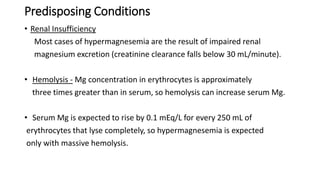
• Plasma [Mg 2+ ] is closely regulated between 1.7 and 2.1 mEq/L (0.7–1
mmol/L or 1.7–2.4 mg/dL) through interaction of
• Gastrointestinal tract (absorption)
• Bone (storage), and
• Kidneys (excretion)
• Approximately 50% to 60% of plasma magnesium is unbound and
diffusible.
11. HYPERMAGNESEMIA
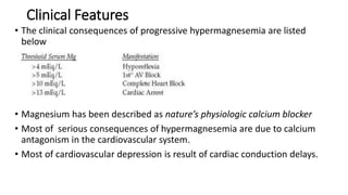
• Hypermagnesemia (serum Mg >2 mEq/L)
• Increases in plasma [Mg 2+ ] are nearly always due to excessive intake
(magnesium-containing antacids or laxatives: magnesium hydroxide,
Milk of Magnesia), kidney impairment (GFR<30ml/min) or both.
• Magnesium sulfate therapy for preeclampsia and eclampsia can result in
maternal and fetal hypermagnesemia.
12. Predisposing Conditions
• Renal Insufficiency
Most cases of hypermagnesemia are the result of impaired renal
magnesium excretion (creatinine clearance falls below 30 mL/minute).
• Hemolysis - Mg concentration in erythrocytes is approximately
three times greater than in serum, so hemolysis can increase serum Mg.
• Serum Mg is expected to rise by 0.1 mEq/L for every 250 mL of
erythrocytes that lyse completely, so hypermagnesemia is expected
only with massive hemolysis.
13. Clinical Features
• The clinical consequences of progressive hypermagnesemia are listed
below
• Magnesium has been described as nature’s physiologic calcium blocker
• Most of serious consequences of hypermagnesemia are due to calcium
antagonism in the cardiovascular system.
• Most of cardiovascular depression is result of cardiac conduction delays.
14. ECG signs may include
• Prolongation of the P–R interval and
• Widening of the QRS complex.
• Severe hypermagnesemia can lead to respiratory and cardiac arrest.
15. Treatment of Hypermagnesemia
• Mild hypermagnesemia-
• Discontinue source of magnesium intake (most often antacids or
laxatives).
• High hypermagnesemia
• Especially in presence of clinical signs of magnesium toxicity, IV
calcium can temporarily antagonize most of the effects of clinical
toxicity.
• Forced diuresis with a loop diuretic and IV fluid replacement
enhances urinary magnesium excretion in patients with adequate
kidney function.
16. • Dialysis will be necessary for patients with significant kidney
impairment or kidney failure.
• Ventilatory or circulatory support, or both, may be necessary.
17. Anaesthetic Considerations
Hypermagnesemia requires close monitoring of
• ECG,
• Blood pressure, and
• Neuromuscular function.
• Potentiation of vasodilatory and negative inotropic properties of
anesthetics should be expected.
• Dosages of nondepolarizing NMBs should be reduced.
18. HYPOMAGNESEMIA
Hypomagnesemia is a common problem,
• Particularly in critically ill patients.
• Patients undergoing major cardiothoracic or abdominal
operations
• Often associated with deficiencies of other intracellular
components such as potassium and phosphorus.
• Its incidence among patients in ICU may exceed 50%.
19. Deficiencies of magnesium are generally the result of
• Inadequate intake,
• Reduced gastrointestinal absorption,
• Increased renal excretion,
20. Causes of hypomagnesemia
Inadequate intake
Nutritional
Reduced gastrointestinal absorption
Malabsorption syndromes
Small bowel or biliary fistulas
Prolonged nasogastric suctioning
Severe vomiting or diarrhoea
Chronic laxative abuse
Chronic proton pump inhibitor (PPI) use
23. Drugs that cause renal wasting of magnesium include
• Ethanol,
• Theophylline,
• Diuretics,
• Cisplatin,
• Aminoglycosides,
• Cyclosporine,
• Amphotericin B
• Pentamidine
• Granulocyte colony stimulating factor
24. Clinical Manifestations of Hypomagnesemia
Most patients with hypomagnesemia are asymptomatic but
• Weakness,
• Fasciculation,
• Paresthesia,
• Confusion,
• Ataxia, and
• Seizures may be encountered.
25. Hypomagnesemia is frequently associated with
• Hypocalcemia (impaired PTH secretion) and
• Hypokalemia (due to renal K+ wasting).
Cardiac manifestations include
• Arrhythmias and
• Potentiation of digoxin toxicity
• Both are worsened by hypokalemia.
• Hypomagnesemia is associated with an increased incidence of atrial
fibrillation.
• Prolongation of the P–R and QT intervals may also be present.
28. Treatment of Hypomagnesemia
• Asymptomatic hypomagnesemia can
be treated orally or intramuscularly.
• Serious manifestations such as
• Seizures should be treated with
intravenous magnesium sulfate, 1 to 2 g
(8–16 mEq or 4–8 mmol) given over 10
to 60 minutes.
29. Magnesium replacement protocols
Mild, Asymptomatic Hypomagnesemia
• The following guidelines can be used for a serum Mg of 1–1.4 mEq/L with
no apparent complications.
1. Assume a total magnesium deficit of 1–2 mEq/kg.
2. Because 50% of the infused magnesium can be lost in the urine, assume
that the total magnesium requirement is twice the magnesium deficit.
3. Replace 1 mEq/kg for the first 24 hours, and 0.5 mEq/kg daily for the next
3–5 days.
30. Moderate Hypomagnesemia
The following protocol is recommended for a serum Mg <1 mEq/L, or for a
low serum Mg that is accompanied by other electrolyte abnormalities
1. Add 6 g MgSO4 (48 mEq of Mg) to 250 or 500 mL isotonic saline and infuse
over 3 hours.
2. Follow with 5 g MgSO4 (40 mEq of Mg) in 250 or 500 mL isotonic saline
infused over the next 6 hours.
3. Continue with 5 g MgSO4 every 12 hours (by continuous infusion) for the
next 5 days
31. Life-Threatening Hypomagnesemia
• The following is recommended for hypomagnesemia associated with
serious cardiac arrhythmias (e.g., torsade de pointes) or generalized
seizures:
1. Infuse 2 g MgSO4 (16 mEq of Mg) intravenously over 2–5 minutes.
2. Follow with 5 g MgSO4 (40 mEq of Mg) in 250 or 500 mL isotonic
saline infused over the next 6 hours.
3. Continue with 5 g MgSO4 every 12 hours (by continuous infusion) for
the next 5 days.
32. Monitoring Replacement Therapy
• Serum Mg levels will rise after initial magnesium bolus, but will begin to fall
after 15 minutes.
• Therefore, it is important to follow bolus dose with a continuous
magnesium infusion.
• Serum Mg levels may normalize after 1 to 2 days, but it will take several
days to replenish the total body magnesium stores.
• The magnesium retention test can be valuable for identifying end-point of
potassium replacement therapy; i.e., magnesium replacement is continued
until urinary magnesium excretion is ≥80% of the infused magnesium load.
33. Anaesthetic Considerations
• Although no specific anaesthetic interactions are described,
coexistent electrolyte disturbances such as
• Hypokalemia,
• Hypophosphatemia, and
• Hypocalcemia
are often present and should be corrected before surgery.
34. • Isolated hypomagnesemia should be corrected before elective
procedures because of its potential to cause arrhythmias.
• Magnesium appears to have intrinsic antiarrhythmic properties
and possibly cerebral protective effects.
• It is frequently administered preemptively inpatients undergoing
cardiac surgery.
Serum is favored over plasma for magnesium assays
because the anticoagulant used for plasma samples can be contaminated with citrate or other anions that bind magnesium
Meq/l=mg/dl multiplied by 10 and valency divided by mol wt
Magnesium has an atomic weight of 24 and a valence of 2, so a serum Mg concentration of 1.7 mg/dL is equivalent to (1.7×10)/24×2 = 1.4 mEq/L.
serum Mg is abnormally low, it is not possible to determine if the problem is a decrease in the ionized (active) fraction, or a decrease in the bound fractions (e.g., hypoproteinemia). The level of ionized Mg can be measured with an ion-specific electrode, but this is not routinely available. However, because only a small amount of magnesium resides in plasma, the difference between the ionized and bound magnesium content may not be large enough to be clinically relevant.
serum Mg remains in the normal range one week after starting a Mg-3 diet, while the urinary Mg excretion has dropped to negligible levels. This illustrates the relative value of urinary magnesium excretion in the detection of magnesium deficiency
Pth?
Unbound- active form and readily diffusible across cell membranes
However, hypermagnesemia is not a prominent feature of renal insufficiency unless magnesium intake is increased
Depressed contractility and vasodilation are not prominent.
PR??
QRS???
Why NMB dose is reduced???
IONOTROPIC- CONTRACTILITY
Hypomagnesemia has also been associated with long-term proton pump inhibitor (PPI) therapy, with such cases ascribed to the impaired intestinal absorption of magnesium.
Hypomagnesemia is common after cardiopulmonary bypass because of hemodilution and the frequent use of albumin, transfusion, and other magnesium-lowering constituents in the priming solution.
Acidosis causes hypocalcemia and hypomagnesimia
Hypercalcemia due to hyperparathyrodism inhibits reabsorption of Mg from kidneys leads to hypomagnesium
Aldosterone- Na reabsorption and K & Mg excretion
Alcoholism causes reduced intestinal absorption and increased urinary losses.
Electrolyte abnormalities are seen in burn patients.
Normal PR interval(beginning of P wave to QRS complex)-0.12 – 0.2 sec(Prolonged PR -1ST Degree AV block
Normal QT INTERVAL 0.35 to 0.43 sec
Hypomagnesmia (hypokalemia, hypercalcemia) causes increased sensitivity to digoxin making toxicity more likely