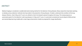
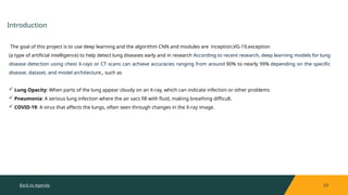
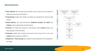
Lung Disease Detection Using Deep
Learning: Pneumonia and COVID-
19
ABHISHEK C B
KIRAN R
KIRAN T L
MAHAMAD BUDEN SAB K N
PRESENTED BY
GUIDE NAME
SANDEEP SIR.
AGENDA
2 Abstract
3 Introduction
4 Problem statement and motivation
5 Software requirments
6 Architecture
7 Pnemonia detection and covid detection
8 Deep learning and Models
9 Datasets used
10 Methodology
11 Conclusion
Back to Agenda 03
ABSTRACT
Medical imaging is considered a suitable alternative testing method for the detection of lung diseases. Many researchers have been working
to develop various detection methods that have aided in the prevention of lung diseases. To better understand the condition of the lung
disease infection, chest X-Ray and CT scans are utilized to check the disease’s spread throughout the lungs. This study proposes an
automated system for the detection multi lung diseases in X-Ray and CT scans. A customized convolutional neural network (CNN) and two
pre-trained deep learning models with a new image enhancement model are proposed for image classification.
Back to Agenda 03
Introduction
The goal of this project is to use deep learning and the algorithm CNN and modules are inception,VG-19,exception
(a type of artificial intelligence) to help detect lung diseases early and in research According to recent research, deep learning models for lung
disease detection using chest X-rays or CT scans can achieve accuracies ranging from around 90% to nearly 99% depending on the specific
disease, dataset, and model architecture,, such as
 Lung Opacity: When parts of the lung appear cloudy on an X-ray, which can indicate infection or other problems
 Pneumonia: A serious lung infection where the air sacs fill with fluid, making breathing difficult.
 COVID-19: A virus that affects the lungs, often seen through changes in the X-ray image.
Problem statement Lung disease detection using deep learning based on the x-ray images
APPLICATION
 Early Detection: Identifies diseases like pneumonia, tuberculosis, and lung cancer early for
timely treatment.
 Improved Accuracy: Differentiates between similar lung diseases with high precision.
 Automated Screening: Quickly processes large numbers of images for mass screenings.
 Affordable in Low-Resource Areas: Provides diagnostic support where access to
radiologists is limited.
Back to Agenda 04
PROBLEM STATEMENT & MOTIVATION
Back to Agenda
SOFTWARE REQUIRMENTS:
05
OPERATING SYSTEM:
I. Windwos
PROGRAMING LAUNGAUGE:
II. Python .3
PYTHON LIBRARIES FOR DEEP LEARNING AND IMAGE PROCESSING
i. TensorFlow or PyTorch: For building and training deep learning models
ii. Keras: A high-level API for TensorFlow, simplifying model development.
iii. OpenCV: For image preprocessing and augmentation
iv. PIL (Pillow): For basic image manipulation.
v. NumPy: For numerical operations on arrays (image data is often handled in arrays)
vi. Matplotlib and Seaborn: For data visualization, including plotting loss, accuracy, and confusion matrices
Dataset Management.
vii.Pandas: For data handling and manipulation (useful for metadata, CSVs, etc.).
viii.h5py: If your datasets are stored in HDF5 format.
Back to Agenda 05
Figure 3. Taxonomy of lung disease detection using deep learning.
ARCHITECTURE:
Back to Agenda
ARCHITECTURE:
06
.
FIG:. Image processing-based classification model
Back to Agenda
PNEMONIA DETECTION.
07
The proposed model is designed exclusively for the classification and prediction
of pneumonia by utilizing CXR radiographs. The technique works on the basis of
neural network (NN) architecture, which uses several neurons to concatenate,
identify and extract significant features from a set of images.
Pneumonia is an inflammatory condition of the lung affecting primarily the small
air sacs known as alveoli.Symptoms typically include some combination of
productive or dry cough, chest pain, fever and difficulty breathing. The severity of
the condition is variable. Pneumonia is usually caused by infection with viruses or
bacteria and less commonly by other microorganisms, certain medications or
conditions such as autoimmune diseases.Risk factors include cystic fibrosis,
chronic obstructive pulmonary disease (COPD), asthma, diabetes, heart failure, a
history of smoking
Back to Agenda
COVID-19 DETECTION.
07
 COVID-19. COVID-19 infected people have symptoms that are related to
pneumonia, and the virus affects the body's respiratory organs, making
breathing difficult.
 Infections in the lungs can range from a simple cold to a life-threatening
condition. Symptoms of the respiratory system often accompany infections
caused by coronaviruses.
 Individuals may have minor, self-limiting illnesses with adverse effects like
influenza on rare occasions. Fever, cough, and difficulty breathing are among
the symptoms of respiratory issues, weariness, and a sore throat . The use of
X-rays and computed tomography scans is one of the fundamental approaches
to diagnosing COVID-19.
Back to Agenda
DEEP LEARNING AND MODELS
08
inception v3:
 Inception-v3 is made up of three Inception modules and has 48 layers. Inception-v3 is often trained on the ImageNet
database, which contains over a million images. The pretrained network can classify images into 1,000 object
categories
VGG19:
 VGG-16 has 16 convolutional and 3 fully connected layers. VGG-16 had 138 million parameters. A deeper version, VGG-
19, was also constructed along with VGG-16
Xception:
 Xception has 71 hidden layers and 23 million parameters.
ResNet-50:
 At 50 layers deep and featuring 25.5 million parameters. , ResNet-50 was pretrained on more than a million images
from the ImageNet dataset
Back to Agenda
DATA SETS USED
09
•Lung X-Ray Image Dataset
•A publicly available dataset that includes 3,475 X-ray images, divided into normal, lung opacity, and viral pneumonia
•Kaggle
•A repository that contains medical image datasets, such as the pneumonia dataset and the tuberculosis dataset
•SIRM and Radiopaedia
•A publicly available dataset that includes CT scans and X-rays of COVID-19, pneumonia, and normal cases
Back to Agenda
METHODOLOGY:
10
 Data Collection: We used open-source datasets of chest X-ray and CT scan images for
diseases like pneumonia and COVID-19.
 Preprocessing: Images were resized, normalized, and augmented to enhance model
training.
 Model Selection: Four CNN architectures—ResNet-50, Inception V3, VGG19, and
Xception—were employed with transfer learning.
 Training: The models were trained on a split dataset, utilizing Adam optimizer and
early stopping to avoid overfitting.
 Evaluation: Models were assessed using metrics like accuracy and F1 score, with
Xception performing best for COVID-19.
 Deployment: A Flask web app was created for real-time predictions from uploaded X-
ray images.
Back to Agenda
CONCLUSION:
11
Diseases affect all living beings, and without proper care, even minor illnesses can become life-threatening. Recent deadly diseases like cancer,
tuberculosis, and especially COVID-19 have posed significant challenges, particularly in densely populated and resource-limited areas due to the
high cost and limited availability of testing kits. To address this, computer vision can be used to detect COVID-19 from X-ray and CT scan images.
In this study, we used CNN models (ResNet-50, Inception_v3, VGG19, and Xception) and found that Xception achieved the highest accuracy for
detecting COVID-19. Additionally, VGG19 and Xception performed best in identifying viral and bacterial pneumonia from X-ray images. We also
introduced a Flask app to automate and deploy the detection system for remote use

lung disease detection using deep learning

  • 1.
    Lung Disease DetectionUsing Deep Learning: Pneumonia and COVID- 19 ABHISHEK C B KIRAN R KIRAN T L MAHAMAD BUDEN SAB K N PRESENTED BY GUIDE NAME SANDEEP SIR.
  • 2.
    AGENDA 2 Abstract 3 Introduction 4Problem statement and motivation 5 Software requirments 6 Architecture 7 Pnemonia detection and covid detection 8 Deep learning and Models 9 Datasets used 10 Methodology 11 Conclusion
  • 3.
    Back to Agenda03 ABSTRACT Medical imaging is considered a suitable alternative testing method for the detection of lung diseases. Many researchers have been working to develop various detection methods that have aided in the prevention of lung diseases. To better understand the condition of the lung disease infection, chest X-Ray and CT scans are utilized to check the disease’s spread throughout the lungs. This study proposes an automated system for the detection multi lung diseases in X-Ray and CT scans. A customized convolutional neural network (CNN) and two pre-trained deep learning models with a new image enhancement model are proposed for image classification.
  • 4.
    Back to Agenda03 Introduction The goal of this project is to use deep learning and the algorithm CNN and modules are inception,VG-19,exception (a type of artificial intelligence) to help detect lung diseases early and in research According to recent research, deep learning models for lung disease detection using chest X-rays or CT scans can achieve accuracies ranging from around 90% to nearly 99% depending on the specific disease, dataset, and model architecture,, such as  Lung Opacity: When parts of the lung appear cloudy on an X-ray, which can indicate infection or other problems  Pneumonia: A serious lung infection where the air sacs fill with fluid, making breathing difficult.  COVID-19: A virus that affects the lungs, often seen through changes in the X-ray image.
  • 5.
    Problem statement Lungdisease detection using deep learning based on the x-ray images APPLICATION  Early Detection: Identifies diseases like pneumonia, tuberculosis, and lung cancer early for timely treatment.  Improved Accuracy: Differentiates between similar lung diseases with high precision.  Automated Screening: Quickly processes large numbers of images for mass screenings.  Affordable in Low-Resource Areas: Provides diagnostic support where access to radiologists is limited. Back to Agenda 04 PROBLEM STATEMENT & MOTIVATION
  • 6.
    Back to Agenda SOFTWAREREQUIRMENTS: 05 OPERATING SYSTEM: I. Windwos PROGRAMING LAUNGAUGE: II. Python .3 PYTHON LIBRARIES FOR DEEP LEARNING AND IMAGE PROCESSING i. TensorFlow or PyTorch: For building and training deep learning models ii. Keras: A high-level API for TensorFlow, simplifying model development. iii. OpenCV: For image preprocessing and augmentation iv. PIL (Pillow): For basic image manipulation. v. NumPy: For numerical operations on arrays (image data is often handled in arrays) vi. Matplotlib and Seaborn: For data visualization, including plotting loss, accuracy, and confusion matrices Dataset Management. vii.Pandas: For data handling and manipulation (useful for metadata, CSVs, etc.). viii.h5py: If your datasets are stored in HDF5 format.
  • 7.
    Back to Agenda05 Figure 3. Taxonomy of lung disease detection using deep learning. ARCHITECTURE:
  • 8.
    Back to Agenda ARCHITECTURE: 06 . FIG:.Image processing-based classification model
  • 9.
    Back to Agenda PNEMONIADETECTION. 07 The proposed model is designed exclusively for the classification and prediction of pneumonia by utilizing CXR radiographs. The technique works on the basis of neural network (NN) architecture, which uses several neurons to concatenate, identify and extract significant features from a set of images. Pneumonia is an inflammatory condition of the lung affecting primarily the small air sacs known as alveoli.Symptoms typically include some combination of productive or dry cough, chest pain, fever and difficulty breathing. The severity of the condition is variable. Pneumonia is usually caused by infection with viruses or bacteria and less commonly by other microorganisms, certain medications or conditions such as autoimmune diseases.Risk factors include cystic fibrosis, chronic obstructive pulmonary disease (COPD), asthma, diabetes, heart failure, a history of smoking
  • 10.
    Back to Agenda COVID-19DETECTION. 07  COVID-19. COVID-19 infected people have symptoms that are related to pneumonia, and the virus affects the body's respiratory organs, making breathing difficult.  Infections in the lungs can range from a simple cold to a life-threatening condition. Symptoms of the respiratory system often accompany infections caused by coronaviruses.  Individuals may have minor, self-limiting illnesses with adverse effects like influenza on rare occasions. Fever, cough, and difficulty breathing are among the symptoms of respiratory issues, weariness, and a sore throat . The use of X-rays and computed tomography scans is one of the fundamental approaches to diagnosing COVID-19.
  • 11.
    Back to Agenda DEEPLEARNING AND MODELS 08 inception v3:  Inception-v3 is made up of three Inception modules and has 48 layers. Inception-v3 is often trained on the ImageNet database, which contains over a million images. The pretrained network can classify images into 1,000 object categories VGG19:  VGG-16 has 16 convolutional and 3 fully connected layers. VGG-16 had 138 million parameters. A deeper version, VGG- 19, was also constructed along with VGG-16 Xception:  Xception has 71 hidden layers and 23 million parameters. ResNet-50:  At 50 layers deep and featuring 25.5 million parameters. , ResNet-50 was pretrained on more than a million images from the ImageNet dataset
  • 12.
    Back to Agenda DATASETS USED 09 •Lung X-Ray Image Dataset •A publicly available dataset that includes 3,475 X-ray images, divided into normal, lung opacity, and viral pneumonia •Kaggle •A repository that contains medical image datasets, such as the pneumonia dataset and the tuberculosis dataset •SIRM and Radiopaedia •A publicly available dataset that includes CT scans and X-rays of COVID-19, pneumonia, and normal cases
  • 13.
    Back to Agenda METHODOLOGY: 10 Data Collection: We used open-source datasets of chest X-ray and CT scan images for diseases like pneumonia and COVID-19.  Preprocessing: Images were resized, normalized, and augmented to enhance model training.  Model Selection: Four CNN architectures—ResNet-50, Inception V3, VGG19, and Xception—were employed with transfer learning.  Training: The models were trained on a split dataset, utilizing Adam optimizer and early stopping to avoid overfitting.  Evaluation: Models were assessed using metrics like accuracy and F1 score, with Xception performing best for COVID-19.  Deployment: A Flask web app was created for real-time predictions from uploaded X- ray images.
  • 14.
    Back to Agenda CONCLUSION: 11 Diseasesaffect all living beings, and without proper care, even minor illnesses can become life-threatening. Recent deadly diseases like cancer, tuberculosis, and especially COVID-19 have posed significant challenges, particularly in densely populated and resource-limited areas due to the high cost and limited availability of testing kits. To address this, computer vision can be used to detect COVID-19 from X-ray and CT scan images. In this study, we used CNN models (ResNet-50, Inception_v3, VGG19, and Xception) and found that Xception achieved the highest accuracy for detecting COVID-19. Additionally, VGG19 and Xception performed best in identifying viral and bacterial pneumonia from X-ray images. We also introduced a Flask app to automate and deploy the detection system for remote use