Download to read offline


![Chapter 13 | EUROPEAN MR/CT REGISTRY 133132 Chapter 13 | EUROPEAN MR/CT REGISTRY
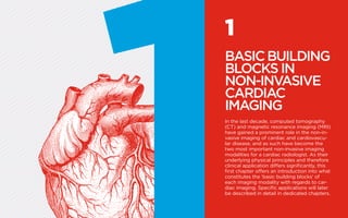
THE HEART REVEALED RADIOLOGY IN THE DIAGNOSIS AND MANAGEMENT OF CARDIAC CONDITIONSTHE HEART REVEALED RADIOLOGY IN THE DIAGNOSIS AND MANAGEMENT OF CARDIAC CONDITIONS
than 80% of all indications. Around 20%
of indications are equally distributed
between congenital heart disease and
valve disease. Therefore, the strength of
CMR is the differential diagnosis and the
strength of Cardiac CT the fast and relia-
ble rule out of CAD, especially in patients
with a low intermediate pre-test probabil-
ity (PTP)1
.
An estimate of the PTP can be achieved
only by taking the age, gender and symp-
toms into account (Table 1). According to
the current guidelines for the manage-
ment of patients with suspected stable
CAD, only patients with an intermediate
pre-test probability between 15–85%
should be referred to a non-invasive imag-
ing test, patients with a high PTP > 85%
of CAD should be referred directly to ICA
and in patients with a low PTP < 15% other
causes of chest pain should be evaluated
first1, 2
.
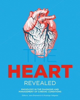
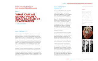
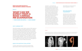
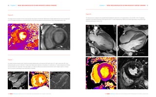
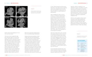
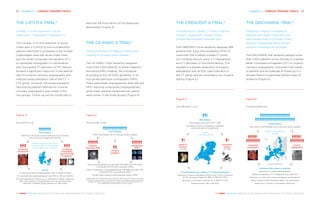
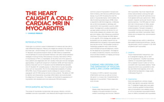
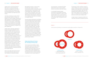
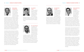
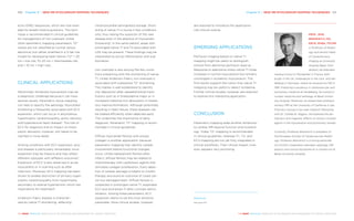
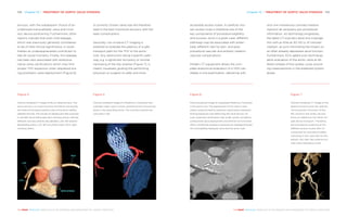
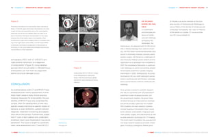
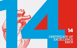
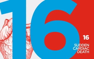
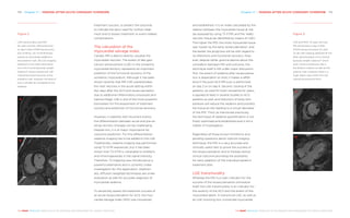
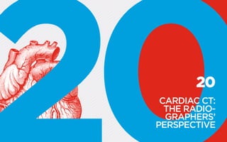
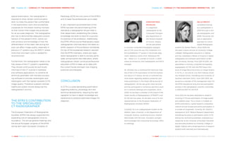
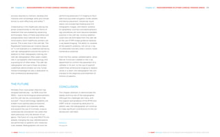
MAJOR COMPLICATIONS
IN CMR/CCT
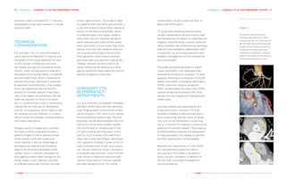
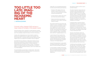
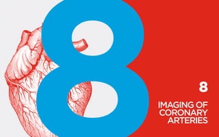
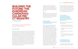
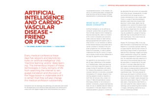
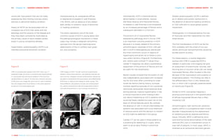
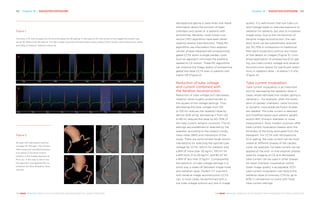
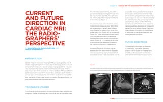
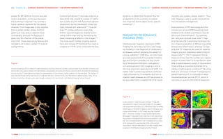
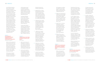
CCT and CMR are very safe methods.
According to the documented cases in the
ESCR Cardiac MR/CT Registry, the overall
adverse event rate in CCT was as low as
0.44% and in CMR slightly higher with
0.5%, because it includes stress MRI with a
pharmacological stressor like adenosine in
91%, regadenoson in 7% or dobutamine in
2% (Figure 3) to evaluate myocardial per-
fusion in suspected or known CAD with an
intrinsically higher risk3
. However, severe
adverse events such as an allergic shock
or resuscitation occurred only in 1:10,000
patients.
CLINICAL CONSEQUENCES
One of the major goals of using differ-
ent non-invasive imaging methods is to
avoid unnecessary ICAs, which could be
achieved in approximately 1/3 of CMR
examinations and approximately 37% of
CCTs. 22% of all CCTs and 6% of CMRs
had a direct impact on interventional
procedures. A direct referral to the cath
lab was only necessary in 2.5–4.9%. All
other patients could be discharged or
only required a change of drug regime
(Table 2).
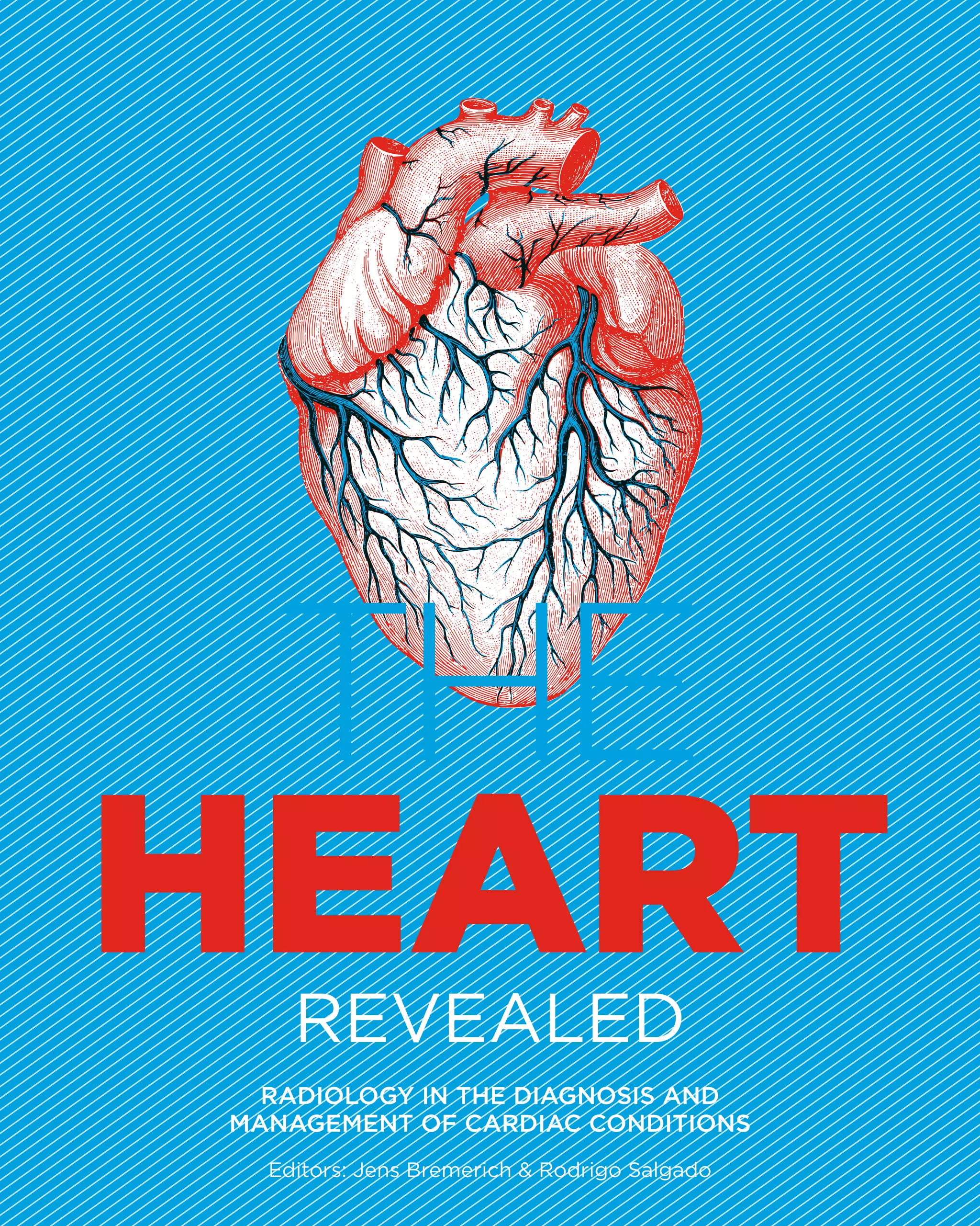
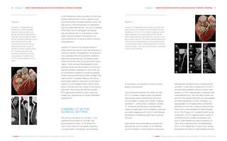
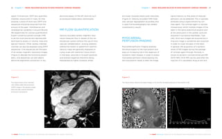
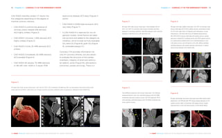
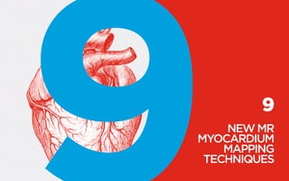
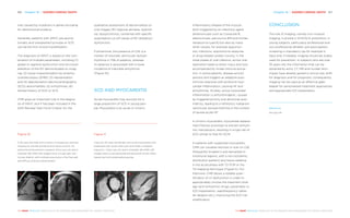
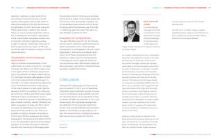
RADIATION EXPOSURE
In clinical trials, CCT can be performed
under certain circumstances (low and
stable heart rate, latest CT equipment etc.)
with a radiation exposure as low as < 1 mSv.
However, in a clinical standard scenario
this can’t be always achieved, especially if
not only the heart, but also the aorta, i.e.
pre-interventionally before transcatheter
aortic valve implantation (TAVI), has to be
assessed. Nevertheless, the results of the
ESCR Cardiac MR/CT Registry revealed
that even in a clinical scenario dose reduc-
tion protocols, like ‘step-and-shoot’ or the
so-called ‘flash-mode’ are used in more
than 50% of all patients, resulting in a
Table 2
CLINICAL CONSEQUENCES CARDIAC MR CARDIAC CT
No further invasive diagnostics 32.5% 36.6%
Impact on interventional
procedure
6% 21.7%
Direct referral to catheter
laboratory
2.5% 4.9%
Patient discharge 4.4% 3.3%
Change of drug regime 5.6% 2.4%
Multiple clinical consequences for one imaging study possible
Figure 3
Distribution of the different pharmacological substances
used in stress-MRI according to the documented cases
in the ESCR MR/CT Registry. Figure from the MR/CT Reg-
istry booklet 2018.
Table 1
Pre-test probability of obstructive CHD in patients with stable chest pain to estimate the need for further diagnostic tests on
the cardiology care level
TYPICAL ANGINA PECTORIS ATYPICAL ANGINA PECTORIS NON-ANGINAL CHEST PAIN
Age* [years] Men Women Men Women Men Women
30–39 59% 28% 29% 10% 18% 5%
40–49 69% 37% 38% 14% 25% 8%
50–59 77% 47% 49% 20% 34% 12%
60–69 84% 58% 59% 28% 44% 17%
70–79 89% 68% 69% 37% 54% 24%
>80 93% 76% 78% 47% 65% 32%
This data is based on the following definition of anginal symptoms: (A) squeezing pain located either retrosternally or in neck,
shoulder, jaw or arm, (B) aggravated by physical exertion or emotional stress, (C) improved with rest and/or nitro-glycerine
within 5 minutes. The combined presence of the following features define typical angina pectoris if three of the features are
present and atypical angina pectoris if two are present, while one or none of the features defines non-cardiac chest pain.
* The calculated probabilities for the age groups represent the estimates for patients aged 35, 45, 55, 65, 75, and 85 years,
respectively (modified from reference 1).
ESCR Cardiac MR/CT Registry
Stress-MR (n=27165)
(01/01/2018)
91%
Stress-Adenosin
7%
Stress-Regadenoson
2%
Stress-Dobutamin](https://image.slidesharecdn.com/idor2018-bookweb-181107152308/85/Libro-del-Dia-Internacional-Radiologia-2018-67-320.jpg)
![Chapter 13 | EUROPEAN MR/CT REGISTRY 135134 Chapter 13 | EUROPEAN MR/CT REGISTRY
THE HEART REVEALED RADIOLOGY IN THE DIAGNOSIS AND MANAGEMENT OF CARDIAC CONDITIONSTHE HEART REVEALED RADIOLOGY IN THE DIAGNOSIS AND MANAGEMENT OF CARDIAC CONDITIONS
tomography angiography (CCTA) as the
first line method in suspected stable coro-
nary artery disease, which is therefore also
covered by the National Health Service
(NHS). On the other hand, unlike the NHS,
CMR and CCT is not covered by the com-
pulsory health insurance in Germany and
an affected patient has to apply for health
insurance coverage individually. Therefore,
these valuable methods can be provided
in Germany and other European countries
mainly in inpatients during their hospital
treatment, and during their ambulatory
treatment only if privately insured or as a
self-paying patient.
CONCLUSION
According to the results of the ESCR Car-
diac MR/CT Registry, non-invasive imaging
techniques, especially CMR and CCT have
the potential to replace unnecessary inva-
sive catheterisation for diagnosing cardio-
vascular diseases. Invasive cardiac cathe-
terisation should only be used to guide and
perform cardiovascular interventions in the
near future. The non-invasive methods CMR
and CCT have proven their widespread use
in clinical routine by radiologists alone or
together with cardiologists and its bene-
fits for our patients. Especially cardiovas-
cular CT helped up to 2/3 of the patients
to avoid invasive cardiac catheterisation.
The ESCR cardiovascular MR/CT Registry
is one example of how carefully collected
and documented medical data provides
evidence that new radiological techniques
help to improve medical care and especially
to ensure optimal patient care and maybe
replace other less cost-effective and less
gentle methods in the near future.
Furthermore, the documentation and
being a pre-requisite for accreditation and
certification of medical personnel and
institutions help to maintain certain quality
standards in performing these new diag-
nostic tools. We hope that these new and
very beneficial methods for the patients
will be reimbursed by more and more
health services throughout Europe in the
near future.
The ESCR Cardiac MR/CT Registry may
also serve us to build the future by estab-
lishing a database for big data analysis,
deep learning and the use of artificial intel-
ligence to improve diagnosis.
References
See page 236
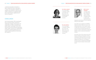
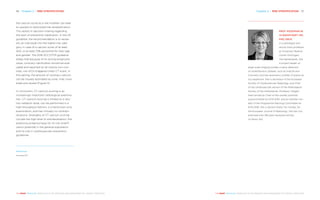
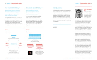
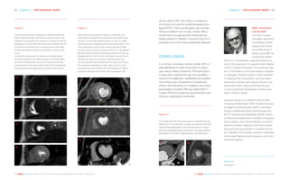
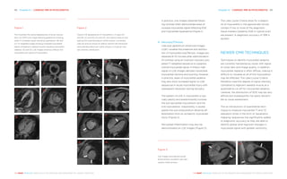
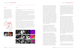
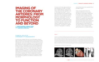
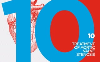
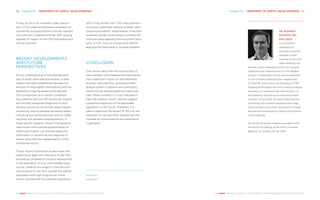
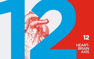
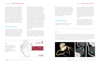
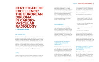
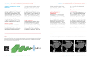
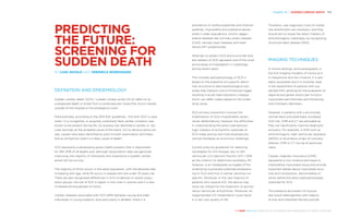
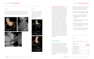
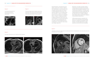
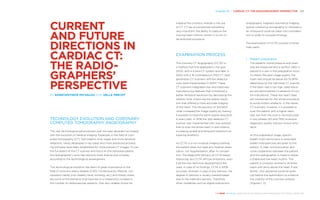
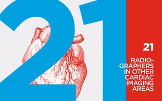
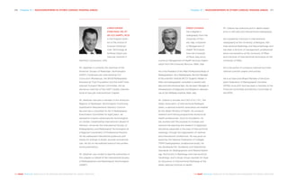
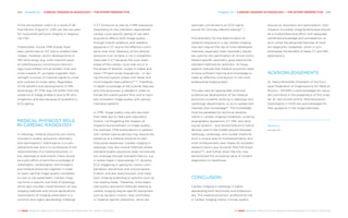
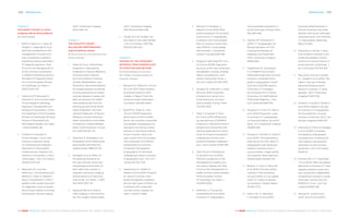
median radiation exposure of 4.21 mSv and
lower (Figure 4), which is within the range
of a median radiation exposure with ICA.
WHO IS PERFORMING
AND REPORTING IT?
Most CMRs and CCTs are performed and
reported on by radiologists (Figure 5)
according to the ESCR MR/CT Registry
data. But up to 1/3 of the examinations
are reported on together, in consensus
reading between cardiologists and radiol-
ogists, which might be a very good way to
keep high-quality standards in less experi-
enced or not yet certified imaging centres.
WILL MY INSURANCE
COMPANY PAY FOR
CARDIAC MR AND/
OR CARDIAC CT?
Despite the proven value and support by
national and international guidelines to
perform non-invasive imaging techniques
in diagnosing cardiovascular disease
rather than using invasive procedures like
ICA, the reimbursement policy of health
insurance systems and companies varies
substantially throughout Europe. The most
progressive approach was introduced in
the UK in 2016 by the National Institute
for Health and Care Excellence (NICE).
On the one hand the NICE-Guidelines
recommend using coronary computed
Figure 4
‘Box-plots’ of the mean effective dose for the most
commonly used four acquisition techniques in cardiac
computed tomography (CCT). It demonstrates that even
in a clinical scenario, dose reduction protocols, like ‘step-
and-shoot’ or the so-called ‘flash-mode’ are used in more
than 50% of all patients, resulting in a median radiation
exposure of 4.21 mSv or lower, which is within the range
of a median radiation exposure with invasive coronary
angiography (ICA).
Figure 5
Pie charts of the distribution of radiologists reporting
on cardiac CT and cardiac MRI and consensus reading
together with cardiologists according to the ESCR Car-
diac MR/CT Registry (from the 2018 Booklet).
ESCR Cardiac MR/CT Registry
Who did the CMR reporting
(01/01/2018)
CT
MR
82.2%
Radiologist
17.0%
Consensus Reading
0.6%
Cardiologist
0.2%
Seperate Reading
71.7%
Radiologist
27.7%
Consensus Reading
0.5%
Cardiologist
0.1%
Seperate Reading
16.6%
11.93 mSV
Standard
retrospective
18.8%
8.51 mSV
Retrospective
tube current
27.4%
4.21 mSV
(IQR: 1.89 – 4.72 mSv)
step and shoot
26.8%
3.21 mSV
(IQR: 1.81 – 6.51 mSv)
Flash ECG
effectivedose[mSv]
0
20
40](https://image.slidesharecdn.com/idor2018-bookweb-181107152308/85/Libro-del-Dia-Internacional-Radiologia-2018-68-320.jpg)


![References 235234 References
THE HEART REVEALED RADIOLOGY IN THE DIAGNOSIS AND MANAGEMENT OF CARDIAC CONDITIONSTHE HEART REVEALED RADIOLOGY IN THE DIAGNOSIS AND MANAGEMENT OF CARDIAC CONDITIONS
With Aortic Stenosis. Circ
Cardiovasc Imaging. 2018
Mar;11(3):e007146. doi: 10.1161/
CIRCIMAGING.117.007146.
Salgado RA et al. Preproce-
dural CT evaluation of tran-
scatheter aortic valve replace-
ment: what the radiologist
needs to know. Radiographics.
2014 Oct;34(6):1491-514. doi:
10.1148/rg.346125076.
Chapter 11:
A NEW VALVE: NON-INVASIVE
IMAGING OF PROSTHETIC
HEART VALVES
By Ricardo P.J. Budde
1 Sucha D, Symersky P, Tanis
W, Mali WPThM, Leiner T,
Herwerden van LA, Budde
RPJ. Multimodality imaging
assessment of prosthetic
heart valves. Circulation
Cardiovasc Imaging. 2015
Sep;8(9):e003703. doi: 10.1161/
CIRCIMAGING.115.003703.
2 Faure ME, Swart LE, Dijkshoorn
ML, Bekkers JA, Straten van M,
Nieman K, Parizel PP, Krestin
GP, Budde RPJ. Advanced CT
acquisition protocol with a
third generation dual-source
CT scanner and iterative
reconstruction technique for
comprehensive prosthetic
heart valve assessment.
Eur Radiol. 2017 Dec 12. doi:
10.1007/s00330-017-5163-7.
[Epub ahead of print].
3 Sucha D, Chamuleau SAJ,
Symersky P, Meijs MFL,
Brink van den RBA, Mol de
BAJM, Mali WPThM, Habets
J, Herwerden van LA, Budde
RPJ. Baseline MDCT findings
after prosthetic heart valve
implantation provide impor-
tant complementary informa-
tion to echocardiography for
follow-up purposes. Eur Radiol.
2016;26:997-1006.
4 Swart LE, Scholtens AM, Tanis
W, Nieman K, Bogers AJJC,
Verzijlbergen FJ, Krestin GP,
Roos-Hesselink JW, Budde
RPJ. 18F-FDG PET/CT and
CT angiography in prosthetic
heart valve endocarditis: from
guidelines to clinical practice.
Eur Heart J. 2018 Jan 16. doi:
10.1093/eurheartj/ehx784.
[Epub ahead of print].
Chapter 12:
AN OFTEN-OVERLOOKED
CONNECTION: THE HEART-
BRAIN AXIS
By Birgitta Velthuis
1 Qiu C, Fratiglioni L. A major
role for cardiovascular burden
in age-related cognitive
decline. Nat Rev Cardiol.
2015;12(5):267-77.
2 Fernández-Friera L, Peñalvo
JL, Fernández-Ortiz A, et al.
Prevalence, vascular distribu-
tion, and multiterritorial extent
of subclinical atherosclerosis
in a middle-aged cohort: The
PESA (Progression of Early
Subclinical Atherosclerosis)
Study. Circulation. 2015 Jun
16;131(24):2104-13.
3 Tarkin JM; Dweck MR;
Evans NR, et al. Imaging
atherosclerosis. Circ Res.
2016;118:750-769.
4 Piepoli MF, Hoes AW, Agewall
S, et al. 2016 European
guidelines on cardiovascular
disease prevention in clinical
practice. EHJ 2016;37:2315-81.
5 Maroules CD, Khera A, Ayers C,
et al. Cardiovascular outcome
associations among cardio-
vascular magnetic resonance
measures of arterial stiffness:
the Dallas heart study. Journal
of Cardiovascular Magnetic
Resonance 2014;16:33.
6 Silverman MG, Blaha MJ,
Krumholz HM, et al. Impact of
coronary artery calcium on
coronary heart disease events
in individuals at the extremes
of traditional risk factor bur-
den: the Multi-Ethnic Study of
Atherosclerosis. Eur Heart J.
2014;35(33):2232-41.
7 Zoet GA, Benschop L,
Boersma E, et al. CREW
consortium. Prevalence of
subclinical coronary artery
disease assessed by coro-
nary Computed Tomography
angiography in 45- to 55-year-
old women with a history of
preeclampsia. Circulation.
2018;137(8):877-79.
8 Aengevaeren VL, Mosterd A,
Braber TL, et al. Relationship
between lifelong exercise vol-
ume and coronary atheroscle-
rosis in athletes. Circulation.
2017;136(2):138-48.
9 Hur J, Choi BW. Cardiac CT
imaging for ischemic stroke:
current and evolving clinical
applications. Radiology. 2017
Apr;283(1):14-28.
10 Cremers CHP, van der Bilt
IAC, van der Schaaf IC, et al.
Relationship between cardiac
Quantification of myocardial
perfusion using dynamic
64-detector computed
tomography. Invest Radiol
42:815-822.
22 Dweck MR, Williams MC, Moss
AJ, Newby DE, Fayad ZA
(2016) Computed Tomogra-
phy and Cardiac Magnetic
Resonance in Ischemic Heart
Disease. J Am Coll Cardiol
68:2201-2216.
23 Pijls NH, De Bruyne B, Peels
K et al (1996) Measurement
of fractional flow reserve to
assess the functional severity
of coronary-artery stenoses. N
Engl J Med 334:1703-1708.
24 Nakanishi R, Budoff MJ (2016)
Noninvasive FFR derived from
coronary CT angiography in
the management of coronary
artery disease: technology
and clinical update. Vascular
Health and Risk Management
12:269-278.
Chapter 9:
THE COLOURFUL HEART:
NEW MAPPING TECHNIQUES
HELP IN MYOCARDIAL TISSUE
CHARACTERISATION
By Jens Bremerich
1 Kawel-Boehm N, Maceira A,
Valsangiacomo-Buechel ER,
Vogel-Claussen J, Turkbey EB,
Williams R, Plein S, Tee M, Eng
J, Bluemke DA. Normal values
for cardiovascular magnetic
resonance in adults and
children. Journal of Cardio-
vascular Magnetic Resonance
2015;17:29.
2 Treibel TA, Kozor R, Menacho
K, Castelletti S, Bulluck H, Ros-
mini S, Nordin S, Maestrini V,
Fontana M, Moon JC. Left Ven-
tricular Hypertrophy Revisited.
Cell and Matrix Expansion have
disease specific relationships.
Circulation 2017;136:2519–2521.
3 Martinez-Naharro A, Treibel
TA, Abdel-Gadir A, Bulluck H,
Zumbo G, Knight DS, Kotecha
T, Francis R, Hutt DF, Rezk T,
Rosmini S, Quarta CC, Whelan
CJ, Kellman P, Gillmore JD,
Moon JC, Hawkins PN, Fontana
M. Magnetic Resonance in
Transthyretin Cardiac Amy-
loidosis. J Am Coll Cardiol
2017;70:466–77.
4 Swoboda PP, McDiarmid AK,
Erhayiem B, Broadbent DA,
Dobson LE, Garg P, Ferguson
C, Page SP, Greenwood JP,
Plein S. Assessing Myocar-
dial Extracellular Volume by
T1-Mapping to Distinguish
Hypertrophic Cardiomyo-
pathy From Athlete’s Heart.
J Am Coll Cardiol 2016
10;67(18):2189-2190.
5 Sado DM, White SK, Piechnik
SK, Banypersad SM, Trei-
bel T, Captur G, Fontana M,
Maestrini V, Flett AS, Robson
MD, Lachmann RH, Murphy E,
Mehta A, Hughes D, Neu-
bauer S, Elliott PM, Moon JC.
Identification and assessment
of Anderson-Fabry Disease by
Cardiovascular Magnetic Reso-
nance Noncontrast Myocardial
T1-mapping. Circ Cardiovasc
Imaging 2013;6:392-398.
Chapter 10:
NEW SOLUTIONS TO OLD
PROBLEMS: AORTIC VALVE
STENOSIS
By Rodrigo Salgado
Benjamin EJ, Virani SS, Calla-
way CW et al (2018) Heart Dis-
ease and Stroke Statistics-2018
Update: A Report from the
American Heart Association.
Circulation 137:e67-e492.
Baumgartner HC, Hung JC-C,
Bermejo J et al (2017) Recom-
mendations on the echocar-
diographic assessment of
aortic valve stenosis: a focused
update from the European
Association of Cardiovascular
Imaging and the American
Society of Echocardiography.
Eur Heart J Cardiovasc Imag-
ing 18:254-275.
Kodali SK et al. Two-year out-
comes after transcatheter or
surgical aortic-valve replace-
ment. N Engl J Med. 2012
May 3;366(18):1686-95. doi:
10.1056/NEJMoa1200384.
Lean MB et al. Transcatheter
aortic-valve implantation for
aortic stenosis in patients
who cannot undergo surgery.
N Engl J Med. 2010 Oct
21;363(17):1597-607. doi:
10.1056/NEJMoa1008232.
Makkar RR et al. Transcathe-
ter aortic-valve replacement
for inoperable severe aortic
stenosis. N Engl J Med. 2012
May 3;366(18):1696-704. doi:
10.1056/NEJMoa1202277.
Pawade T et al. Computed
Tomography Aortic Valve
Calcium Scoring in Patients](https://image.slidesharecdn.com/idor2018-bookweb-181107152308/85/Libro-del-Dia-Internacional-Radiologia-2018-118-320.jpg)
![References 237236 References
THE HEART REVEALED RADIOLOGY IN THE DIAGNOSIS AND MANAGEMENT OF CARDIAC CONDITIONSTHE HEART REVEALED RADIOLOGY IN THE DIAGNOSIS AND MANAGEMENT OF CARDIAC CONDITIONS
Hayashi M, Shimizu W, Albert
CM (2015). The spectrum of
epidemiology underlying sud-
den cardiac death. Circulation
research, 116(12), 1887-1906.
Suzuki T, Nazarian S, Jer-
osch-Herold M, Chugh SS
(2016). Imaging for assessment
of sudden death risk: current
role and future prospects. EP
Europace, 18(10), 1491-1500.
Yousuf O, Chrispin J, Tomaselli
GF, Berger RD (2015). Clinical
management and prevention
of sudden cardiac death.
Circulation research, 116(12),
2020-2040.
Authors/Task Force Members,
Priori SG, Blomström-Lun-
dqvist C, Mazzanti A, Blom
N, Borggrefe M, Hindricks G
(2015). 2015 ESC Guidelines for
the management of patients
with ventricular arrhythmias
and the prevention of sudden
cardiac death: The Task
Force for the Management
of Patients with Ventricular
Arrhythmias and the Preven-
tion of Sudden Cardiac Death
of the European Society of
Cardiology (ESC) Endorsed
by: Association for European
Paediatric and Congenital Car-
diology (AEPC). EP Europace,
17(11), 1601-1687.
Chapter 17:
WHEN THE ACTION IS OVER:
IMAGING AFTER ACUTE
CORONARY SYNDROME
By Christian Loewe
F. Saremi. Cardiac MR Imaging
in Acute Coronary Syndrome:
Application and Image
Interpretation. Radiology
2017:282(1):17-32.
Eitel I, Desch S, Fuernau G, et
al. Prognostic significance and
determinants of myocardial
salvage assessed by cardio-
vascular magnetic resonance
in acute reperfused myocardial
infarction. J Am Coll Cardiol
2010;55(22):2470–2479.
Eitel I, de Waha S, Wöhrle
J, et al. Comprehensive
prognosis assessment by
CMR imaging after ST-seg-
ment elevation myocardial
infarction. J Am Coll Cardiol
2014;64(12):1217–1226.
Kramer CM, Rogers WJ Jr,
Mankad S, Theobald TM,
Pakstis DL, Hu YL. Contractile
reserve and contrast uptake
pattern by magnetic resonance
imaging and functional recov-
ery after reperfused myo-
cardial infarction. J Am Coll
Cardiol 2000;36(6):1835–1840.
Larose E, Rodés-Cabau J,
Pibarot P, et al. Predicting
late myocardial recovery and
outcomes in the early hours
of ST-segment elevation
myocardial infarction tradi-
tional measures compared
with microvascular obstruc-
tion, salvaged myocardium,
and necrosis characteristics
by cardiovascular magnetic
resonance. J Am Coll Cardiol
2010;55(22):2459–2469.
Chapter 18:
INFLUENCE OF PROTOCOL
OPTIMISATION ON RADIATION
EXPOSURE IN CARDIAC IMAGING
By Valentin Sinitsyn and Maria Glazkova,
on behalf of EuroSafe Imaging
1 Hirshfeld JW Jr, Ferrari VA,
Bengel FM, Bergersen L,
Chambers CE. Et al. ACC/HRS/
NASCI/SCAI/SCCT Expert
Consensus Document on Opti-
mal Use of Ionizing Radiation in
Cardiovascular Imaging – Best
Practices for Safety and Effec-
tiveness. J Am Coll Cardiol.
2018. S0735-1097(18)33224-8.
2 Hedgire SS, Baliyan V, Gho-
shhajra BB, Kalra MK. Recent
advances in cardiac computed
tomography dose reduction
strategies: a review of scientific
evidence and technical devel-
opments. J Med Imaging (Bell-
ingham). 2017 Jul;4(3):031211.
3 Hedgire S, Ghoshhajra B,
Kalra M. Dose optimization in
cardiac CT. Phys Med. 2017
Sep;41:97-103.
4 ESR iGuide: Clinical Decision
Support using European Imag-
ing Referral Guidelines: https://
www.myESR.org/ESRiGuide
Chapter 19:
CURRENT AND FUTURE
DIRECTION IN CARDIAC MRI: THE
RADIOGRAPHERS’ PERSPECTIVE
By Aideen Fallon, Alison Fletcher and
Vasilis Syrgiamiotis
1 Haaf P, Gard P, Messroghli DR,
Broadbent DA, Greenwood JP,
Plein S. Cardiac T1 Mapping
and Extracellular Volume
(ECV) in clinical practice: a
dysfunction and cerebral per-
fusion in patients with aneurys-
mal subarachnoid hemorrhage.
Neurocrit Care 2016;24:202-6.
Chapter 13:
BUILDING THE FUTURE: THE
EUROPEAN CARDIOVASCULAR
MR/CT REGISTRY
By Matthias Gutberlet
1 Albus C, Barkhausen J, Fleck E,
Haasenritter J, Lindner O, Sil-
ber S, on behalf of the German
National Disease Manage-
ment Guideline ‘Chronic CHD’
development group: Clinical
practice guideline: The diagno-
sis of chronic coronary heart
disease. Dtsch Arztebl Int
2017; 114: 712–9. DOI: 10.3238/
arztebl.2017.0712.
2 Bundesärztekammer (BÄK),
Kassenärztliche Bundesv-
ereinigung (KBV), Arbeits-
gemeinschaft der Wissen-
schaftlichen Medizinischen
Fachgesellschaften (AWMF):
Nationale Versorgungs Leitlinie
Chronische KHK – Leitlinien-
report, 4. Auflage. Version
1. http://doi.org/ 10.6101/
AZQ/000264 (last accessed
on 26 June 2017).
3 ESCR Cardiac MR/CT Booklet
2018.
https://www.mrct-registry.org/
images/ESCR_2018_CardiacM-
RCTbooklet.pdf
Chapter 14:
CERTIFICATE OF EXCELLENCE:
THE EUROPEAN DIPLOMA IN
CARDIOVASCULAR RADIOLOGY
By Karl-Friedrich Kreitner
1 ESR European Training Curric-
ulum for Subspecialisation in
Radiology: https://www.myesr.
org/media/2840
2 European Diploma in Cardio-
vascular Imaging: http://www.
escr.org/cms/website.php?id=/
en/education/diploma.htm
Chapter 15:
ARTIFICIAL INTELLIGENCE AND
CARDIOVASCULAR DISEASE –
FRIEND OR FOE?
By Tim Leiner, Jelmer M. Wolterink and
Ivana Išgum
Langs G, Röhrich S, Hof-
manninger J, Prayer F, Pan J,
Herold C, Prosch H. Machine
learning: from radiomics to
discovery and routine. Radiol-
oge 2018 Jun 19. doi: 10.1007/
s00117-018-0407-3 [Epub
ahead of print].
Choy G, Khalilzadeh O,
Michalski M, Do S, Samir AE,
Pianykh OS, Geis JR, Pand-
haripande PV, Brink JA, Dreyer
KJ. Current Applications and
Future Impact of Machine
Learning in Radiology. Radiol-
ogy 2018 Jun 26. doi: 10.1148/
radiol.2018171820 [Epub ahead
of print].
Litjens G, Kooi T, Bejnordi
BE, et al. A survey on deep
learning in medical image
analysis. Med Image Anal
2017;42:60-88.
Dreyer KJ and Geis JR. When
machines think: Radiolo-
gy’s next frontier. Radiology
2017;285:713-718.
Gillies RJ, Kinahan PE, Hricak
H. Radiomics: Images are more
than pictures, they are data.
Radiology 2016;278:563-77.
Chapter 16:
PREDICTING THE FUTURE:
SCREENING FOR SUDDEN DEATH
By Luigi Natale and Veronica Bordonaro
AlJaroudi WA, Flamm SD,
Saliba W, Wilkoff BL, Kwon D
(2013). Role of CMR imag-
ing in risk stratification for
sudden cardiac death. JACC:
Cardiovascular Imaging, 6(3),
392-406.
Wu KC (2017). Sudden cardiac
death substrate imaged by
magnetic resonance imaging:
From Investigational tool to
clinical applications. Circula-
tion: Cardiovascular Imaging,
10(7), e005461.
Kim SS, Ko SM, Choi SI, Choi
BH, Stillman AE (2016). Sudden
cardiac death from structural
heart diseases in adults: imag-
ing findings with cardiovas-
cular computed tomography
and magnetic resonance. The
International Journal of Cardio-
vascular Imaging, 32(1), 21-43.
Shriki JE, Shinbane JS, Rashid
MA, Hindoyan A, Withey JG,
DeFrance A, Wilcox A (2012).
Identifying, characterizing, and
classifying congenital anom-
alies of the coronary arteries.
Radiographics, 32(2), 453-468.](https://image.slidesharecdn.com/idor2018-bookweb-181107152308/85/Libro-del-Dia-Internacional-Radiologia-2018-119-320.jpg)
![References 239238 References
THE HEART REVEALED RADIOLOGY IN THE DIAGNOSIS AND MANAGEMENT OF CARDIAC CONDITIONSTHE HEART REVEALED RADIOLOGY IN THE DIAGNOSIS AND MANAGEMENT OF CARDIAC CONDITIONS
comprehensive review. Journal
of Cardiovascular Magnetic
Resonance 2016, 18:89.
2 Dyverfeldt P, Bissell M, Barker
AJ, Bolger AF, Carlhäll C-J,
Ebbers T, Francios CJ, Frydry-
chowicz A, Geiger J, Giese D,
Hope MD, Kilner PJ, Kozerke S,
Myerson S, Neubauer S, Wie-
ben O, Markl M. 4D flow cardi-
ovascular magnetic resonance
consensus statement. Journal
of Cardiovascular Magnetic
Resonance 2015, 17:72.
3 Messroghli DR, Moon J, Fer-
reira VM, Grosse-Wortmann L,
He T, Kellman P, Mascherbauer
J, Nezafat R, Salerno M, Schel-
bert EB, Taylor AJ, Thompson
R, Ugander M, van Heeswijk
RB, Friedrich MG (2017)
Clinical recommendations
for cardiovascular magnetic
resonance mapping of T1, T2,
T2* and extracellular volume:
A consensus statement by the
Society for Cardiovascular
Magnetic Resonance (SCMR)
endorsed by the European
Association for Cardiovascular
Imaging (EACVI), Journal of
Cardiovascular Magnetic Reso-
nance, 19:75.
Chapter 21:
THE ROLE OF RADIOGRAPHERS
IN OTHER AREAS OF CARDIAC
IMAGING
By Christopher Steelman and Diego
Catania
1 Moscucci M, ed. Baim & Gross-
man’s Cardiac Catheterization,
Angiography, and Intervention.
Philadelphia, PA: Wolters Klu-
wer Health; 2013.
2 Belenkov IN, Samko AN,
Batyraliev TA, Pershukov IV.
[Coronary angioplasty: view
through 30 years]. Kardi-
ologiia. 2007;47(9):4-14.
3 The Nobel Foundation. Werner
Forssmann - Biographical.
https://www.nobelprize.org/
nobel_prizes/medicine/laure-
ates/1956/forssmann-bio.html
Accessed June 17, 2018.
4 Delewi R, Vlastra W, Rohling
WJ, et al. Anxiety levels of
patients undergoing cor-
onary procedures in the
catheterization laboratory.
International Journal of Car-
diology. 2017;228:926-930.
doi:10.1016/j.ijcard.2016.11.043.
5 Astin F, Jones K, Thompson
DR. Prevalence and patterns
of anxiety and depression in
patients undergoing elective
percutaneous transluminal
coronary angioplasty. Heart &
Lung: The Journal of Acute and
Critical Care. 2005;34(6):393-
401. doi:10.1016/j.
hrtlng.2005.05.002.
6 European Qualifications
Framework (EQF) Level 6
Benchmarking Document:
Radiographers. European
Federation of Radiographer
Societies website
https://www.efrs.eu/pub-
lications#publication_1261
Accessed June 17, 2018.
7 The Cath Lab of the Future.
DAIC. https://www.dicardiol-
ogy.com/article/cath-lab-fu-
ture. Published June 17, 2014.
Accessed June 17, 2018.
8 Kim KP, Miller DL. Minimising
radiation exposure to phy-
sicians performing fluoro-
scopically guided cardiac
catheterisation procedures: a
review. Radiation Protection
Dosimetry. 2009;133(4):227-
233. doi:10.1093/rpd/ncp052.
9 Good practices in interven-
tional fluoroscopy. https://
www.iaea.org/resources/rpop/
health-professionals/interven-
tional-procedures/good-prac-
tices-in-interventional-fluoros-
copy. Published August 7, 2017.
Accessed June 18, 2018.
10 European Federation of
Radiographer Societies. Con-
tinuous professional devel-
opment recommendations
and guidance notes. https://
www.efrs.eu/publications/
see/2018_EFRS_Radiogra-
pher_CPD_recommenda-
tions_and_guidance_notes?-
file=1253 Published January
2018. Accessed June 17, 2018.
11 Cardiovascular Credentialing
International
http://www.cci-online.org/.
Accessed June 18, 2018.
12 The Cath Lab of the Future.
DAIC. https://www.dicardiol-
ogy.com/article/cath-lab-fu-
ture. Published June 17, 2014.
Accessed June 18, 2018.
Chapter 22:
CARDIAC IMAGING IN
RADIOLOGY – THE EFOMP
PERSPECTIVE
By Mika Kortesniemi and Touko
Kaasalainen
1 Desjardins B, Kazerooni EA.
ECG-gated cardiac CT. AJR
Am J Roentgenol. 2004
Apr;182(4):993-1010.
2 Mangla A, Oliveros E, Williams
KA Sr, Kalra DK. Cardiac
Imaging in the Diagnosis of
Coronary Artery Disease.
Curr Probl Cardiol. 2017
Oct;42(10):316-366.
3 Evans S, Christofides S, Bram-
billa M. The European Federa-
tion of Organisations for Med-
ical Physics. Policy Statement
No. 7.1: The roles, responsibili-
ties and status of the medical
physicist including the criteria
for the staffing levels in a
Medical Physics Department
approved by EFOMP Council
on 5th February 2016. Phys
Med. 2016 Apr;32(4):533-540.
4 European Union Council Direc-
tive 2013/59/EURATOM. Off J
Eur Union 2014;L 013.
5 Shapiro BP, Young PM, Kan-
tor B, et al. Radiation dose
reduction in CT coronary
angiography. Curr Cariol Rep
2010;12:59-67.
6 Midiri M, La Grutta L, Grasse-
donio E, Toia P, Guglielmi
G. Non invasive imaging of
myocardial infarction with
computed tomography and
magnetic resonance. Curr Vasc
Pharmacol. 2015;13(1):64-77.
7 Dweck MR, Williams MC, Moss
AJ, Newby DE, Fayad ZA.
Computed Tomography and
Cardiac Magnetic Resonance
in Ischemic Heart Disease.
J Am Coll Cardiol. 2016 Nov
15;68(20):2201-2216.
8 Ben-Haim S, Kennedy J, Keidar
Z. Novel Cadmium Zinc Tel-
luride Devices for Myocardial
Perfusion Imaging-Techno-
logical Aspects and Clinical
Applications. Semin Nucl Med.
2016 Jul;46(4):273-285.
9 Mordi IR, Badar AA, Irving RJ,
Weir-McCall JR, Houston JG,
Lang CC. Efficacy of noninva-
sive cardiac imaging tests in
diagnosis and management of
stable coronary artery disease.
Vasc Health Risk Manag. 2017
Nov 21;13:427-437.
10 La Grutta L, Toia P, Maffei E,
Cademartiri F, Lagalla R, Midiri
M. Infarct characterization
using CT. Cardiovasc Diagn
Ther. 2017 Apr;7(2):171-188.
11 Danad I, Raijmakers PG,
Driessen RS, Leipsic J, Raju
R, Naoum C, Knuuti J, Mäki
M, Underwood RS, Min JK,
Elmore K, Stuijfzand WJ, van
Royen N, Tulevski II, Somsen
AG, Huisman MC, van Lingen
AA, Heymans MW, van de Ven
PM, van Kuijk C, Lammertsma
AA, van Rossum AC, Knaapen
P. Comparison of Coronary CT
Angiography, SPECT, PET, and
Hybrid Imaging for Diagnosis
of Ischemic Heart Disease
Determined by Fractional Flow
Reserve. JAMA Cardiol. 2017
Oct 1;2(10):1100-1107.
12 Vanhoenacker PK, Heijen-
brok-Kal MH, Van Heste R,
Decramer I, Van Hoe LR, Wijns
W, Hunink MG. Diagnostic per-
formance of multidetector CT
angiography for assessment
of coronary artery disease:
meta-analysis. Radiology. 2007
Aug;244(2):419-428.
13 Danad I, Fayad ZA, Willemink
MJ, Min JK. New Applications
of Cardiac Computed Tomog-
raphy: Dual-Energy, Spectral,
and Molecular CT Imaging.
JACC Cardiovasc Imaging.
2015 Jun;8(6):710-723.
14 Kalisz K, Buethe J, Saboo
SS, Abbara S, Halliburton S,
Rajiah P. Artifacts at Car-
diac CT: Physics and Solu-
tions. Radiographics. 2016
Nov-Dec;36(7):2064-2083.
15 Caruso D, Eid M, Schoepf
UJ, Jin KN, Varga-Szemes A,
Tesche C, Mangold S, Spandor-
fer A, Laghi A, De Cecco CN.
Dynamic CT myocardial per-
fusion imaging. Eur J Radiol.
2016 Oct;85(10):1893-1899.
16 Branch KR, Haley RD, Bitten-
court MS, Patel AR, Hulten E,
Blankstein R. Myocardial com-
puted tomography perfusion.
Cardiovasc Diagn Ther. 2017
Oct;7(5):452-462.
17 Ko SM, Choi JW, Song MG,
Shin JK, Chee HK, Chung HW,
Kim DH. Myocardial perfusion
imaging using adenosine-in-
duced stress dual-energy
computed tomography of the
heart: comparison with cardiac
magnetic resonance imaging
and conventional coronary](https://image.slidesharecdn.com/idor2018-bookweb-181107152308/85/Libro-del-Dia-Internacional-Radiologia-2018-120-320.jpg)
![References 241240 References
THE HEART REVEALED RADIOLOGY IN THE DIAGNOSIS AND MANAGEMENT OF CARDIAC CONDITIONSTHE HEART REVEALED RADIOLOGY IN THE DIAGNOSIS AND MANAGEMENT OF CARDIAC CONDITIONS
angiography. Eur Radiol. 2011
Jan;21(1):26-35.
18 Jin KN, De Cecco CN, Caruso
D, Tesche C, Spandorfer A,
Varga-Szemes A, Schoepf
UJ. Myocardial perfusion
imaging with dual energy
CT. Eur J Radiol. 2016
Oct;85(10):1914-1921.
19 Carrascosa P, Capunay C.
Myocardial CT perfusion imag-
ing for ischemia detection.
Cardiovasc Diagn Ther. 2017
Apr;7(2):112-128.
20 Pontone G, Muscogiuri G,
Andreini D, Guaricci AI,
Guglielmo M, Mushtaq S,
Baggiano A, Conte E, Beltrama
V, Annoni A, Formenti A,
Mancini E, Rabbat MG, Pepi M.
The New Frontier of Cardiac
Computed Tomography
Angiography: Fractional Flow
Reserve and Stress Myocar-
dial Perfusion. Curr Treat
Options Cardiovasc Med. 2016
Dec;18(12):74.
21 Cademartiri F, Seitun S,
Clemente A, La Grutta L, Toia
P, Runza G, Midiri M, Maffei
E. Myocardial blood flow
quantification for evaluation
of coronary artery disease
by computed tomography.
Cardiovasc Diagn Ther. 2017
Apr;7(2):129-150.
22 Krishnamurthy R, Cheong B,
Muthupillai R. Tools for cardio-
vascular magnetic resonance
imaging. Cardiovasc Diagn
Ther. 2014;4:104-125.
23 Green JJ, Berger JS, Kramer
CM, Salerno M. Prognos-
tic value of late gadolinium
enhancement in clinical
outcomes for hypertrophic
cardiomyopathy. J Am Coll
Cardio Img 2012;5:370-377.
24 White SK, Sado DM, Flett
AS, Moon JC. Characterising
the myocardial interstitial
space: the clinical relevance of
non-invasive imaging. Heart
2012;98:773-779.
25 Taylor AJ, Salerno M, Dhar-
makumar R, et al. T1 mapping:
Basic techniques and clinical
applications. J Am Coll Cardio
ImgJACC: Cardiovasc Imaging
2016;9:67-81.
26 Bull S, White SK, Piechnik SK
et al. Human non-contrast T1
values and correlation with his-
tology in diffuse fibrosis. Heart
2013;99:932-937.
27 Kim PK, Hong YJ, Im DJ, et al.
Myocardial T1 and T2 map-
ping: Techniques and clinical
applications. Korean J Radiol.
2017;18:113-131.
28 Ruzsics B, Gebregziabher M,
Lee H, Brothers RL, All-
mendinger T, Vogt S, Costello
P, Schoepf UJ. Coronary CT
angiography: automatic cardi-
ac-phase selection for image
reconstruction. Eur Radiol.
2009 Aug;19(8):1906-1913.
29 Hassan A, Nazir SA, Alkadhi H.
Technical challenges of coro-
nary CT angiography: today
and tomorrow. Eur J Radiol.
2011 Aug;79(2):161-171.
30 Boll DT, Merkle EM, Paulson
EK, Mirza RA, Fleiter TR. Calci-
fied vascular plaque speci-
mens: assessment with cardiac
dual-energy multidetector CT
in anthropomorphically mov-
ing heart phantom. Radiology.
2008 Oct;249(1):119-126.
31 Van der Werf NR, Willemink
MJ, Willems TP, Vliegenthart R,
Greuter MJW, Leiner T. Influ-
ence of heart rate on coronary
calcium scores: a multi-manu-
facturer phantom study. Int J
Cardiovasc Imaging. 2017 Dec
28. doi: 10.1007/s10554-017-
1293-x. [Epub ahead of print]
32 Christofides S, Isidoro J, Peszn-
yak C, Cremers F, Figueira R,
van Swol C, Evans S, Torresin
A. The European Federation
of Organisations for Medical
Physics Policy Statement No.
10.1: Recommended Guidelines
on National Schemes for Con-
tinuing Professional Develop-
ment of Medical Physicists.
Phys Med. 2016 Jan;32(1):7-11.
33 Empir project, Metrology for
multi-modality imaging of
impaired tissue perfusion,
Euramet 15HLT05 Perfusim-
aging, 2016-2019, link: http://
www.ptb.de/emrp/perfusim-
aging-project.html (accessed
5-Jun-2018).
Further reading
Greenwood JP, Maredia N,
Younger JF, et al. Cardiovas-
cular magnetic resonance and
single-photon emission com-
puted tomography for diagno-
sis of coronary heart disease
(CE-MARC): a prospective trial.
Lancet 2012;379:453-460.
Halliburton SS, Tanabe Y,
Partovi S, Rajiah P. The role
of advanced reconstruction
algorithms in cardiac CT.
Cardiovasc Diagn Ther. 2017
Oct;7(5):527-538.
Hsiao EM, Rybicki FJ, Steigner
M. CT coronary angiography:
256-slice and 320-detector
row scanners. Curr Cardiol
Rep. 2010 Jan;12(1):68-75.](https://image.slidesharecdn.com/idor2018-bookweb-181107152308/85/Libro-del-Dia-Internacional-Radiologia-2018-121-320.jpg)


The document titled 'The Heart Revealed: Radiology in the Diagnosis and Management of Cardiac Conditions' discusses the vital role of cardiac imaging techniques, primarily computed tomography (CT) and magnetic resonance imaging (MRI), in diagnosing and managing cardiovascular diseases. It highlights advancements in these non-invasive techniques and their importance in improving patient outcomes, particularly concerning coronary artery disease. The publication includes contributions from various experts on topics such as risk stratification, imaging trials, and new technologies like artificial intelligence in cardiovascular disease.


![CARDIOVASCULAR SYSTEM new [Autosaved].pptx](https://cdn.slidesharecdn.com/ss_thumbnails/cardiovascularsystemnewautosaved-240711150130-2b5a8369-thumbnail.jpg?width=640&height=640&fit=bounds)


![ONFH[AVN HIP] -TRIPLE REGIME -A NOVAL SURGICAL CONCEPT .pptx](https://cdn.slidesharecdn.com/ss_thumbnails/onfhavnhip2026koaconcalicutdrgokuldevdrmashraf-260210064517-213ec005-thumbnail.jpg?width=640&height=640&fit=bounds)


![CTEV [ clubfoot] DR ARUN LAL ,DR MOHAMED ASHRAF travancore medical college k...](https://cdn.slidesharecdn.com/ss_thumbnails/ctevclubfootdrarunlaldrmohamedashraftravancoremedicalcollegekollamkeralaindia-260208063247-18fc466c-thumbnail.jpg?width=640&height=640&fit=bounds)


