Download to read offline
![www.ejpmr.com 474
Yadav et al. European Journal of Pharmaceutical and Medical Research
LERI’S DISEASE
Keshri Singh Yadav*1
, Subhash Chandra Yadav2
, Ankur Yadav3
, Nirdesh Chouhan3
and Asif Hussain4
1
Junior Resident, P.G. Department of Medicine, S.N. Medical College, Agra, India.
2
Assistant Professor, P.G. Department of Medicine, S.N. Medical College, Agra, India.
3
Junior Residents, P.G. Department of Medicine, S.N. Medical College, Agra, India.
4
Junior Resident Department of Orthopedics, S.N. Medical College, Agra, India.
Article Received on 12/01/2016 Article Revised on 04/02/2016 Article Accepted on 24/02/2016
INTRODUCTION
Melorheostosis is a relatively rare chronic sclerosing
bone disorder also known as Leri’s disease, candle bone
disease,or melting wax syndrome. The disease was first
described by Leri and Joanny[1]
in 1922. The disease
affects men and women equally. Most common
presentation is pain & most common bone part is
diaphysis of long bones of lower limb of one side with
rare involvement of axial skeleton. Radiological pictures
shows flowing hyperosteosis’ resembling hardened wax
which has dripped down the side of candle.
CASE REPORT
A 35 years male presented to us in status epilepticus in
emergency department. Patient has history of fever of
high grade, headache and projectile vomiting from five
days and became unconceous after seizure( GTCS type)
from two days. Patient have history of right leg pain (dull
aching) since 20 years of age & then develop mild
swelling & limitation of knee & elbow joint movements
which was gradually progressed so much that patient was
unable to walk from 7 to 8 months.There was no relevant
family history or trauma or any other musculoskeletal
disorder.
On examination blood pressure of the patient was 100/80
mmHg,pulse rate 94 per minute, regular ,pallor present
but icterus , cyanosis,and, clubbing were absent & paedal
oedema was present.
CNS- Patient was in postictal phase with GCS-
E4V4M3. Signs of meningeal irritation were present.
Deep tendon reflexes were diminished and planter reflex
were bilaterally equivocal, tone in right upper & lower
limb increased due to flexion deformity at both knee and
elbow joint and pupils were bilaterally semidialated and
normaly reacting to light. On respiratory system
examination, bilaterally vesicular breath sound present.
Per abdomen- soft, non tender, no organomegaly present.
There was no sign of inflamation in upper & lower limb.
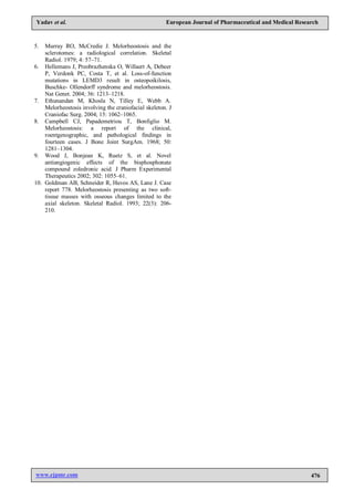
There was hard bony swelling over right hand & wrist
joint and great toe & tibia along with hyperpigmentation
of skin overlying deformed right hand and right lower
limb (fig-1&2) .
Figure 1 Right leg showing multiple bony swellings
Haematological examination Hb 12.3mg/dl,TLC
12000/cumm,N85L14E01.Ceribrospinal fluid protein
244mg/dl,glucose 23mg/dl,N05L95,RBC 03. blood sugar
114mg/ dl,serum creatinine 1.2 mg/dl, blood urea
45mg/dl,Na 141 meq/l,K 4.5 meq/l,ca 9.9 meq/l, AST
45.0 U/L, ALT 55 U/L ,urine 2-4 pus cells no albumin
and sugar.Body mass index of the patient was 15.2
SJIF Impact Factor 3.628
Case Report
ISSN 3294-3211
EJPMR
EUROPEAN JOURNAL OF PHARMACEUTICAL
AND MEDICAL RESEARCH
www.ejpmr.com
ejpmr, 2016,3(3), 474-476
*Author for Correspondence: Dr. Keshri Singh Yadav
Junior resident, P.G. Department of Medicine, S.N. Medical College, Agra, India.
ABSTRACT
Melorheostosis or leri’s disease is a rare bone disorder which is characterized by subperiosteal sclerosis of bones.
The most common bone part is diaphysis of long bones of lower limb of one side with rare involvement of axial
skeleton. Some times bones of the hands and feet may involved. Radiological pictures shows flowing hyperosteosis’
resembling hardened wax which has dripped down the side of the candle.although patient presented with seizure and
diagnosed as tubercular meningitis but we are reporting this case because of its rarety, in a young male.
KEY WORDS: Melorheostosis, Subperiosteal sclerosis, Candle dripping, flowing wax.](https://image.slidesharecdn.com/leridisease-170412035817/85/Leri-disease-1-320.jpg)
![www.ejpmr.com 474
Yadav et al. European Journal of Pharmaceutical and Medical Research
LERI’S DISEASE
Keshri Singh Yadav*1
, Subhash Chandra Yadav2
, Ankur Yadav3
, Nirdesh Chouhan3
and Asif Hussain4
1
Junior Resident, P.G. Department of Medicine, S.N. Medical College, Agra, India.
2
Assistant Professor, P.G. Department of Medicine, S.N. Medical College, Agra, India.
3
Junior Residents, P.G. Department of Medicine, S.N. Medical College, Agra, India.
4
Junior Resident Department of Orthopedics, S.N. Medical College, Agra, India.
Article Received on 12/01/2016 Article Revised on 04/02/2016 Article Accepted on 24/02/2016
INTRODUCTION
Melorheostosis is a relatively rare chronic sclerosing
bone disorder also known as Leri’s disease, candle bone
disease,or melting wax syndrome. The disease was first
described by Leri and Joanny[1]
in 1922. The disease
affects men and women equally. Most common
presentation is pain & most common bone part is
diaphysis of long bones of lower limb of one side with
rare involvement of axial skeleton. Radiological pictures
shows flowing hyperosteosis’ resembling hardened wax
which has dripped down the side of candle.
CASE REPORT
A 35 years male presented to us in status epilepticus in
emergency department. Patient has history of fever of
high grade, headache and projectile vomiting from five
days and became unconceous after seizure( GTCS type)
from two days. Patient have history of right leg pain (dull
aching) since 20 years of age & then develop mild
swelling & limitation of knee & elbow joint movements
which was gradually progressed so much that patient was
unable to walk from 7 to 8 months.There was no relevant
family history or trauma or any other musculoskeletal
disorder.
On examination blood pressure of the patient was 100/80
mmHg,pulse rate 94 per minute, regular ,pallor present
but icterus , cyanosis,and, clubbing were absent & paedal
oedema was present.
CNS- Patient was in postictal phase with GCS-
E4V4M3. Signs of meningeal irritation were present.
Deep tendon reflexes were diminished and planter reflex
were bilaterally equivocal, tone in right upper & lower
limb increased due to flexion deformity at both knee and
elbow joint and pupils were bilaterally semidialated and
normaly reacting to light. On respiratory system
examination, bilaterally vesicular breath sound present.
Per abdomen- soft, non tender, no organomegaly present.
There was no sign of inflamation in upper & lower limb.
There was hard bony swelling over right hand & wrist
joint and great toe & tibia along with hyperpigmentation
of skin overlying deformed right hand and right lower
limb (fig-1&2) .
Figure 1 Right leg showing multiple bony swellings
Haematological examination Hb 12.3mg/dl,TLC
12000/cumm,N85L14E01.Ceribrospinal fluid protein
244mg/dl,glucose 23mg/dl,N05L95,RBC 03. blood sugar
114mg/ dl,serum creatinine 1.2 mg/dl, blood urea
45mg/dl,Na 141 meq/l,K 4.5 meq/l,ca 9.9 meq/l, AST
45.0 U/L, ALT 55 U/L ,urine 2-4 pus cells no albumin
and sugar.Body mass index of the patient was 15.2
SJIF Impact Factor 3.628
Case Report
ISSN 3294-3211
EJPMR
EUROPEAN JOURNAL OF PHARMACEUTICAL
AND MEDICAL RESEARCH
www.ejpmr.com
ejpmr, 2016,3(3), 474-476
*Author for Correspondence: Dr. Keshri Singh Yadav
Junior resident, P.G. Department of Medicine, S.N. Medical College, Agra, India.
ABSTRACT
Melorheostosis or leri’s disease is a rare bone disorder which is characterized by subperiosteal sclerosis of bones.
The most common bone part is diaphysis of long bones of lower limb of one side with rare involvement of axial
skeleton. Some times bones of the hands and feet may involved. Radiological pictures shows flowing hyperosteosis’
resembling hardened wax which has dripped down the side of the candle.although patient presented with seizure and
diagnosed as tubercular meningitis but we are reporting this case because of its rarety, in a young male.
KEY WORDS: Melorheostosis, Subperiosteal sclerosis, Candle dripping, flowing wax.](https://image.slidesharecdn.com/leridisease-170412035817/75/Leri-disease-1-2048.jpg)
![www.ejpmr.com 475
Yadav et al. European Journal of Pharmaceutical and Medical Research
kg/mm2
. HIV ELISA, Hbs Ag, anti HCV were non
reactive. On the basis of above investigation diagnosis of
tubercular meningitis was made and treatment started
accordingly along with antiepileptics.
Figure 2 Right hand showing multiple bony swelling
X-ray of right leg (Fig-3) showed dense irregular cortical
hyperostosis , which looks like candle wax, extending
along tibia of right leg resulting in deformity of bone. X-
ray of right hand showed irregular cortical thickening of
metacarpals and phalanges . X-ray picture of left upper
and lower limb appear normal.
Figure 3 X ray both legs showing subperiosteal bony
growths
The orthopedic and radiology experts were consulted and
diagnosis of Leri disease with tubercular meningitis was
made. Antitubercular treatment was given along with
bishphosphonate. Some operative treatment was adviced
by orthopedicians but patient refuses to take treatment.
DISCUSSION
Melorheostosis is a rare chronic bone disorder which was
first described in 1922 by Leri and Joanny[1]
. Male and
female are equally affected, and no hereditary features
have been discovered. The onset of this rare diseases is
insidious, and the first symptom is usually dull aching
pain due to subperiosteal bone formation. Skin become
rough, hard and in 17% of cases that may include
hyperpigmentation. Melorheostosis mainly affects, the
long bones of the upper and lower limb, and also short
bones of hand and foot, but rarely the axial skeleton.[2,3]
Melorheostosis may present in a monostotic, polyostotic,
or monomelic form. The monomelic form is most
common.[4]
The most accepted hypothesis was given by Murray and
McCredie1979.[5]
was that, embryonic infection of nerve
root causes neural scarring and segmental bone sclerosis
responsible for melorheostosis. One possible etiology of
melorheostosis is a loss of function mutation in the
LEMD3 gene (12q12–12q14.3), a protein involved in
bone morphogenic protein and tumor growth factor-β
signaling.[6]
It is associated with vascular malformations, soft tissue
masses adjacent to the affected bone and scleroderma of
the overlying skin.[7]
Routine laboratory findings usually
are normal. Histological findings are usually nonspecific
and often show dense bone formation, a mixture of
mature and immature bone elements.[7]
Osteoclastic
activity is not a prominent feature; however, osteoblastic
activity along the margins of osteons is common.[8]
Treatment is mainly symptomatic. Most patient receives
nonoperative treatment. Operative treatment consists of
tendon lengthening, excision of hyperostotic bone,
osteotomies ,sympathectomy and amputation[3]
.Bisphosphonate are commonly use.[3]
Potential causes of
the bone pain in melorheostosis include increased
osteoclastic bone resorption and activation of pain
receptors, raised intraosseous pressure and increased
vascularity secondary to hyperosteosis and soft tissue
involvement around joints. Thus, bisphosphonate
treatment via a number of mechanisms would be
expected to reduce inflammatory bone pain and
symptoms in melorheostosis. Bisphosphonates inhibit
osteoclast mediated bone resorption by direct and
indirect actions on osteoblasts and macrophages and
bone vascularity. They have been shown to decrease
bone pain, slow progression of bone lesion.[9]
The
prognosis of a patient with melorheostosis is variable and
depends on the anatomical location, extension into the
soft tissues, and soft tissue changes. Melorheostosis does
not shorten life span, however, morbidity may be
considerable. The disease exhibits a slow, chronic
course, with periods of exacerbation and arrest.
Recurrence usually is expected after operative
excision.[10]
REFERENCES
1. Leri A, Joanny J. Une affection non décrite des os
hyperostose “en coulée” sur toute la longeur d'un
member ou “melorhéostose.” Bull Mem Soc Med
Hosp Paris. 1922; 46: 1141–1145.
2. Greenspan A, Azouz EM. Bone dysplasia series:
melorheostosis: review and update. Can Assoc
Radiol J. 1999; 50: 324–330.
3. Freyschmidt J.: Melorheostosis: a review of 23
cases. Eur Radiol 2001; 11: 474–9
4. Zeiller SC, Vaccaro AR, Wimberley DW, Albert TJ,
Harrop JS, Hilibrand AS. Severe myelopathy
resulting from melorheostosis of the cervicothoracic
spine: a case report. J Bone Joint Surg Am. 2005;
87: 2759–2762.](https://image.slidesharecdn.com/leridisease-170412035817/85/Leri-disease-2-320.jpg)

This document presents a case report of a 35-year-old male patient who presented with status epilepticus and was diagnosed with tuberculous meningitis and Leri's disease. Leri's disease, also known as melorheostosis, is a rare bone disorder characterized by subperiosteal bone growth resembling hardened wax dripping down the side of a candle. The patient had a 20-year history of right leg pain and deformities. Imaging showed irregular bone growths along the right tibia and bones of the right hand. The patient was treated for tuberculous meningitis and with bisphosphonates for pain from the Leri's disease.


![CTEV [ clubfoot] DR ARUN LAL ,DR MOHAMED ASHRAF travancore medical college k...](https://cdn.slidesharecdn.com/ss_thumbnails/ctevclubfootdrarunlaldrmohamedashraftravancoremedicalcollegekollamkeralaindia-260208063247-18fc466c-thumbnail.jpg?width=640&height=640&fit=bounds)

