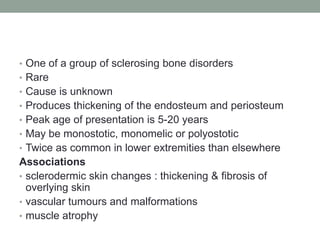
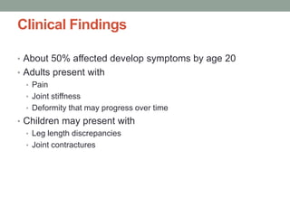
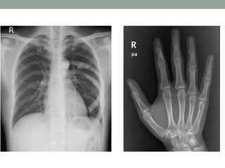
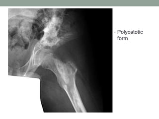
MELORHEOSTOSIS
Dr. Nikhil Murkey
• One of a group of sclerosing bone disorders
• Rare
• Cause is unknown
• Produces thickening of the endosteum and periosteum
• Peak age of presentation is 5-20 years
• May be monostotic, monomelic or polyostotic
• Twice as common in lower extremities than elsewhere
Associations
• sclerodermic skin changes : thickening & fibrosis of
overlying skin
• vascular tumours and malformations
• muscle atrophy
Clinical Findings
• About 50% affected develop symptoms by age 20
• Adults present with
• Pain
• Joint stiffness
• Deformity that may progress over time
• Children may present with
• Leg length discrepancies
• Joint contractures
Radiologic features
• Patterns described include:
1. Resembling osteoma(s)
2. Candle-wax appearance (classic)
3. Resembling myositis ossificans
4. Resembling osteopathia striata
5. Mixed
The classic appearance is one of thick undulating ridges of
bone, reminiscent of molten wax. The abnormality appears
confined to sclertomes, and can be seen apparently flowing
across joints to the next bone.
• Usually low signal on MRI Enhance with Gadolinium
• Bone scan is markedly positive
• WAVY NEW BONE
FORMATION. Fibula
• MELORHEOSTOSIS: DISTRIBUTION ALONG ONE SIDE OF THE BONE. A.
Humerus. B. Hand. Note the extensive involvement along the lateral aspect of the
hand and humerus.
• ENDOSTEAL
INVOLVEMENT.
A. Femur. Note
the endosteal
involvement of
the femur
encroaching on
the medullary
space.
• B. Tibia. Note
that the
medullary canal
appears
compromised
owing to
endosteal bone
formation
• Polyostotic
form
• Differential Diagnosis
• Osteopathia striata
• Longitudinal dense striations
• Osteopoikilosis
• Punctate, rounded bone islands surrounding joints
• Osteosarcoma
• Bone destruction
Not infrequently features of melorheostosis, osteopathia
striata and osteopoikilosis may co-exist in so-called overlap
syndromes.

Melorheostosis

  • 1.
  • 2.
    • One ofa group of sclerosing bone disorders • Rare • Cause is unknown • Produces thickening of the endosteum and periosteum • Peak age of presentation is 5-20 years • May be monostotic, monomelic or polyostotic • Twice as common in lower extremities than elsewhere Associations • sclerodermic skin changes : thickening & fibrosis of overlying skin • vascular tumours and malformations • muscle atrophy
  • 3.
    Clinical Findings • About50% affected develop symptoms by age 20 • Adults present with • Pain • Joint stiffness • Deformity that may progress over time • Children may present with • Leg length discrepancies • Joint contractures
  • 4.
    Radiologic features • Patternsdescribed include: 1. Resembling osteoma(s) 2. Candle-wax appearance (classic) 3. Resembling myositis ossificans 4. Resembling osteopathia striata 5. Mixed The classic appearance is one of thick undulating ridges of bone, reminiscent of molten wax. The abnormality appears confined to sclertomes, and can be seen apparently flowing across joints to the next bone. • Usually low signal on MRI Enhance with Gadolinium • Bone scan is markedly positive
  • 5.
    • WAVY NEWBONE FORMATION. Fibula
  • 6.
    • MELORHEOSTOSIS: DISTRIBUTIONALONG ONE SIDE OF THE BONE. A. Humerus. B. Hand. Note the extensive involvement along the lateral aspect of the hand and humerus.
  • 7.
    • ENDOSTEAL INVOLVEMENT. A. Femur.Note the endosteal involvement of the femur encroaching on the medullary space. • B. Tibia. Note that the medullary canal appears compromised owing to endosteal bone formation
  • 9.
  • 10.
    • Differential Diagnosis •Osteopathia striata • Longitudinal dense striations • Osteopoikilosis • Punctate, rounded bone islands surrounding joints • Osteosarcoma • Bone destruction Not infrequently features of melorheostosis, osteopathia striata and osteopoikilosis may co-exist in so-called overlap syndromes.