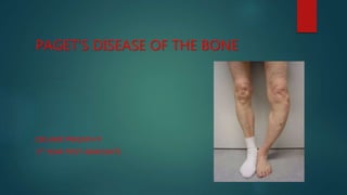
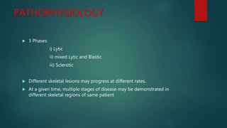
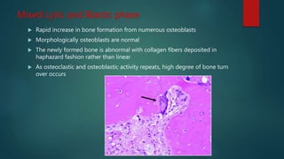
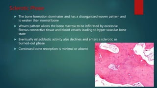
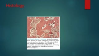
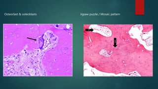
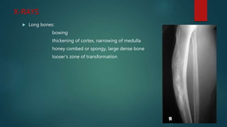
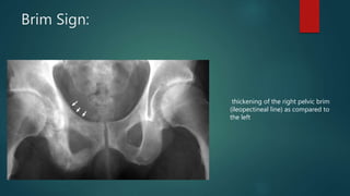
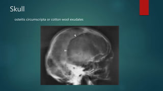
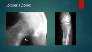
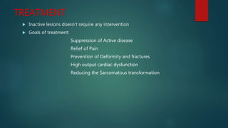
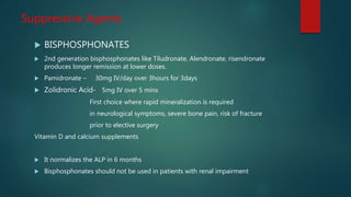
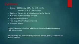
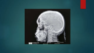
PAGET’S DISEASE OF THE BONE
DR.HARI PRASATH P
1ST YEAR POST GRADUATE
INTRODUCTION
The condition was initially described by Dr. James Paget in 1877
 Also called as Osteitis Deformans
 Partial or complete involvement of a single or multiple bones by
exaggerated rates of resorptive and osteogenic activity leading to bony
thickening and deformity.
 Schmorl believed that approximately 3% of everyone above 40 years had
osteitis deformans
INTRODUCTION (Cont)
 It has a predilection for the axial skeleton
 Pelvis>tibia > Femur > Skull>spine >clavicle
 But any bone may be affected
 Paget disease is common in Europe, North America
 It is rare in Asia and Africa
ETIOLOGY
 UNKNOWN
 Occasionally hereditary influence is noted on chromosome 18q
 On electron microscopy of bone biopsies has demonstrated nuclear
inclusions,
 similar to those found in viral diseases (Paramyxoviridae family) are found
in the highly nucleated osteoclasts
 Endocrine and metabolic disturbances are unlikely because despite
extensive involvement , many bones are free of disease
PATHOPHYSIOLOGY
 3 Phases:
i) Lytic
ii) mixed Lytic and Blastic
iii) Sclerotic
 Different skeletal lesions may progress at different rates.
 At a given time, multiple stages of disease may be demonstrated in
different skeletal regions of same patient
LYTIC PHASE
 Disease begins with lytic phase
 The bone is resorbed by osteoclasts that are more numerous, larger, and
have more nuclei (upto 100)
 Bone turnover rate increased as much as 20times normal
Mixed Lytic and Blastic phase
 Rapid increase in bone formation from numerous osteoblasts
 Morphologically osteoblasts are normal
 The newly formed bone is abnormal with collagen fibers deposited in
haphazard fashion rather than linear
 As osteoclastic and osteoblastic activity repeats, high degree of bone turn
over occurs
Sclerotic Phase
 The bone formation dominates and has a disorganized woven pattern and
is weaker than normal bone
 Woven pattern allows the bone marrow to be infiltrated by excessive
fibrous connective tissue and blood vessels leading to hyper vascular bone
state
 Eventually osteoblastic activity also declines and enters a sclerotic or
burned-out phase
 Continued bone resorption is minimal or absent
Histology
Osteoclast & osteoblasts Jigsaw puzzle / Mosaic pattern
1
2
Complications
 Fractures and bony deformity
 Secondary osteoarthritis ( when pagets disease around a joint)
 Neurological complications – nerve root compression and cauda equina
syndrome
 Skull involvement- deafness
vertigo
tinnitus
dental malocclusion
basilar invagination
cranial nerve disorders
Sarcomatous degeneration - Osteosarcoma
 Increased bone vascularity – high output cardiac failure
SURGICAL COMPLICATIONS:
 Highly vascular marrow – Profuse bleeding
 Structurally weak bone
 Spinal / Epidural Anesthesia may be difficult
Investigations
 Serum Alkaline phosphatase , BSAP
 Serum Acid phosphatase
 Urinary Markers – hydroxyproline, deoxypyridinoline, C-telopeptide, N-
telopeptide
 Serum calcium and phosphate levels will be normal
X-RAYS
 Long bones:
bowing
thickening of cortex, narrowing of medulla
honey combed or spongy, large dense bone
looser’s zone of transformation
Brim Sign:
thickening of the right pelvic brim
(ileopectineal line) as compared to
the left
Skull
osteitis circumscripta or cotton wool exudates
tam o' shanter sign
widening of the diploic space and an
overall enlargement of the cranium
Pagets Spine
Ivory Vertebra
Picture frame sign
Blade of Grass or Candle flame sign:
begins as a subchondral area of lucency with advancing
tip of V-shaped osteolysis, extending towards the
diaphysis
Looser’s Zone:
Scintigraphy
Technetium 99 scan shows increased
uptake in right hemipelvis and spine
DIFFERENTIAL DIAGNOSIS:
 Osteomalacia
 Fibrous Dysplasia
 Multiple Myeloma
 osteopetrosis
TREATMENT
 Inactive lesions doesn’t require any intervention
 Goals of treatment:
Suppression of Active disease
Relief of Pain
Prevention of Deformity and fractures
High output cardiac dysfunction
Reducing the Sarcomatous transformation
Suppressive Agents
 BISPHOSPHONATES
 2nd generation bisphosphonates like Tiludronate, Alendronate, risendronate
produces longer remission at lower doses.
 Pamidronate – 30mg IV/day over 3hours for 3days
 Zolidronic Acid- 5mg IV over 5 mins
First choice where rapid mineralization is required
in neurological symptoms, severe bone pain, risk of fracture
prior to elective surgery
Vitamin D and calcium supplements
 It normalizes the ALP in 6 months
 Bisphosphonates should not be used in patients with renal impairment
Calcitonin
 Dosage – 100 IU / day SC/IM for 6-18 months
reduced to 50 IU / day x 3/week
 Calcitonin therapy can temporarily arrest active disease
 ALP, urine Hydroxyproline is reduced
 Positive Calcium balance
 High output heart failure is improved
 Bone pain relieved
Surgical treatment is reserved for fractures, correction of bone deformity,
THR, Spinal surgery
Preoperatively and postoperatively calcitonin therapy gives good results and
reduces bleeding
CASE
Alkaline phosphatase – 1510 IU/L
Sr. calcium – 8.7mg/dl
Sr. Phosphorus – 3.1 mg/dl
THANK YOU

Pagetsdiseaseofthebone

  • 1.
    PAGET’S DISEASE OFTHE BONE DR.HARI PRASATH P 1ST YEAR POST GRADUATE
  • 2.
    INTRODUCTION The condition wasinitially described by Dr. James Paget in 1877  Also called as Osteitis Deformans  Partial or complete involvement of a single or multiple bones by exaggerated rates of resorptive and osteogenic activity leading to bony thickening and deformity.  Schmorl believed that approximately 3% of everyone above 40 years had osteitis deformans
  • 3.
    INTRODUCTION (Cont)  Ithas a predilection for the axial skeleton  Pelvis>tibia > Femur > Skull>spine >clavicle  But any bone may be affected  Paget disease is common in Europe, North America  It is rare in Asia and Africa
  • 4.
    ETIOLOGY  UNKNOWN  Occasionallyhereditary influence is noted on chromosome 18q  On electron microscopy of bone biopsies has demonstrated nuclear inclusions,  similar to those found in viral diseases (Paramyxoviridae family) are found in the highly nucleated osteoclasts  Endocrine and metabolic disturbances are unlikely because despite extensive involvement , many bones are free of disease
  • 5.
    PATHOPHYSIOLOGY  3 Phases: i)Lytic ii) mixed Lytic and Blastic iii) Sclerotic  Different skeletal lesions may progress at different rates.  At a given time, multiple stages of disease may be demonstrated in different skeletal regions of same patient
  • 6.
    LYTIC PHASE  Diseasebegins with lytic phase  The bone is resorbed by osteoclasts that are more numerous, larger, and have more nuclei (upto 100)  Bone turnover rate increased as much as 20times normal
  • 7.
    Mixed Lytic andBlastic phase  Rapid increase in bone formation from numerous osteoblasts  Morphologically osteoblasts are normal  The newly formed bone is abnormal with collagen fibers deposited in haphazard fashion rather than linear  As osteoclastic and osteoblastic activity repeats, high degree of bone turn over occurs
  • 8.
    Sclerotic Phase  Thebone formation dominates and has a disorganized woven pattern and is weaker than normal bone  Woven pattern allows the bone marrow to be infiltrated by excessive fibrous connective tissue and blood vessels leading to hyper vascular bone state  Eventually osteoblastic activity also declines and enters a sclerotic or burned-out phase  Continued bone resorption is minimal or absent
  • 9.
  • 10.
    Osteoclast & osteoblastsJigsaw puzzle / Mosaic pattern 1 2
  • 11.
    Complications  Fractures andbony deformity  Secondary osteoarthritis ( when pagets disease around a joint)  Neurological complications – nerve root compression and cauda equina syndrome  Skull involvement- deafness vertigo tinnitus dental malocclusion basilar invagination cranial nerve disorders Sarcomatous degeneration - Osteosarcoma  Increased bone vascularity – high output cardiac failure
  • 12.
    SURGICAL COMPLICATIONS:  Highlyvascular marrow – Profuse bleeding  Structurally weak bone  Spinal / Epidural Anesthesia may be difficult
  • 13.
    Investigations  Serum Alkalinephosphatase , BSAP  Serum Acid phosphatase  Urinary Markers – hydroxyproline, deoxypyridinoline, C-telopeptide, N- telopeptide  Serum calcium and phosphate levels will be normal
  • 14.
    X-RAYS  Long bones: bowing thickeningof cortex, narrowing of medulla honey combed or spongy, large dense bone looser’s zone of transformation
  • 15.
    Brim Sign: thickening ofthe right pelvic brim (ileopectineal line) as compared to the left
  • 16.
    Skull osteitis circumscripta orcotton wool exudates
  • 17.
    tam o' shantersign widening of the diploic space and an overall enlargement of the cranium
  • 18.
  • 19.
    Blade of Grassor Candle flame sign: begins as a subchondral area of lucency with advancing tip of V-shaped osteolysis, extending towards the diaphysis
  • 20.
  • 21.
    Scintigraphy Technetium 99 scanshows increased uptake in right hemipelvis and spine
  • 22.
    DIFFERENTIAL DIAGNOSIS:  Osteomalacia Fibrous Dysplasia  Multiple Myeloma  osteopetrosis
  • 23.
    TREATMENT  Inactive lesionsdoesn’t require any intervention  Goals of treatment: Suppression of Active disease Relief of Pain Prevention of Deformity and fractures High output cardiac dysfunction Reducing the Sarcomatous transformation
  • 24.
    Suppressive Agents  BISPHOSPHONATES 2nd generation bisphosphonates like Tiludronate, Alendronate, risendronate produces longer remission at lower doses.  Pamidronate – 30mg IV/day over 3hours for 3days  Zolidronic Acid- 5mg IV over 5 mins First choice where rapid mineralization is required in neurological symptoms, severe bone pain, risk of fracture prior to elective surgery Vitamin D and calcium supplements  It normalizes the ALP in 6 months  Bisphosphonates should not be used in patients with renal impairment
  • 25.
    Calcitonin  Dosage –100 IU / day SC/IM for 6-18 months reduced to 50 IU / day x 3/week  Calcitonin therapy can temporarily arrest active disease  ALP, urine Hydroxyproline is reduced  Positive Calcium balance  High output heart failure is improved  Bone pain relieved Surgical treatment is reserved for fractures, correction of bone deformity, THR, Spinal surgery Preoperatively and postoperatively calcitonin therapy gives good results and reduces bleeding
  • 26.
    CASE Alkaline phosphatase –1510 IU/L Sr. calcium – 8.7mg/dl Sr. Phosphorus – 3.1 mg/dl
  • 28.