7 | Carbohydrates and
Glycobiology
© 2017 W. H. Freeman and Company
Chapter 8 Opener
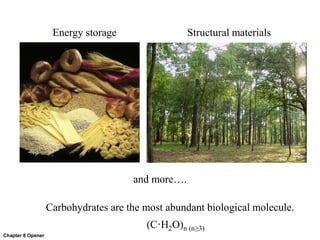
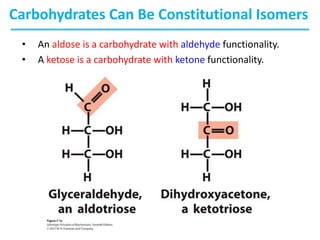
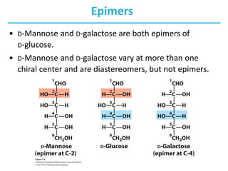
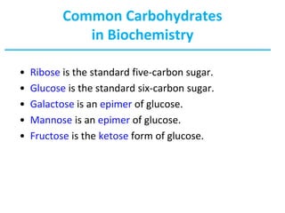
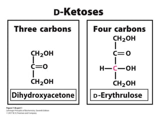
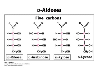
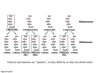
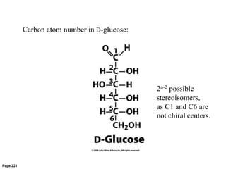
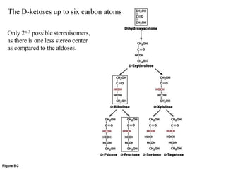
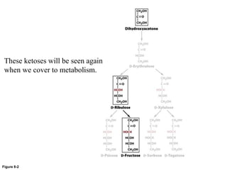
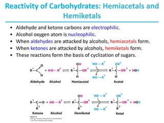
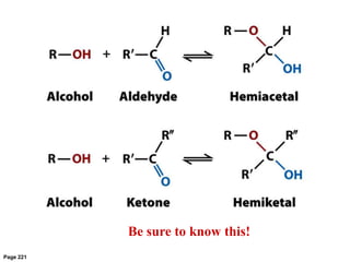
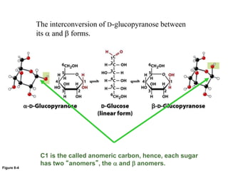
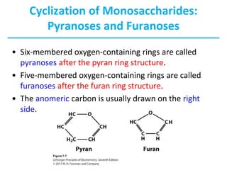
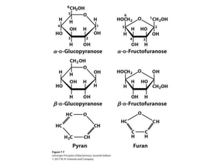
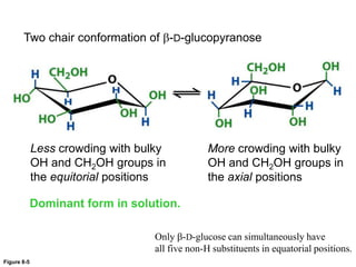
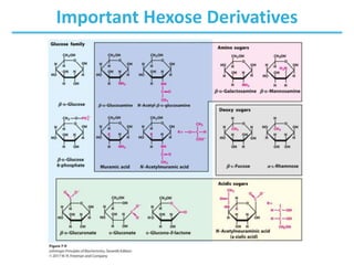
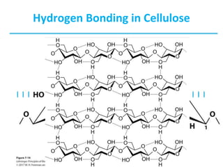
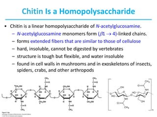
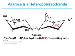
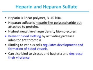
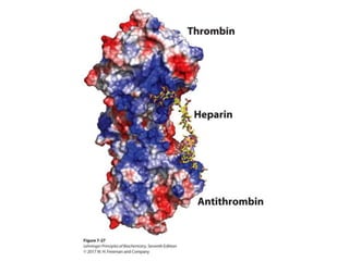
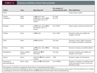
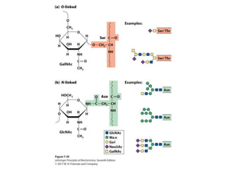
Carbohydrates are the most abundant biological molecule.
(C·H2O)n (n≥3)
Energy storage Structural materials
and more….
Carbohydrates
• Named so because many have formula Cn(H2O)n
• Produced from CO2 and H2O via photosynthesis in plants
• Range from as small as glyceraldehyde (Mw = 90 g/mol) to as
large as amylopectin (Mw > 200,000,000 g/mol)
• Fulfill a variety of functions, including:
– energy source and energy storage
– structural component of cell walls and exoskeletons
– informational molecules in cell-cell signaling
• Can be covalently linked with proteins and lipids
• Basic nomenclature:
– number of carbon atoms in the carbohydrate +
-ose
– example: three carbons = triose
• Common functional groups:
– All carbohydrates initially had a carbonyl
functional group.
– aldehydes = aldose
– ketones = ketose
Carbohydrates
Carbohydrates Can Be Constitutional Isomers
• An aldose is a carbohydrate with aldehyde functionality.
• A ketose is a carbohydrate with ketone functionality.
Enantiomers Are Mirror Images
Carbohydrates Can Be Stereoisomers
• Enantiomers
– stereoisomers that are nonsuperimposable mirror images
• In sugars that contain many chiral centers, only the
one that is most distant from the carbonyl carbon is
designated as d (right) or l (left).
• d and l isomers of a sugar are enantiomers.
– For example, l- and d-glucose have the same water solubility.
• Most hexoses in living organisms are d stereoisomers.
• Some simple sugars occur in the l form, such as
l-arabinose.
Drawing Monosaccharides
• Chiral compounds can be
drawn using perspective
formulas.
• However, chiral
carbohydrates are usually
represented by Fischer
projections.
• Horizontal bonds are
pointing toward you;
vertical bonds are
projecting away from you.
Carbohydrates Can Be Stereoisomers
• Epimers are stereoisomers that differ at only one chiral center.
• Epimers are NOT mirror images, and therefore are NOT
enantiomers.
• Epimers are diastereomers; diastereomers have different
physical properties (i.e., water solubility, melting temp).
• example: D-Threose is the C-2 epimer of D-erythrose.
• Both are D sugars because they both have the same
orientation around the last chiral carbon in the chain.
Diastereomers
• Diastereomers: stereoisomers that are not mirror
images
• Diastereomers have different physical properties.
– For example, water solubilities of threose and erythrose are
different.
Epimers
• D-Mannose and D-galactose are both epimers of
D-glucose.
• D-Mannose and D-galactose vary at more than one
chiral center and are diastereomers, but not epimers.
Common Carbohydrates
in Biochemistry
• Ribose is the standard five-carbon sugar.
• Glucose is the standard six-carbon sugar.
• Galactose is an epimer of glucose.
• Mannose is an epimer of glucose.
• Fructose is the ketose form of glucose.
Figure 8-1
The D-aldoses with up to six carbon atoms
D sugars have the same
absolute configuration at the
asymmetric center farthest
from their carbonyl group.
Figure 8-1 part 1
CHO
HOCH
HOCH
CH2OH
CHO
HCOH
HOCH
CH2OH
L-Erythrose L-Threose
2n-2 possible
stereoisomers
Here, n = 4,
so there are 4 stereoisomers
D sugars
L sugars
Figure 8-1 part 2
Ribose is the most common aldo-pentose
Figure 8-1 part 3
Glucose and mannose are “epimers”, as they differ by at only one chiral center.
Page 221
2n-2 possible
stereoisomers,
as C1 and C6 are
not chiral centers.
Carbon atom number in D-glucose:
Figure 8-2
Only 2n-3 possible stereoisomers,
as there is one less stereo center
as compared to the aldoses.
The D-ketoses up to six carbon atoms
Figure 8-2
These ketoses will be seen again
when we cover to metabolism.
Reactivity of Carbohydrates: Hemiacetals and
Hemiketals
• Aldehyde and ketone carbons are electrophilic.
• Alcohol oxygen atom is nucleophilic.
• When aldehydes are attacked by alcohols, hemiacetals form.
• When ketones are attacked by alcohols, hemiketals form.
• These reactions form the basis of cyclization of sugars.
Page 221
Be sure to know this!
Cyclization of
Monosaccharides
• The nucleophilic alcohol attacks
the electrophilic carbonyl
carbon, allowing formation of a
hemiacetal.
• As a result, the linear
carbohydrate forms a ring
structure.
• At the completion of this
structure, the carbonyl carbon is
reduced to an alcohol
• The orientation of the alcohol
around the carbon is variable
and transient.
Figure 8-4
The interconversion of D-glucopyranose between
its a and b forms.
C1 is the called anomeric carbon, hence, each sugar
has two “anomers”, the a and b anomers.
Cyclization of Monosaccharides
• Pentoses and hexoses readily undergo intramolecular
cyclization.
• The former carbonyl carbon becomes a new chiral
center, called the anomeric carbon.
– When the former carbonyl oxygen becomes a hydroxyl group,
the position of this group determines if the anomer is a or b.
– If the hydroxyl group is on the opposite side (trans) of the ring
as the CH2OH moiety, the configuration is a.
– If the hydroxyl group is on the same side (cis) of the ring as
the CH2OH moiety, the configuration is b.
Cyclization of Monosaccharides:
Pyranoses and Furanoses
• Six-membered oxygen-containing rings are called
pyranoses after the pyran ring structure.
• Five-membered oxygen-containing rings are called
furanoses after the furan ring structure.
• The anomeric carbon is usually drawn on the right
side.
Conformational Formulas of Cyclized
Monosaccharides
• Cyclohexane rings have “chair” or “boat” conformations.
• Pyranose rings favor “chair” conformations.
• Multiple “chair” conformations are possible but require energy
for interconversion (~46 kJ/mole).
Figure 8-5
Two chair conformation of b-D-glucopyranose
Less crowding with bulky
OH and CH2OH groups in
the equitorial positions
More crowding with bulky
OH and CH2OH groups in
the axial positions
Dominant form in solution.
Only b-D-glucose can simultaneously have
all five non-H substituents in equatorial positions.
Important Hexose Derivatives
Box 8-2a
Often seen along with table sugar:
Box 8-2b
Also seen at the table, and in diet sodas:
Box 8-2c
Sometimes used with aspartame, acts synergistically to make both
compounds sweeter.
Since 1879
Since 1981
Carcinogen ?
(problem for phenylketonuria)
Causes cancer in rats in
VERY high dosages.
Still room for improvement…
Reducing Sugars
• Reducing sugars have a free anomeric carbon.
– Aldehyde can reduce Cu2+ to Cu+ (Fehling’s test).
– Aldehyde can reduce Ag+ to Ag0 (Tollens’ test).
– It allows detection of reducing sugars, such as glucose.
– Modern detection techniques use colorimetric and
electrochemical tests.
Colorimetric Glucose Analysis
• Enzymatic methods are used to
quantify reducing sugars such as
glucose.
– The enzyme glucose oxidase
catalyzes the conversion of
glucose to glucono--lactone and
hydrogen peroxide.
– Hydrogen peroxide oxidizes
organic molecules into highly
colored compounds.
– Concentrations of such
compounds is measured
colorimetrically.
O
OH
OH
OH
O
H
CH2OH
O
OH
OH O
O
H
CH2OH
NH2
NH2
OCH3
OCH3
NH
NH
OCH3
OCH3
b-D-Glucose
-D-Gluconolactone
Glucose oxidase
O2
H2O2
Peroxidase
2 H2O
Reduced
o-dianisidine
(faint orange)
Oxidized
o-dianisidine
(bright orange)
• Electrochemical detection is
used in portable glucose
sensors.
The Glycosidic Bond
• Two sugar molecules can be joined via a glycosidic bond
between an anomeric carbon and a hydroxyl carbon.
• The glycosidic bond (an acetal) between monomers is more
stable and less reactive than the hemiacetal at the second
monomer.
– The second monomer, with the hemiacetal, is reducing.
– The anomeric carbon involved in the glycosidic linkage is
nonreducing.
• Disacharides can be named by the organization and linkage
or a common name.
– The disaccharide formed upon condensation of two glucose
molecules via a 1  4 bond is described as α-D-glucopyranosyl-
(14)-D-glucopyranose.
– The common name for this disaccharide is maltose.
Nonreducing Disaccharides
• Two sugar molecules can be
also joined via a glycosidic
bond between two anomeric
carbons.
• The product has two acetal
groups and no hemiacetals.
• There are no reducing ends;
this is a nonreducing sugar.
Polysaccharides
• Natural carbohydrates are usually found as polymers.
• These polysaccharides can be:
– homopolysaccharides (one monomer unit)
– heteropolysaccharides (multiple monomer units)
– linear (one type of glycosidic bond)
– branched (multiple types of glycosidic bonds)
• Polysaccharides do not have a defined molecular weight.
– This is in contrast to proteins because, unlike proteins,
no template is used to make polysaccharides.
– Polysaccharides are often in a state of flux; monomer
units are added and removed as needed by the
organism.
Homopolymers of Glucose:
Glycogen
• Glycogen is a branched homopolysaccharide of
glucose.
– Glucose monomers form (a1  4) linked chains.
– There are branch points with (a1  6) linkers every
8–12 residues.
– Molecular weight reaches several millions.
– It functions as the main storage polysaccharide in
animals.
Homopolymers of Glucose:
Starch
• Starch is a mixture of two homopolysaccharides of
glucose.
• Amylose is an unbranched polymer of (a1  4)
linked residues.
• Amylopectin is branched like glycogen, but the
branch points with (a1  6) linkers occur every
24–30 residues.
• Molecular weight of amylopectin is up to 200
million.
• Starch is the main storage polysaccharide in plants.
Glycosidic Linkages in Glycogen and Starch
Mixture of Amylose and
Amylopectin in Starch
Metabolism of Glycogen and Starch
• Glycogen and starch are insoluble due to their high
molecular weight and often form granules in cells.
• Granules contain enzymes that synthesize and
degrade these polymers.
• Glycogen and amylopectin have one reducing end
but many nonreducing ends.
• Enzymatic processing occurs simultaneously in many
nonreducing ends.
Homopolymers of Glucose:
Cellulose
• Cellulose is a linear homopolysaccharide of glucose.
– Glucose monomers form (b1  4) linked chains.
– Hydrogen bonds form between adjacent monomers.
– There are additional H-bonds between chains.
– Structure is tough and water insoluble.
– It is the most abundant polysaccharide in nature.
– Cotton is nearly pure fibrous cellulose.
Hydrogen Bonding in Cellulose
Cellulose Metabolism
• The fibrous structure and water insolubility make cellulose a
difficult substrate to act upon.
• Most animals cannot use cellulose as a fuel source because they
lack the enzyme to hydrolyze (b1 4) linkages.
• Fungi, bacteria, and protozoa secrete cellulase, which allows
them to use wood as source of glucose.
• Ruminants and termites live symbiotically with microorganisms
that produce cellulase and are able to absorb the freed glucose
into their bloodstreams.
• Cellulases hold promise in the fermentation of biomass into
biofuels.
Q: What do these two have in common?
A: They both have a “sugary” shell
80% sucrose
10% glucose
10% fructose
100% N-acetylglucosamine.
Chitin Is a Homopolysaccharide
• Chitin is a linear homopolysaccharide of N-acetylglucosamine.
– N-acetylglucosamine monomers form (b1  4)-linked chains.
– forms extended fibers that are similar to those of cellulose
– hard, insoluble, cannot be digested by vertebrates
– structure is tough but flexible, and water insoluble
– found in cell walls in mushrooms and in exoskeletons of insects,
spiders, crabs, and other arthropods
Agar and Agarose
• Agar is a branched heteropolysaccharide composed
of agarose and agaropectin.
• Agar serves as a component of cell wall in some
seaweeds.
• Agar solutions form gels that are commonly used in
the laboratory as a surface for growing bacteria.
• Agarose solutions form gels that are commonly
used in the laboratory for separation DNA by
electrophoresis.
Agarose Is a Heteropolysaccharide
Glycosaminoglycans
• Linear polymers of repeating disaccharide units
• One monomer is either:
– N-acetyl-glucosamine or
– N-acetyl-galactosamine
• Negatively charged
– uronic acids (C6 oxidation)
– sulfate esters
• Extended hydrated molecule
– minimizes charge repulsion
• Forms meshwork with fibrous proteins to form extracellular
matrix
– connective tissue
– lubrication of joints
Heparin and Heparan Sulfate
• Heparin is linear polymer, 3–40 kDa.
• Heparan sulfate is heparin-like polysaccharide but
attached to proteins.
• Highest negative-charge density biomolecules
• Prevent blood clotting by activating protease
inhibitor antithrombin
• Binding to various cells regulates development and
formation of blood vessels.
• Can also bind to viruses and bacteria and decrease
their virulence
TABLE 7-2 Structures and Roles of Some Polysaccharides
Primer Typea Repeating unitb
Size (number of
monosaccharide units) Roles/significance
Starch Energy storage: in plants
Amylose
Amylopectin
Homo-
Homo-
(α1S4) Glc, linear
(α1S4) Glc, with (α1S6)
Glc branches every 24–30
residues
50–5,000
Up to 106
Glycogen Homo- (α1S4) Glc, with (α1S6)
Glc branches every 8–12
residues
Up to 50,000 Energy storage: in bacteria and animal cells
Cellulose Homo- (β1S4) Glc Up to 15,000 Structural: in plants, gives rigidity and
strength to cell walls
Chitin Homo- (β1S4) GlcNAc Very large Structural: in insects, spiders, crustaceans,
gives rigidity and strength to exoskeletons
Dextran Homo- (α1S6) Glc, with (α1S3)
branches
Wide range Structural: in bacteria, extracellular adhesive
Peptidoglycan Hetero-; peptides
attached
4)Mur2Ac(β1S4)
GlcNAc(β1
Very large Structural: in bacteria, gives rigidity and
strength to cell envelope
Agarose Hetero- 3)D-Gal (β1S4)3,6- anhydro-
L-Gal(α1
1,000 Structural: in algae, cell wall material
Hyaluronan (a
glycosaminoglycan)
Hetero-; acidic 4)GlcA (β1S3) GlcNAc(β1 Up to 100,000 Structural: in vertebrates, extracellular matrix
of skin and connective tissue; viscosity and
lubrication in joints
aEach polymer is classified as a homopolysaccharide (homo-) or heteropolysaccharide (hetero-).
bThe abbreviated names for the peptidoglycan, agarose, and hyaluronan repeating units indicate that the polymer contains repeats of this disaccharide unit. For example,
in peptidoglycan, the GlcNAc of one disaccharide unit is (β1S4)-linked to the first residue of the next disaccharide unit.
Glycoconjugates: Glycoprotein
• A protein with small oligosaccharides attached
– Carbohydrate ia attached via its anomeric carbon to amino
acids on the protein.
• Common connections occur at Ser, Thr, and Asn.
– About half of mammalian proteins are glycoproteins.
– Only some bacteria glycosylate a few of their proteins.
– Carbohydrates play role in protein-protein recognition.
– Viral proteins are heavily glycosylated; this helps evade the
immune system.
Glycoconjugates: Glycolipids
• Lipids with covalently bound oligosaccharide
– They are parts of plant and animal cell membranes.
– In vertebrates, ganglioside carbohydrate
composition determines blood groups.
– In gram-negative bacteria, lipopolysaccharides
cover the peptidoglycan layer.
Glycoconjugates: Proteoglycans
• Sulfated glucoseaminoglycans attached to a large rod-
shaped protein in cell membrane
– syndecans: protein has a single transmembrane
domain
– glypicans: protein is anchored to a lipid membrane
– interact with a variety of receptors from
neighboring cells and regulate cell growth
Proteoglycans
• Different glycosaminoglycans are linked to the core
protein.
• Linkage from anomeric carbon of xylose to serine
hydroxyl
• Our tissues have many different core proteins;
aggrecan is the best studied.
Proteoglycan Aggregates
• Hyaluronan and aggrecan form huge (Mr > 2 • 108)
noncovalent aggregates.
• They hold a lot of water (1000 its weight) and
provide lubrication.
• Very low friction material
• Covers joint surfaces: articular cartilage
– reduced friction
– load balancing
Extracellular Matrix (ECM)
• Material outside the cell
• Strength, elasticity, and physical barrier in tissues
• Main components
– proteoglycan aggregates
– collagen fibers
– elastin (a fibrous protein)
• ECM is a barrier for tumor cells seeking to invade new
tissues.
– Some tumor cells secrete heparinase that degrades ECM.
Interaction of the Cells with ECM
• Some integral membrane proteins are proteoglycans.
– syndecans
• Other integral membrane proteins are receptors for extracellular
proteoglycans.
– integrins
• These proteins link cellular cytoskeleton to the ECM and transmit
signals into the cell to regulate:
– cell growth
– cell mobility
– apoptosis
– wound healing
Oligosaccharides in Recognition
Glycoconjugates: Analysis
Chapter 7: Summary
• structures of some important monosaccharides
• structures and properties of disaccharides
• structures and biological roles of polysaccharides
• functions of glycosylaminoglycans as structural
components of the extracellular matrix
• functions glycoconjugates in regulating a variety of
biological functions
In this chapter, we learned about:

Lehninger_Ch07.ppt

  • 1.
    7 | Carbohydratesand Glycobiology © 2017 W. H. Freeman and Company
  • 2.
    Chapter 8 Opener Carbohydratesare the most abundant biological molecule. (C·H2O)n (n≥3) Energy storage Structural materials and more….
  • 3.
    Carbohydrates • Named sobecause many have formula Cn(H2O)n • Produced from CO2 and H2O via photosynthesis in plants • Range from as small as glyceraldehyde (Mw = 90 g/mol) to as large as amylopectin (Mw > 200,000,000 g/mol) • Fulfill a variety of functions, including: – energy source and energy storage – structural component of cell walls and exoskeletons – informational molecules in cell-cell signaling • Can be covalently linked with proteins and lipids
  • 4.
    • Basic nomenclature: –number of carbon atoms in the carbohydrate + -ose – example: three carbons = triose • Common functional groups: – All carbohydrates initially had a carbonyl functional group. – aldehydes = aldose – ketones = ketose Carbohydrates
  • 5.
    Carbohydrates Can BeConstitutional Isomers • An aldose is a carbohydrate with aldehyde functionality. • A ketose is a carbohydrate with ketone functionality.
  • 6.
  • 7.
    Carbohydrates Can BeStereoisomers • Enantiomers – stereoisomers that are nonsuperimposable mirror images • In sugars that contain many chiral centers, only the one that is most distant from the carbonyl carbon is designated as d (right) or l (left). • d and l isomers of a sugar are enantiomers. – For example, l- and d-glucose have the same water solubility. • Most hexoses in living organisms are d stereoisomers. • Some simple sugars occur in the l form, such as l-arabinose.
  • 8.
    Drawing Monosaccharides • Chiralcompounds can be drawn using perspective formulas. • However, chiral carbohydrates are usually represented by Fischer projections. • Horizontal bonds are pointing toward you; vertical bonds are projecting away from you.
  • 9.
    Carbohydrates Can BeStereoisomers • Epimers are stereoisomers that differ at only one chiral center. • Epimers are NOT mirror images, and therefore are NOT enantiomers. • Epimers are diastereomers; diastereomers have different physical properties (i.e., water solubility, melting temp). • example: D-Threose is the C-2 epimer of D-erythrose. • Both are D sugars because they both have the same orientation around the last chiral carbon in the chain.
  • 10.
    Diastereomers • Diastereomers: stereoisomersthat are not mirror images • Diastereomers have different physical properties. – For example, water solubilities of threose and erythrose are different.
  • 11.
    Epimers • D-Mannose andD-galactose are both epimers of D-glucose. • D-Mannose and D-galactose vary at more than one chiral center and are diastereomers, but not epimers.
  • 12.
    Common Carbohydrates in Biochemistry •Ribose is the standard five-carbon sugar. • Glucose is the standard six-carbon sugar. • Galactose is an epimer of glucose. • Mannose is an epimer of glucose. • Fructose is the ketose form of glucose.
  • 16.
    Figure 8-1 The D-aldoseswith up to six carbon atoms D sugars have the same absolute configuration at the asymmetric center farthest from their carbonyl group.
  • 17.
    Figure 8-1 part1 CHO HOCH HOCH CH2OH CHO HCOH HOCH CH2OH L-Erythrose L-Threose 2n-2 possible stereoisomers Here, n = 4, so there are 4 stereoisomers D sugars L sugars
  • 18.
    Figure 8-1 part2 Ribose is the most common aldo-pentose
  • 19.
    Figure 8-1 part3 Glucose and mannose are “epimers”, as they differ by at only one chiral center.
  • 20.
    Page 221 2n-2 possible stereoisomers, asC1 and C6 are not chiral centers. Carbon atom number in D-glucose:
  • 21.
    Figure 8-2 Only 2n-3possible stereoisomers, as there is one less stereo center as compared to the aldoses. The D-ketoses up to six carbon atoms
  • 22.
    Figure 8-2 These ketoseswill be seen again when we cover to metabolism.
  • 23.
    Reactivity of Carbohydrates:Hemiacetals and Hemiketals • Aldehyde and ketone carbons are electrophilic. • Alcohol oxygen atom is nucleophilic. • When aldehydes are attacked by alcohols, hemiacetals form. • When ketones are attacked by alcohols, hemiketals form. • These reactions form the basis of cyclization of sugars.
  • 24.
    Page 221 Be sureto know this!
  • 25.
    Cyclization of Monosaccharides • Thenucleophilic alcohol attacks the electrophilic carbonyl carbon, allowing formation of a hemiacetal. • As a result, the linear carbohydrate forms a ring structure. • At the completion of this structure, the carbonyl carbon is reduced to an alcohol • The orientation of the alcohol around the carbon is variable and transient.
  • 26.
    Figure 8-4 The interconversionof D-glucopyranose between its a and b forms. C1 is the called anomeric carbon, hence, each sugar has two “anomers”, the a and b anomers.
  • 27.
    Cyclization of Monosaccharides •Pentoses and hexoses readily undergo intramolecular cyclization. • The former carbonyl carbon becomes a new chiral center, called the anomeric carbon. – When the former carbonyl oxygen becomes a hydroxyl group, the position of this group determines if the anomer is a or b. – If the hydroxyl group is on the opposite side (trans) of the ring as the CH2OH moiety, the configuration is a. – If the hydroxyl group is on the same side (cis) of the ring as the CH2OH moiety, the configuration is b.
  • 28.
    Cyclization of Monosaccharides: Pyranosesand Furanoses • Six-membered oxygen-containing rings are called pyranoses after the pyran ring structure. • Five-membered oxygen-containing rings are called furanoses after the furan ring structure. • The anomeric carbon is usually drawn on the right side.
  • 30.
    Conformational Formulas ofCyclized Monosaccharides • Cyclohexane rings have “chair” or “boat” conformations. • Pyranose rings favor “chair” conformations. • Multiple “chair” conformations are possible but require energy for interconversion (~46 kJ/mole).
  • 31.
    Figure 8-5 Two chairconformation of b-D-glucopyranose Less crowding with bulky OH and CH2OH groups in the equitorial positions More crowding with bulky OH and CH2OH groups in the axial positions Dominant form in solution. Only b-D-glucose can simultaneously have all five non-H substituents in equatorial positions.
  • 32.
  • 33.
    Box 8-2a Often seenalong with table sugar:
  • 34.
    Box 8-2b Also seenat the table, and in diet sodas:
  • 35.
    Box 8-2c Sometimes usedwith aspartame, acts synergistically to make both compounds sweeter.
  • 36.
    Since 1879 Since 1981 Carcinogen? (problem for phenylketonuria) Causes cancer in rats in VERY high dosages. Still room for improvement…
  • 37.
    Reducing Sugars • Reducingsugars have a free anomeric carbon. – Aldehyde can reduce Cu2+ to Cu+ (Fehling’s test). – Aldehyde can reduce Ag+ to Ag0 (Tollens’ test). – It allows detection of reducing sugars, such as glucose. – Modern detection techniques use colorimetric and electrochemical tests.
  • 38.
    Colorimetric Glucose Analysis •Enzymatic methods are used to quantify reducing sugars such as glucose. – The enzyme glucose oxidase catalyzes the conversion of glucose to glucono--lactone and hydrogen peroxide. – Hydrogen peroxide oxidizes organic molecules into highly colored compounds. – Concentrations of such compounds is measured colorimetrically. O OH OH OH O H CH2OH O OH OH O O H CH2OH NH2 NH2 OCH3 OCH3 NH NH OCH3 OCH3 b-D-Glucose -D-Gluconolactone Glucose oxidase O2 H2O2 Peroxidase 2 H2O Reduced o-dianisidine (faint orange) Oxidized o-dianisidine (bright orange) • Electrochemical detection is used in portable glucose sensors.
  • 39.
    The Glycosidic Bond •Two sugar molecules can be joined via a glycosidic bond between an anomeric carbon and a hydroxyl carbon. • The glycosidic bond (an acetal) between monomers is more stable and less reactive than the hemiacetal at the second monomer. – The second monomer, with the hemiacetal, is reducing. – The anomeric carbon involved in the glycosidic linkage is nonreducing. • Disacharides can be named by the organization and linkage or a common name. – The disaccharide formed upon condensation of two glucose molecules via a 1  4 bond is described as α-D-glucopyranosyl- (14)-D-glucopyranose. – The common name for this disaccharide is maltose.
  • 41.
    Nonreducing Disaccharides • Twosugar molecules can be also joined via a glycosidic bond between two anomeric carbons. • The product has two acetal groups and no hemiacetals. • There are no reducing ends; this is a nonreducing sugar.
  • 42.
    Polysaccharides • Natural carbohydratesare usually found as polymers. • These polysaccharides can be: – homopolysaccharides (one monomer unit) – heteropolysaccharides (multiple monomer units) – linear (one type of glycosidic bond) – branched (multiple types of glycosidic bonds) • Polysaccharides do not have a defined molecular weight. – This is in contrast to proteins because, unlike proteins, no template is used to make polysaccharides. – Polysaccharides are often in a state of flux; monomer units are added and removed as needed by the organism.
  • 44.
    Homopolymers of Glucose: Glycogen •Glycogen is a branched homopolysaccharide of glucose. – Glucose monomers form (a1  4) linked chains. – There are branch points with (a1  6) linkers every 8–12 residues. – Molecular weight reaches several millions. – It functions as the main storage polysaccharide in animals.
  • 45.
    Homopolymers of Glucose: Starch •Starch is a mixture of two homopolysaccharides of glucose. • Amylose is an unbranched polymer of (a1  4) linked residues. • Amylopectin is branched like glycogen, but the branch points with (a1  6) linkers occur every 24–30 residues. • Molecular weight of amylopectin is up to 200 million. • Starch is the main storage polysaccharide in plants.
  • 46.
    Glycosidic Linkages inGlycogen and Starch
  • 47.
    Mixture of Amyloseand Amylopectin in Starch
  • 48.
    Metabolism of Glycogenand Starch • Glycogen and starch are insoluble due to their high molecular weight and often form granules in cells. • Granules contain enzymes that synthesize and degrade these polymers. • Glycogen and amylopectin have one reducing end but many nonreducing ends. • Enzymatic processing occurs simultaneously in many nonreducing ends.
  • 49.
    Homopolymers of Glucose: Cellulose •Cellulose is a linear homopolysaccharide of glucose. – Glucose monomers form (b1  4) linked chains. – Hydrogen bonds form between adjacent monomers. – There are additional H-bonds between chains. – Structure is tough and water insoluble. – It is the most abundant polysaccharide in nature. – Cotton is nearly pure fibrous cellulose.
  • 50.
  • 51.
    Cellulose Metabolism • Thefibrous structure and water insolubility make cellulose a difficult substrate to act upon. • Most animals cannot use cellulose as a fuel source because they lack the enzyme to hydrolyze (b1 4) linkages. • Fungi, bacteria, and protozoa secrete cellulase, which allows them to use wood as source of glucose. • Ruminants and termites live symbiotically with microorganisms that produce cellulase and are able to absorb the freed glucose into their bloodstreams. • Cellulases hold promise in the fermentation of biomass into biofuels.
  • 52.
    Q: What dothese two have in common? A: They both have a “sugary” shell 80% sucrose 10% glucose 10% fructose 100% N-acetylglucosamine.
  • 53.
    Chitin Is aHomopolysaccharide • Chitin is a linear homopolysaccharide of N-acetylglucosamine. – N-acetylglucosamine monomers form (b1  4)-linked chains. – forms extended fibers that are similar to those of cellulose – hard, insoluble, cannot be digested by vertebrates – structure is tough but flexible, and water insoluble – found in cell walls in mushrooms and in exoskeletons of insects, spiders, crabs, and other arthropods
  • 54.
    Agar and Agarose •Agar is a branched heteropolysaccharide composed of agarose and agaropectin. • Agar serves as a component of cell wall in some seaweeds. • Agar solutions form gels that are commonly used in the laboratory as a surface for growing bacteria. • Agarose solutions form gels that are commonly used in the laboratory for separation DNA by electrophoresis.
  • 55.
    Agarose Is aHeteropolysaccharide
  • 56.
    Glycosaminoglycans • Linear polymersof repeating disaccharide units • One monomer is either: – N-acetyl-glucosamine or – N-acetyl-galactosamine • Negatively charged – uronic acids (C6 oxidation) – sulfate esters • Extended hydrated molecule – minimizes charge repulsion • Forms meshwork with fibrous proteins to form extracellular matrix – connective tissue – lubrication of joints
  • 60.
    Heparin and HeparanSulfate • Heparin is linear polymer, 3–40 kDa. • Heparan sulfate is heparin-like polysaccharide but attached to proteins. • Highest negative-charge density biomolecules • Prevent blood clotting by activating protease inhibitor antithrombin • Binding to various cells regulates development and formation of blood vessels. • Can also bind to viruses and bacteria and decrease their virulence
  • 62.
    TABLE 7-2 Structuresand Roles of Some Polysaccharides Primer Typea Repeating unitb Size (number of monosaccharide units) Roles/significance Starch Energy storage: in plants Amylose Amylopectin Homo- Homo- (α1S4) Glc, linear (α1S4) Glc, with (α1S6) Glc branches every 24–30 residues 50–5,000 Up to 106 Glycogen Homo- (α1S4) Glc, with (α1S6) Glc branches every 8–12 residues Up to 50,000 Energy storage: in bacteria and animal cells Cellulose Homo- (β1S4) Glc Up to 15,000 Structural: in plants, gives rigidity and strength to cell walls Chitin Homo- (β1S4) GlcNAc Very large Structural: in insects, spiders, crustaceans, gives rigidity and strength to exoskeletons Dextran Homo- (α1S6) Glc, with (α1S3) branches Wide range Structural: in bacteria, extracellular adhesive Peptidoglycan Hetero-; peptides attached 4)Mur2Ac(β1S4) GlcNAc(β1 Very large Structural: in bacteria, gives rigidity and strength to cell envelope Agarose Hetero- 3)D-Gal (β1S4)3,6- anhydro- L-Gal(α1 1,000 Structural: in algae, cell wall material Hyaluronan (a glycosaminoglycan) Hetero-; acidic 4)GlcA (β1S3) GlcNAc(β1 Up to 100,000 Structural: in vertebrates, extracellular matrix of skin and connective tissue; viscosity and lubrication in joints aEach polymer is classified as a homopolysaccharide (homo-) or heteropolysaccharide (hetero-). bThe abbreviated names for the peptidoglycan, agarose, and hyaluronan repeating units indicate that the polymer contains repeats of this disaccharide unit. For example, in peptidoglycan, the GlcNAc of one disaccharide unit is (β1S4)-linked to the first residue of the next disaccharide unit.
  • 63.
    Glycoconjugates: Glycoprotein • Aprotein with small oligosaccharides attached – Carbohydrate ia attached via its anomeric carbon to amino acids on the protein. • Common connections occur at Ser, Thr, and Asn. – About half of mammalian proteins are glycoproteins. – Only some bacteria glycosylate a few of their proteins. – Carbohydrates play role in protein-protein recognition. – Viral proteins are heavily glycosylated; this helps evade the immune system.
  • 65.
    Glycoconjugates: Glycolipids • Lipidswith covalently bound oligosaccharide – They are parts of plant and animal cell membranes. – In vertebrates, ganglioside carbohydrate composition determines blood groups. – In gram-negative bacteria, lipopolysaccharides cover the peptidoglycan layer.
  • 67.
    Glycoconjugates: Proteoglycans • Sulfatedglucoseaminoglycans attached to a large rod- shaped protein in cell membrane – syndecans: protein has a single transmembrane domain – glypicans: protein is anchored to a lipid membrane – interact with a variety of receptors from neighboring cells and regulate cell growth
  • 70.
    Proteoglycans • Different glycosaminoglycansare linked to the core protein. • Linkage from anomeric carbon of xylose to serine hydroxyl • Our tissues have many different core proteins; aggrecan is the best studied.
  • 72.
    Proteoglycan Aggregates • Hyaluronanand aggrecan form huge (Mr > 2 • 108) noncovalent aggregates. • They hold a lot of water (1000 its weight) and provide lubrication. • Very low friction material • Covers joint surfaces: articular cartilage – reduced friction – load balancing
  • 74.
    Extracellular Matrix (ECM) •Material outside the cell • Strength, elasticity, and physical barrier in tissues • Main components – proteoglycan aggregates – collagen fibers – elastin (a fibrous protein) • ECM is a barrier for tumor cells seeking to invade new tissues. – Some tumor cells secrete heparinase that degrades ECM.
  • 75.
    Interaction of theCells with ECM • Some integral membrane proteins are proteoglycans. – syndecans • Other integral membrane proteins are receptors for extracellular proteoglycans. – integrins • These proteins link cellular cytoskeleton to the ECM and transmit signals into the cell to regulate: – cell growth – cell mobility – apoptosis – wound healing
  • 77.
  • 78.
  • 79.
    Chapter 7: Summary •structures of some important monosaccharides • structures and properties of disaccharides • structures and biological roles of polysaccharides • functions of glycosylaminoglycans as structural components of the extracellular matrix • functions glycoconjugates in regulating a variety of biological functions In this chapter, we learned about: