Download to read offline
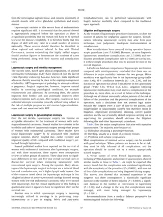

Laparoscopic surgery offers a minimally invasive alternative to traditional open surgical procedures, providing benefits such as reduced postoperative morbidity and shorter recovery times. Despite its advantages, few gynecological surgeries are performed laparoscopically due to the technical complexity and the necessity for extensive retraining for surgeons. A range of laparoscopic procedures, including hysterectomy and myomectomy, can enhance patient outcomes, but challenges in skill acquisition and implementation persist in practice.


