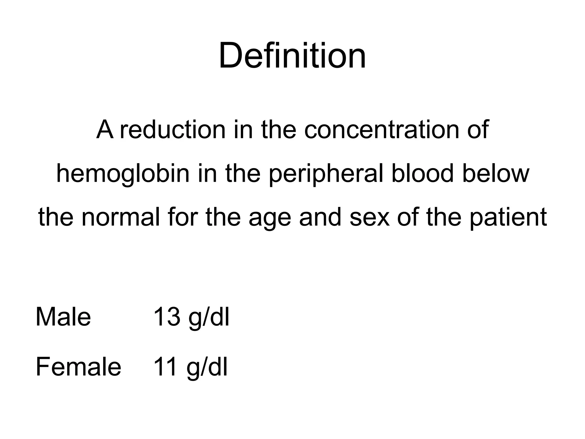
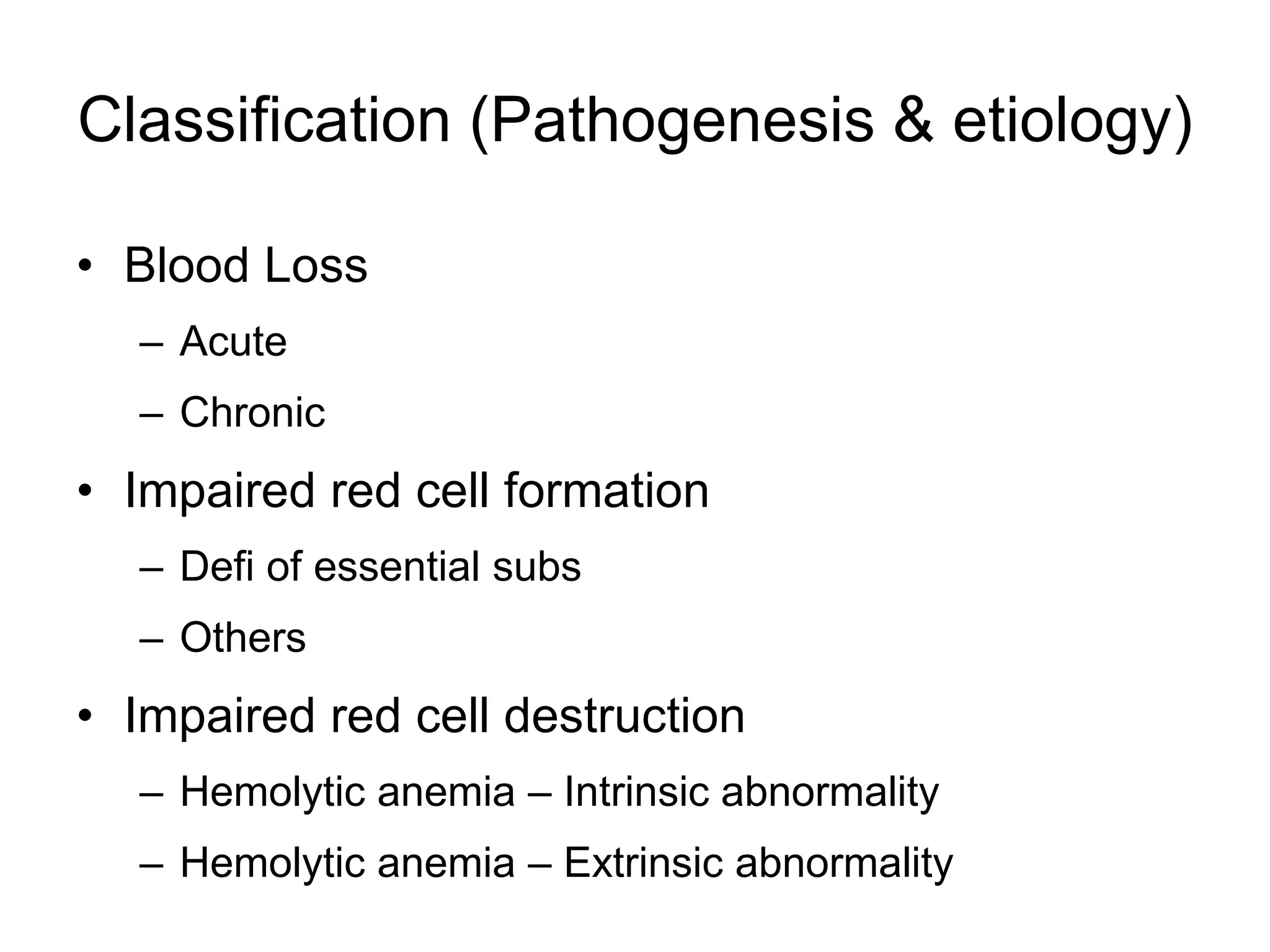
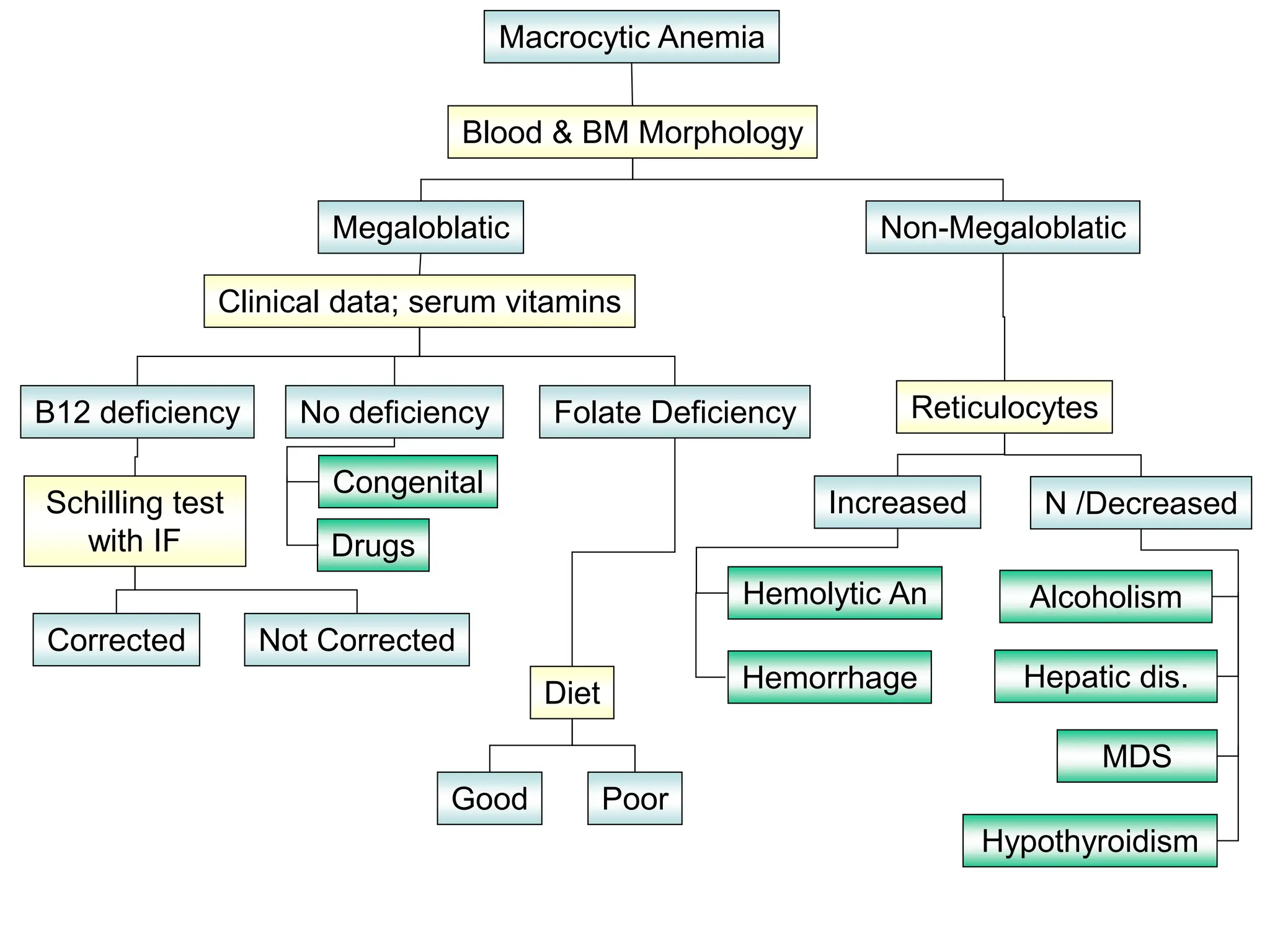
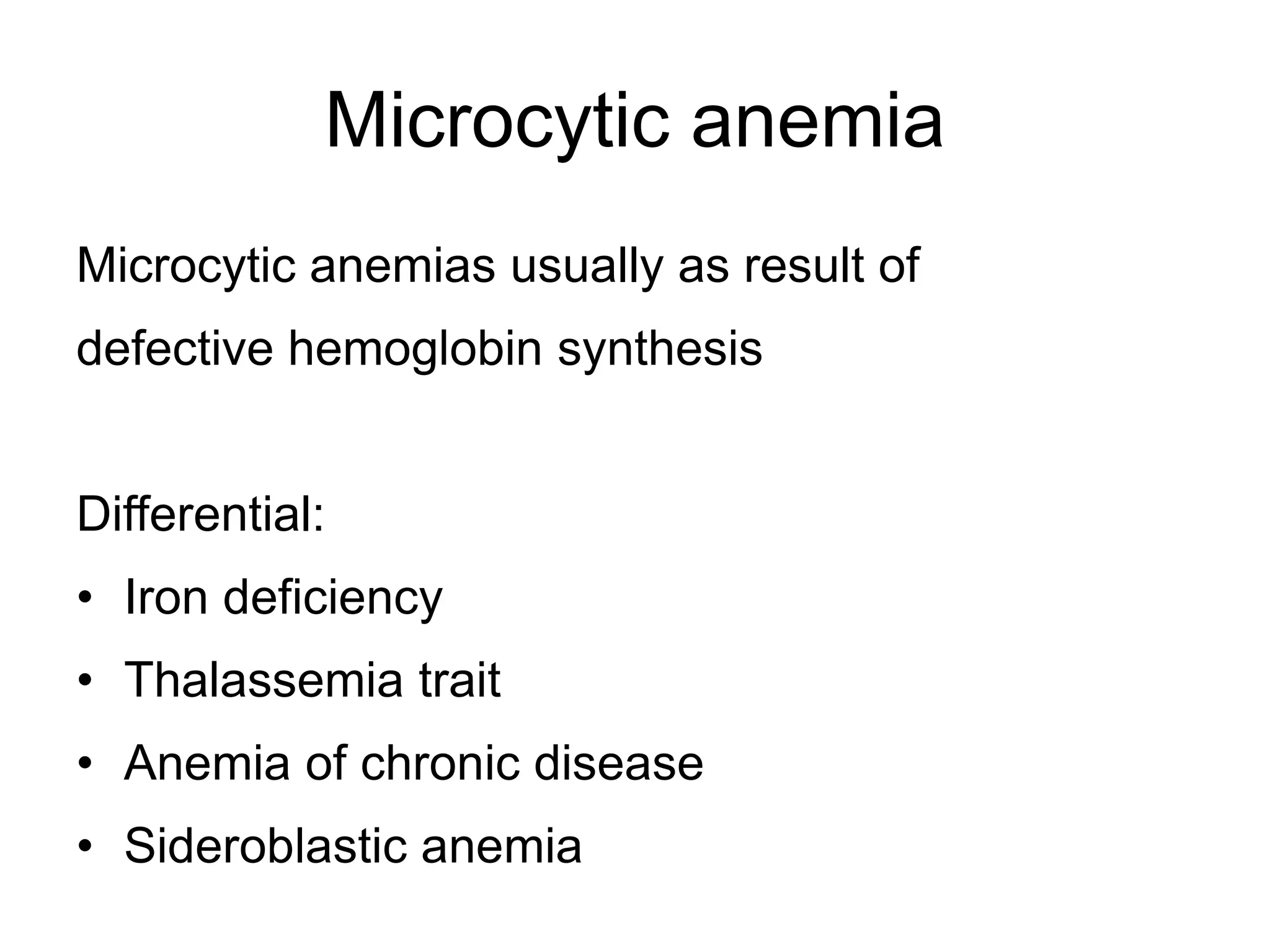
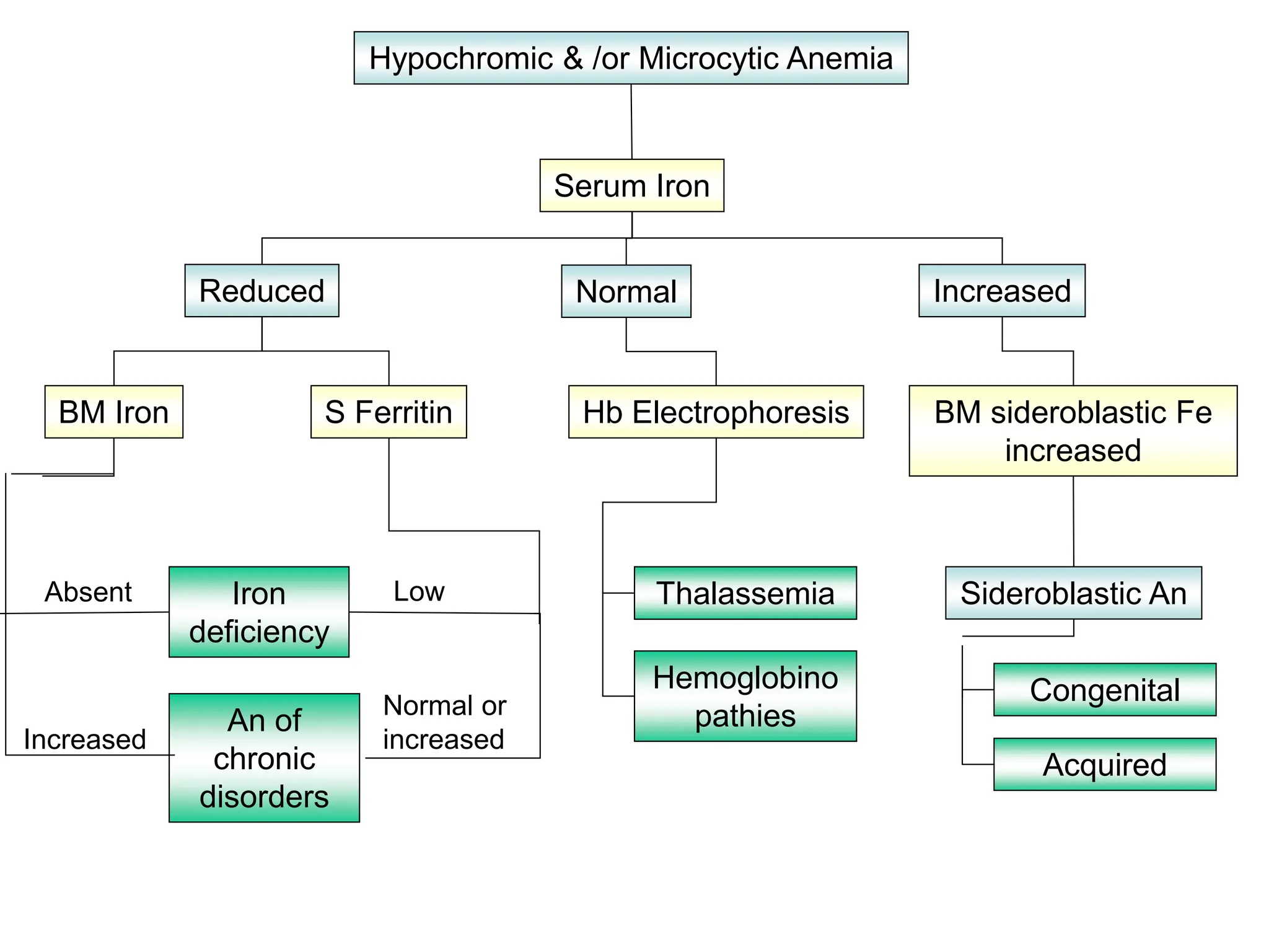
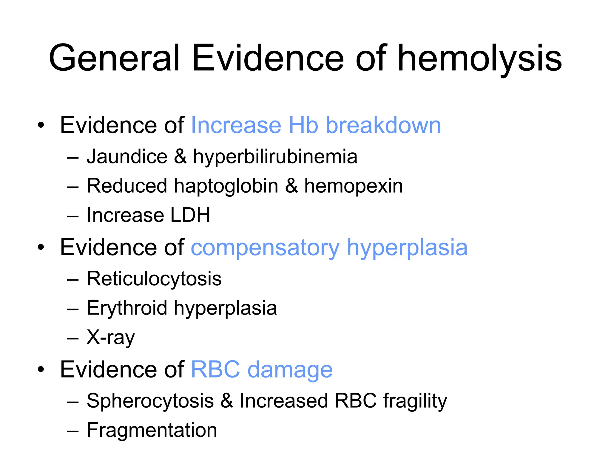
Approach to Anemia
Definition
A reduction in the concentration of
hemoglobin in the peripheral blood below
the normal for the age and sex of the patient
Male 13 g/dl
Female 11 g/dl
Important to Remember:
• Anemia is a symptom and not a disease
• Look for the Primary Cause!
Classification (Pathogenesis & etiology)
• Blood Loss
– Acute
– Chronic
• Impaired red cell formation
– Defi of essential subs
– Others
• Impaired red cell destruction
– Hemolytic anemia – Intrinsic abnormality
– Hemolytic anemia – Extrinsic abnormality
What is the mean corpuscular volume (MCV )
Classify chronic anemia as:
Microcytic (decreased MCV )
Normocytic (normal MCV )
Macrocytic (increased MCV )
Anemia
MCV
Blood smear
MCV >100 MCV 80-100 MCV <80
Macrocytic Normocytic Microcytic
Macrocytic Anemia
Blood & BM Morphology
Hemolytic An Alcoholism
Hemorrhage Hepatic dis.
MDS
Non-Megaloblatic
Clinical data; serum vitamins
Folate Deficiency Reticulocytes
No deficiency
B12 deficiency
N /Decreased
Increased
Hypothyroidism
Congenital
Drugs
Schilling test
with IF
Diet
Corrected Not Corrected
Good Poor
Megaloblatic
Microcytic anemia
Microcytic anemias usually as result of
defective hemoglobin synthesis
Differential:
• Iron deficiency
• Thalassemia trait
• Anemia of chronic disease
• Sideroblastic anemia
Iron Deficiency vs Thalassemia
Hb MCV RDW
Thalassemia Normal/Slight
ly decreased 65 Normal
Fe
deficiency
Decreased 65 Increased
Hypochromic & /or Microcytic Anemia
Serum Iron
Thalassemia Sideroblastic An
Hemoglobino
pathies
Congenital
Acquired
Iron
deficiency
Normal Increased
Reduced
Hb Electrophoresis BM sideroblastic Fe
increased
S Ferritin
BM Iron
An of
chronic
disorders
Low
Normal or
increased
Increased
Absent
Normocytic Anemia
Reticulocytes
BM
Negative
screens
ACD
Hemolytic
An
Normal or decreased
Increased
Post
hemorrhagic
History, Course, PBS, Bile
pigment
Screen for renal,
hepatic, endocrine dis.
Serum
Iron
Normal or
High
Low
Early
IDA
Hypoplastic An
MDS
Infiltration
General Evidence of hemolysis
• Evidence of Increase Hb breakdown
– Jaundice & hyperbilirubinemia
– Reduced haptoglobin & hemopexin
– Increase LDH
• Evidence of compensatory hyperplasia
– Reticulocytosis
– Erythroid hyperplasia
– X-ray
• Evidence of RBC damage
– Spherocytosis & Increased RBC fragility
– Fragmentation

Laboratory Diagnosis and approachof Anemia.ppt

  • 1.
  • 2.
    Definition A reduction inthe concentration of hemoglobin in the peripheral blood below the normal for the age and sex of the patient Male 13 g/dl Female 11 g/dl
  • 3.
    Important to Remember: •Anemia is a symptom and not a disease • Look for the Primary Cause!
  • 4.
    Classification (Pathogenesis &etiology) • Blood Loss – Acute – Chronic • Impaired red cell formation – Defi of essential subs – Others • Impaired red cell destruction – Hemolytic anemia – Intrinsic abnormality – Hemolytic anemia – Extrinsic abnormality
  • 5.
    What is themean corpuscular volume (MCV ) Classify chronic anemia as: Microcytic (decreased MCV ) Normocytic (normal MCV ) Macrocytic (increased MCV )
  • 6.
    Anemia MCV Blood smear MCV >100MCV 80-100 MCV <80 Macrocytic Normocytic Microcytic
  • 7.
    Macrocytic Anemia Blood &BM Morphology Hemolytic An Alcoholism Hemorrhage Hepatic dis. MDS Non-Megaloblatic Clinical data; serum vitamins Folate Deficiency Reticulocytes No deficiency B12 deficiency N /Decreased Increased Hypothyroidism Congenital Drugs Schilling test with IF Diet Corrected Not Corrected Good Poor Megaloblatic
  • 8.
    Microcytic anemia Microcytic anemiasusually as result of defective hemoglobin synthesis Differential: • Iron deficiency • Thalassemia trait • Anemia of chronic disease • Sideroblastic anemia
  • 9.
    Iron Deficiency vsThalassemia Hb MCV RDW Thalassemia Normal/Slight ly decreased 65 Normal Fe deficiency Decreased 65 Increased
  • 10.
    Hypochromic & /orMicrocytic Anemia Serum Iron Thalassemia Sideroblastic An Hemoglobino pathies Congenital Acquired Iron deficiency Normal Increased Reduced Hb Electrophoresis BM sideroblastic Fe increased S Ferritin BM Iron An of chronic disorders Low Normal or increased Increased Absent
  • 11.
    Normocytic Anemia Reticulocytes BM Negative screens ACD Hemolytic An Normal ordecreased Increased Post hemorrhagic History, Course, PBS, Bile pigment Screen for renal, hepatic, endocrine dis. Serum Iron Normal or High Low Early IDA Hypoplastic An MDS Infiltration
  • 12.
    General Evidence ofhemolysis • Evidence of Increase Hb breakdown – Jaundice & hyperbilirubinemia – Reduced haptoglobin & hemopexin – Increase LDH • Evidence of compensatory hyperplasia – Reticulocytosis – Erythroid hyperplasia – X-ray • Evidence of RBC damage – Spherocytosis & Increased RBC fragility – Fragmentation