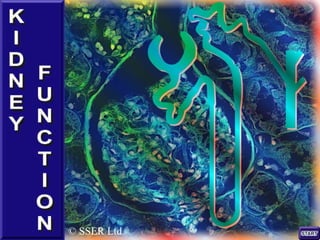
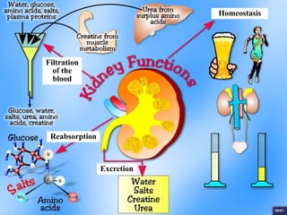
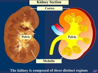
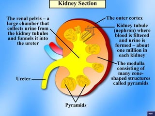
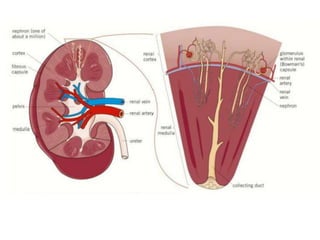
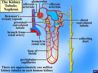
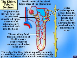
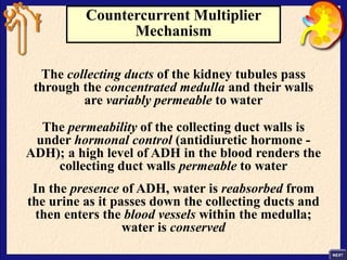
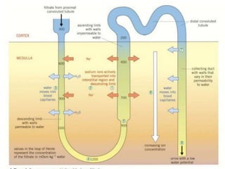
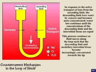
© SSER Ltd.
Filtration
of the
blood
Reabsorption
Excretion
Homeostasis
left kidney
adrenal gland
ureters
The urethra carries urine
away from the bladder
renal artery
renal vein
vena cava aorta
bladder
The Renal
System
The Mammalian Kidney
The human kidneys are two bean-shaped
organs, located against the dorsal body
wall on either side of the backbone
The principal functions of the kidneys are:
• Excretion – the removal of toxic nitrogenous
waste products from the blood; these include
urea and creatine (a waste product of muscle
metabolism)
• Homeostasis – the kidneys regulate the water
content, ion composition and pH of the body
fluids; their role in the regulation of water
content is described as osmoregulation
Kidney Section
Pelvis
Pelvis
Cortex
Medulla
The kidney is composed of three distinct regions
Kidney Section
The outer cortex
Kidney tubule
(nephron) where
blood is filtered
and urine is
formed – about
one million in
each kidney
The medulla
consisting of
many cone-
shaped structures
called pyramids
Pyramids
The renal pelvis – a
large chamber that
collects urine from
the kidney tubules
and funnels it into
the ureter
Ureter
The Kidney Tubule (Nephron)
The kidney tubule or nephron is the
functional unit of the kidney
Each kidney tubule receives its own small supply of
blood from the extensive arteriole branches arising
from the renal artery; each human kidney contains
approximately 1 million tubules
Each kidney tubule is responsible for urine formation
and this involves three basic processes:
• Ultrafiltration of blood
• Selective tubular reabsorption of filtered
materials
• Tubular secretion; the secretion of
substances from the blood capillaries into
the kidney tubules
The Kidney
Tubule;
Nephron
Bowman’s
(renal) capsule
afferent
arteriole
efferent
arteriole
glomerulus
branch from
renal artery
proximal
convoluted
tubule
distal
convoluted
tubule
branch to
renal vein
loop of
Henlé
collecting
duct
peritubular
capillaries
There are approximately one million
kidney tubules in each human kidney
The Kidney
Tubule;
Nephron
Ultrafiltration of the blood
takes place at the glomerulus
The glomerular
filtrate flows into
the proximal
convoluted tubule
where useful
substances are
reabsorbed back
into the blood
The resulting fluid
flows into the loop of
Henlé where a
countercurrent
exchange mechanism
takes place
Water
reabsorption
occurs from the
distal convoluted
tubule and
collecting duct,
allowing for the
production of a
concentrated
urine
The walls of the distal tubules and collecting ducts
are variably permeable to water, depending upon the
presence or absence of antidiuretic hormone (ADH)
Urine formation begins with the filtration of
blood at the glomeruli of the renal corpuscles
Glomerular Filtration
The first step in the formation of urine is
glomerular filtration
The afferent arteriole, which provides the blood
supply to the glomerulus, branches to form a
network of capillaries which reunite to form the
outgoing or efferent arteriole
Ultrafiltration of the blood takes place at the
glomerulus producing a fluid that enters the cavity
of Bowman’s (renal) capsule; this filtered fluid is
called the glomerular filtrate
The driving force behind the filtering process is the
high blood pressure that builds up within the
glomerular capillaries
Blood pressure within the
glomerular capillaries
forces molecules through
the capillary/capsular
membranes
The capillary/capsular
membrane has three layers;
the capillary endothelium,
the basement membrane,
and the inner visceral layer
of Bowman’s capsule
(composed of podocyte cells)
The high blood pressure
(hydrostatic pressure) in
the glomerular capillaries
forces small molecules out
of the blood into the cavity
of Bowman’s capsule
The fluid that flows from the cavity of Bowman’s capsule into the
proximal convoluted tubule is isotonic to the blood plasma
Selective Reabsorption
The glomerular filtrate flows into the
proximal convoluted tubule, and at this
stage the fluid is isotonic with
respect to blood plasma
The principal role of the cells that line the
proximal tubule is the reabsorption of useful
materials back into the bloodstream
The epithelial cells lining the tubule bear
microvilli at their surface and possess numerous
mitochondria; these features facilitate the
reabsorption of water and solutes
lumen
epithelial
cells
microvilli
intercellular
spaces
many
mitochondria
interstitial
fluid
Fluid in the proximal convoluted tubules of the renal tubules contains
filtered molecules that are not intended for complete excretion
Glucose is the body’s main fuel; in healthy individuals, glucose is
completely reabsorbed from the proximal tubules back into the
surrounding blood capillaries
Salt, in the form of sodium and chloride ions, together with
amino acids, are partly reabsorbed into the blood from
the proximal tubule cells
Materials are reabsorbed by diffusion, facilitated diffusion and
active transport
The reabsorption of materials by the epithelial cells increases
their cytoplasmic concentration such that
water follows by osmosis
85% of the filtered fluid is reabsorbed from the proximal
convoluted tubules back into the blood stream
Facilitated
diffusion
Active
transport
Diffusion
Sodium ions in the
lumen of
the proximal
convoluted tubule
Reabsorption of Sodium Ions
• Sodium ions arc actively transported out of the cells lining the
proximal convoluted tubule into blood capillaries which carry them
away.
• The sodium ion concentration of these cells is therefore lowered.
• Sodium ions now diffuse down a concentration gradient from the
lumen of the proximal convoluted tubule into the epithelial lining
cells but only through special carrier proteins by facilitated
diffusion.
• These carrier proteins are of specific types, each of which carries
another molecule (glucose or amino acids or chloride ions, etc.)
along with the sodium ions. This is known as co-transport
• The molecules which have been co-transported into the cells of the
proximal convoluted tubule then diffuse into the blood. As a result,
all the glucose and most other valuable molecules are reabsorbed as
well as water.
Facilitated
diffusion
Facilitated
diffusion
Active
transport
and
diffusion
Active
transport
Diffusion
Glucose
molecules in the
lumen of
the proximal
convoluted tubule
Intercellular
space
The transport of glucose, amino acids and ions into the
epithelial cells creates a gradient of water potential across
the lining of the tubule; water is therefore withdrawn from
the tubule by osmosis, such that approximately 85%
of the filtered fluid is reabsorbed back into the blood
Countercurrent Multiplier
Mechanism
The ‘U’-shaped loop of the kidney tubule is the
loop of Henlé; it consists of a descending limb, a
hairpin bend and an ascending limb that runs in
the opposite direction (counter) to the
descending limb
The purpose of the loops of Henlé is to create a
gradient of hypertonicity (increasing salt
concentration) within the surrounding tissue of the
medulla that becomes increasingly concentrated on
moving from the cortex end towards the pelvis (i.e.
down the area surrounding the loop)
Countercurrent Multiplier
Mechanism
The collecting ducts of the kidney tubules pass
through the concentrated medulla and their walls
are variably permeable to water
The permeability of the collecting duct walls is
under hormonal control (antidiuretic hormone -
ADH); a high level of ADH in the blood renders the
collecting duct walls permeable to water
In the presence of ADH, water is reabsorbed from
the urine as it passes down the collecting ducts and
then enters the blood vessels within the medulla;
water is conserved
In response to the active
transport of ions from the
ascending limb, the
descending limb loses water
by osmosis and becomes
more concentrated; water
loss continues until the
concentrations of the
descending limb and the
interstitial tissue are equal
This process continues as
fluid moves along
the loop such that the
descending limb and
medullary interstitial tissue
become
increasingly concentrated
towards the tip

Kidney.pptx

  • 2.
  • 3.
  • 4.
    left kidney adrenal gland ureters Theurethra carries urine away from the bladder renal artery renal vein vena cava aorta bladder The Renal System
  • 5.
    The Mammalian Kidney Thehuman kidneys are two bean-shaped organs, located against the dorsal body wall on either side of the backbone The principal functions of the kidneys are: • Excretion – the removal of toxic nitrogenous waste products from the blood; these include urea and creatine (a waste product of muscle metabolism) • Homeostasis – the kidneys regulate the water content, ion composition and pH of the body fluids; their role in the regulation of water content is described as osmoregulation
  • 6.
    Kidney Section Pelvis Pelvis Cortex Medulla The kidneyis composed of three distinct regions
  • 7.
    Kidney Section The outercortex Kidney tubule (nephron) where blood is filtered and urine is formed – about one million in each kidney The medulla consisting of many cone- shaped structures called pyramids Pyramids The renal pelvis – a large chamber that collects urine from the kidney tubules and funnels it into the ureter Ureter
  • 9.
    The Kidney Tubule(Nephron) The kidney tubule or nephron is the functional unit of the kidney Each kidney tubule receives its own small supply of blood from the extensive arteriole branches arising from the renal artery; each human kidney contains approximately 1 million tubules Each kidney tubule is responsible for urine formation and this involves three basic processes: • Ultrafiltration of blood • Selective tubular reabsorption of filtered materials • Tubular secretion; the secretion of substances from the blood capillaries into the kidney tubules
  • 10.
    The Kidney Tubule; Nephron Bowman’s (renal) capsule afferent arteriole efferent arteriole glomerulus branchfrom renal artery proximal convoluted tubule distal convoluted tubule branch to renal vein loop of Henlé collecting duct peritubular capillaries There are approximately one million kidney tubules in each human kidney
  • 11.
    The Kidney Tubule; Nephron Ultrafiltration ofthe blood takes place at the glomerulus The glomerular filtrate flows into the proximal convoluted tubule where useful substances are reabsorbed back into the blood The resulting fluid flows into the loop of Henlé where a countercurrent exchange mechanism takes place Water reabsorption occurs from the distal convoluted tubule and collecting duct, allowing for the production of a concentrated urine The walls of the distal tubules and collecting ducts are variably permeable to water, depending upon the presence or absence of antidiuretic hormone (ADH)
  • 13.
    Urine formation beginswith the filtration of blood at the glomeruli of the renal corpuscles
  • 14.
    Glomerular Filtration The firststep in the formation of urine is glomerular filtration The afferent arteriole, which provides the blood supply to the glomerulus, branches to form a network of capillaries which reunite to form the outgoing or efferent arteriole Ultrafiltration of the blood takes place at the glomerulus producing a fluid that enters the cavity of Bowman’s (renal) capsule; this filtered fluid is called the glomerular filtrate The driving force behind the filtering process is the high blood pressure that builds up within the glomerular capillaries
  • 15.
    Blood pressure withinthe glomerular capillaries forces molecules through the capillary/capsular membranes The capillary/capsular membrane has three layers; the capillary endothelium, the basement membrane, and the inner visceral layer of Bowman’s capsule (composed of podocyte cells)
  • 16.
    The high bloodpressure (hydrostatic pressure) in the glomerular capillaries forces small molecules out of the blood into the cavity of Bowman’s capsule The fluid that flows from the cavity of Bowman’s capsule into the proximal convoluted tubule is isotonic to the blood plasma
  • 18.
    Selective Reabsorption The glomerularfiltrate flows into the proximal convoluted tubule, and at this stage the fluid is isotonic with respect to blood plasma The principal role of the cells that line the proximal tubule is the reabsorption of useful materials back into the bloodstream The epithelial cells lining the tubule bear microvilli at their surface and possess numerous mitochondria; these features facilitate the reabsorption of water and solutes
  • 19.
  • 20.
    Fluid in theproximal convoluted tubules of the renal tubules contains filtered molecules that are not intended for complete excretion Glucose is the body’s main fuel; in healthy individuals, glucose is completely reabsorbed from the proximal tubules back into the surrounding blood capillaries Salt, in the form of sodium and chloride ions, together with amino acids, are partly reabsorbed into the blood from the proximal tubule cells Materials are reabsorbed by diffusion, facilitated diffusion and active transport The reabsorption of materials by the epithelial cells increases their cytoplasmic concentration such that water follows by osmosis 85% of the filtered fluid is reabsorbed from the proximal convoluted tubules back into the blood stream
  • 21.
    Facilitated diffusion Active transport Diffusion Sodium ions inthe lumen of the proximal convoluted tubule
  • 22.
    Reabsorption of SodiumIons • Sodium ions arc actively transported out of the cells lining the proximal convoluted tubule into blood capillaries which carry them away. • The sodium ion concentration of these cells is therefore lowered. • Sodium ions now diffuse down a concentration gradient from the lumen of the proximal convoluted tubule into the epithelial lining cells but only through special carrier proteins by facilitated diffusion. • These carrier proteins are of specific types, each of which carries another molecule (glucose or amino acids or chloride ions, etc.) along with the sodium ions. This is known as co-transport • The molecules which have been co-transported into the cells of the proximal convoluted tubule then diffuse into the blood. As a result, all the glucose and most other valuable molecules are reabsorbed as well as water.
  • 23.
  • 24.
    The transport ofglucose, amino acids and ions into the epithelial cells creates a gradient of water potential across the lining of the tubule; water is therefore withdrawn from the tubule by osmosis, such that approximately 85% of the filtered fluid is reabsorbed back into the blood
  • 26.
    Countercurrent Multiplier Mechanism The ‘U’-shapedloop of the kidney tubule is the loop of Henlé; it consists of a descending limb, a hairpin bend and an ascending limb that runs in the opposite direction (counter) to the descending limb The purpose of the loops of Henlé is to create a gradient of hypertonicity (increasing salt concentration) within the surrounding tissue of the medulla that becomes increasingly concentrated on moving from the cortex end towards the pelvis (i.e. down the area surrounding the loop)
  • 27.
    Countercurrent Multiplier Mechanism The collectingducts of the kidney tubules pass through the concentrated medulla and their walls are variably permeable to water The permeability of the collecting duct walls is under hormonal control (antidiuretic hormone - ADH); a high level of ADH in the blood renders the collecting duct walls permeable to water In the presence of ADH, water is reabsorbed from the urine as it passes down the collecting ducts and then enters the blood vessels within the medulla; water is conserved
  • 29.
    In response tothe active transport of ions from the ascending limb, the descending limb loses water by osmosis and becomes more concentrated; water loss continues until the concentrations of the descending limb and the interstitial tissue are equal This process continues as fluid moves along the loop such that the descending limb and medullary interstitial tissue become increasingly concentrated towards the tip