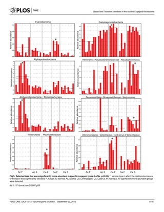
This study investigated the microbiome of three copepod species (Acartia longiremis, Centropages hamatus, Calanus finmarchicus) from the Gulf of Maine over a 3-week period in early summer. The microbiome contained both stable associations and temporal variability. Gammaproteobacteria, especially Pseudoalteromonas species, were consistently abundant across copepod species, suggesting a stable association. However, the microbiome composition also varied between full and starved gut copepods, and over time, influenced by environmental factors like food availability. While some core microbiome was present, temporal changes appeared important in structuring the bacterial communities associated with copepods.
![RESEARCH ARTICLE
Stable Associations Masked by Temporal
Variability in the Marine Copepod
Microbiome
Pia H. Moisander1
*, Andrew D. Sexton2
, Meaghan C. Daley1
1 Department of Biology, University of Massachusetts Dartmouth, North Dartmouth, Massachusetts, United
States of America, 2 Department of Biological Sciences, University of New Hampshire, Durham, New
Hampshire, United States of America
* pmoisander@umassd.edu
Abstract
Copepod-bacteria interactions include permanent and transient epi- and endobiotic associ-
ations that may play roles in copepod health, transfer of elements in the food web, and bio-
geochemical cycling. Microbiomes of three temperate copepod species (Acartia longiremis,
Centropages hamatus, and Calanus finmarchicus) from the Gulf of Maine were investigated
during the early summer season using high throughput amplicon sequencing. The most
prominent stable component of the microbiome included several taxa within Gammaproteo-
bacteria, with Pseudoalteromonas spp. especially abundant across copepod species.
These Gammaproteobacteria appear to be promoted by the copepod association, likely
benefitting from nutrient enriched microenvironments on copepods, and forming a more
important part of the copepod-associated community than Vibrio spp. during the cold-water
season in this temperate system. Taxon-specific associations included an elevated relative
abundance of Piscirickettsiaceae and Colwelliaceae on Calanus, and Marinomonas sp. in
Centropages. The communities in full and voided gut copepods had distinct characteristics,
thus the presence of a food-associated microbiome was evident, including higher abun-
dance of Rhodobacteraceae and chloroplast sequences in the transient communities. The
observed variability was partially explained by collection date that may be linked to factors
such as variable time since molting, gender differences, and changes in food availability
and type over the study period. While some taxon-specific and stable associations were
identified, temporal changes in environmental conditions, including food type, appear to be
key in controlling the composition of bacterial communities associated with copepods in this
temperate coastal system during the early summer.
Introduction
Microbial associations have been reported with numerous marine animals, including inverte-
brates and crustaceans [1, 2], but there is a limited understanding on the stability and function
PLOS ONE | DOI:10.1371/journal.pone.0138967 September 22, 2015 1 / 17
a11111
OPEN ACCESS
Citation: Moisander PH, Sexton AD, Daley MC
(2015) Stable Associations Masked by Temporal
Variability in the Marine Copepod Microbiome. PLoS
ONE 10(9): e0138967. doi:10.1371/journal.
pone.0138967
Editor: Adrianna Ianora, Stazione Zoologica Anton
Dohrn, Naples, ITALY
Received: July 2, 2015
Accepted: September 8, 2015
Published: September 22, 2015
Copyright: © 2015 Moisander et al. This is an open
access article distributed under the terms of the
Creative Commons Attribution License, which permits
unrestricted use, distribution, and reproduction in any
medium, provided the original author and source are
credited.
Data Availability Statement: The sequence data are
submitted to the NCBI database. The numbers are
KT186356-KT186363 for the COI. Illumina
sequences from this project have been deposited to
the NCBI Sequence Read Archive with accession
number SRP059778.
Funding: Funding was provided by National Science
Foundation (OCE 1130495) and the University of
Massachusetts Dartmouth to P.M., and the Shoals
Marine Lab for A.S. and P.M. The funders had no role
in study design, data analysis, decision to publish, or
preparation of the manuscript. Shoals Marine Lab
personnel provided assistance in sample collection.](https://image.slidesharecdn.com/c60e6df5-b2ed-4b19-8b27-a4a1d5040d4f-160113150935/85/journal-pone-0138967-1-320.jpg)
![RESEARCH ARTICLE
Stable Associations Masked by Temporal
Variability in the Marine Copepod
Microbiome
Pia H. Moisander1
*, Andrew D. Sexton2
, Meaghan C. Daley1
1 Department of Biology, University of Massachusetts Dartmouth, North Dartmouth, Massachusetts, United
States of America, 2 Department of Biological Sciences, University of New Hampshire, Durham, New
Hampshire, United States of America
* pmoisander@umassd.edu
Abstract
Copepod-bacteria interactions include permanent and transient epi- and endobiotic associ-
ations that may play roles in copepod health, transfer of elements in the food web, and bio-
geochemical cycling. Microbiomes of three temperate copepod species (Acartia longiremis,
Centropages hamatus, and Calanus finmarchicus) from the Gulf of Maine were investigated
during the early summer season using high throughput amplicon sequencing. The most
prominent stable component of the microbiome included several taxa within Gammaproteo-
bacteria, with Pseudoalteromonas spp. especially abundant across copepod species.
These Gammaproteobacteria appear to be promoted by the copepod association, likely
benefitting from nutrient enriched microenvironments on copepods, and forming a more
important part of the copepod-associated community than Vibrio spp. during the cold-water
season in this temperate system. Taxon-specific associations included an elevated relative
abundance of Piscirickettsiaceae and Colwelliaceae on Calanus, and Marinomonas sp. in
Centropages. The communities in full and voided gut copepods had distinct characteristics,
thus the presence of a food-associated microbiome was evident, including higher abun-
dance of Rhodobacteraceae and chloroplast sequences in the transient communities. The
observed variability was partially explained by collection date that may be linked to factors
such as variable time since molting, gender differences, and changes in food availability
and type over the study period. While some taxon-specific and stable associations were
identified, temporal changes in environmental conditions, including food type, appear to be
key in controlling the composition of bacterial communities associated with copepods in this
temperate coastal system during the early summer.
Introduction
Microbial associations have been reported with numerous marine animals, including inverte-
brates and crustaceans [1, 2], but there is a limited understanding on the stability and function
PLOS ONE | DOI:10.1371/journal.pone.0138967 September 22, 2015 1 / 17
a11111
OPEN ACCESS
Citation: Moisander PH, Sexton AD, Daley MC
(2015) Stable Associations Masked by Temporal
Variability in the Marine Copepod Microbiome. PLoS
ONE 10(9): e0138967. doi:10.1371/journal.
pone.0138967
Editor: Adrianna Ianora, Stazione Zoologica Anton
Dohrn, Naples, ITALY
Received: July 2, 2015
Accepted: September 8, 2015
Published: September 22, 2015
Copyright: © 2015 Moisander et al. This is an open
access article distributed under the terms of the
Creative Commons Attribution License, which permits
unrestricted use, distribution, and reproduction in any
medium, provided the original author and source are
credited.
Data Availability Statement: The sequence data are
submitted to the NCBI database. The numbers are
KT186356-KT186363 for the COI. Illumina
sequences from this project have been deposited to
the NCBI Sequence Read Archive with accession
number SRP059778.
Funding: Funding was provided by National Science
Foundation (OCE 1130495) and the University of
Massachusetts Dartmouth to P.M., and the Shoals
Marine Lab for A.S. and P.M. The funders had no role
in study design, data analysis, decision to publish, or
preparation of the manuscript. Shoals Marine Lab
personnel provided assistance in sample collection.](https://image.slidesharecdn.com/c60e6df5-b2ed-4b19-8b27-a4a1d5040d4f-160113150935/75/journal-pone-0138967-1-2048.jpg)
![of such associations at the microbial community level. Stable symbioses with microbes form in
many terrestrial insects and ticks [3], where the symbiotic bacteria may aid the host in diges-
tion, uptake of nutrients, reproduction, immune response, and other defenses [4]. In terrestrial
insect arthropods and some marine animal symbioses, the symbionts are often passed to the
next generation vertically (maternally), and the host and symbiont commonly undergo co-
evolution [5–7], as evidence of the importance of the permanent co-existence. Stable chemo-
lithoautotrophic symbioses are common in deep-sea invertebrates [2, 8], but little is known
about potential symbioses in copepods, marine pelagic crustaceans (Arthropoda). In addition
to stable symbioses, insects and invertebrates hosting symbionts also contain microbes that are
present only temporarily either as ecto- or endobionts or simply passing through the gut [9].
Copepods are known to host microbial communities [1] that appear to attach at greatest
abundances near the copepod mouth and anus, and egg sacs of females. Cultivation-based
studies have suggested an abundance of Gammaproteobacteria, especially Vibrio spp. both in
the surfaces and guts of copepods, while other groups such as Flavobacterium, Cytophaga, and
Pseudomonas have also been reported [10–13]. To date only few cultivation-independent stud-
ies using high-throughput sequencing methods have been conducted on the marine copepod
microbiome [14, 15], and little is known about temporal variability of the microbiome and fac-
tors controlling it.
Microbial communities in ecto- and endobiotic associations of copepods could potentially
be linked with the surrounding environmental conditions, especially the availability and type
of food [16]. Much of the microbiome could be passively recruited from the environment, thus
environmental controls should play an important role in controlling the composition and func-
tion. Additionally, copepods could contain host-specific associations, or symbioses, and a natu-
ral “core microbiome” dependent on the copepod environment that is independent of food. It
could be argued that a majority of the copepod-associated microorganisms are mostly from
food and random recruitment from the environment, and that the relationship with the cope-
pod is either neutral or parasitic. However, microscopic and cultivation data indicate that cope-
pods also contain intestinal, presumably non-transient flora [17], suggesting the community in
copepods is not purely under environmental control and might form an important ecological
association.
The goal of this study was to address the nature and variability of the microbiome on tem-
perate marine copepods. In the Gulf of Maine (GoM), many fish species rely on copepods as
food during their larval stages, thus copepods play an important role in trophic transfer
towards commercially important fish. This study focused on three common copepod genera
with distinct ecological niches in the Gulf of Maine, Acartia spp. (primarily A. longiremis), Cen-
tropages sp. (primarily C. hamatus), and Calanus finmarchicus. With a body length of 2–3 mm,
C. finmarchicus is the largest of the three, and is an important food source for fishes and whales
in the northwestern Atlantic Ocean [18–20] with its range covering the North Atlantic from
coastal waters to the open ocean. The smaller copepods A. longiremis and C. hamatus are pri-
marily estuarine and coastal species.
The major goal of this study was to provide a description of the microbiome in these impor-
tant North Atlantic copepods, to investigate whether there is a “core microbiome” suggesting
stable associations, whether such core microbiome varies among these three copepods with dif-
ferent ecological niches, and whether the microbiome experiences temporal changes. The study
was conducted over a 3-week period in early summer in the Gulf of Maine located in the
Northwestern Atlantic Ocean.
Stable and Transient Members in the Marine Copepod Microbiome
PLOS ONE | DOI:10.1371/journal.pone.0138967 September 22, 2015 2 / 17
Competing Interests: The authors have declared
that no competing interests exist.](https://image.slidesharecdn.com/c60e6df5-b2ed-4b19-8b27-a4a1d5040d4f-160113150935/85/journal-pone-0138967-2-320.jpg)
![Materials and Methods
Samples were collected between June 4 and 23, 2011, off the Shoals Marine Laboratory on
Appledore Island in the Gulf of Maine (42.9892°N, 70.6150°W) (S1 Table). Sampling was con-
ducted under permits for sampling marine organisms in the Gulf of Maine waters (State of
Maine Department of Marine Resources special license ME 2011-56-01 and New Hampshire
Fish and Game Department permit No. MFD 1117). No endangered or protected species were
sampled in this study. Most of the samples were collected between 9:30 am and noon. Cope-
pods were collected by towing a 200-μm zooplankton net behind a small boat at a slow speed
for approximately 10 min. The sample was diluted with surface seawater immediately upon
collection and poured into clean polyethylene containers. Surface water temperature was mea-
sured using a temperature probe (YSI) and salinity was measured with a refractometer. Upon
returning to the lab, the containers were kept at 4°C and copepods picked out of the plankton
samples under a dissecting microscope for molecular and microscopic analyses. Copepods
were identified to genus level, rinsed with 0.2 μm filtered seawater 2–3 times, then 1–10 live
copepods were placed in a tube with a mixture of 0.1—and 0.5—μm diameter sterile glass
beads (S1 Table) (these samples were termed ‘full gut copepods’). To study the influence of gut
contents on the overall microbiome, some copepods underwent a starvation period for removal
of gut contents. Copepods were picked to filtered seawater in petri dishes that were kept at 4°C
for 24–48 h, and the live copepods were then picked and preserved into tubes as above (these
samples were termed ‘starved copepods’). The relatively low holding temperature minimized
secondary bacterial growth during incubation, and was sufficient in keeping the copepods
alive. Hereafter these ‘starved’ copepods are referred to with ‘S’ and copepods with ‘full gut’,
preserved shortly after sampling, are referred to with ‘F’ (e.g. Acartia S and Acartia F, respec-
tively). Parallel water samples were collected from the same general area where the zooplank-
ton tows were conducted, by filling a polycarbonate bottle below the surface. The seawater was
filtered through a 0.2 μm Supor filter (Pall Gelman, Port Washington, NY) folded into a bead
beater tube with sterile glass beads (as above). Copepods and filtered seawater samples (S1
Table) were immediately placed in liquid nitrogen, and kept there until moved to -80°C for
long-term storage. Very few Calanus individuals were obtained in the samples, and only a set
of ‘Full gut’ individuals was included in the study.
DNA was extracted using the Qiagen Blood and Tissue kit with modifications (S1 Text).
Polymerase chain reaction was used to amplify the mitochondrial cytochrome oxidase gene
(COI) to confirm the identity of copepods, using primers LCO1490F and HCO2198R [21] (S1
Text, S2 Table). The GenBank accession numbers for the COI sequences from this study are
KT186356-KT186363.
For characterization of the microbial community, 16S rRNA gene was amplified using a
paired end amplicon sequencing procedure for Illumina MiSeq (S1 Text) employing the
V3-V4 region of the 16S rRNA gene [22]. The multiplexed sample was sequenced at the Tufts
University Core Facility using Illumina MiSeq V3 600 cycles paired end method. The samples
for this study were included in two separate sequencing runs, each in a set of a total of 96 sam-
ples multiplexed (pooled with samples from other studies). PCR with negative controls with
nuclease free water added in place of DNA template were run in parallel and sequenced,
although bands were not visible when the amplified products from these controls were run on
the gel electrophoresis.
The paired end sequences were first combined and initial downstream analyses conducted
in QIIME [23, 24]. Operational taxonomic unit (OTU) in this study was defined as group of
sequences that had a 97% or higher identity. OTUs that formed >1% of the sequences in the
negatives (no template controls) were removed from sample data, assuming any sequences
Stable and Transient Members in the Marine Copepod Microbiome
PLOS ONE | DOI:10.1371/journal.pone.0138967 September 22, 2015 3 / 17](https://image.slidesharecdn.com/c60e6df5-b2ed-4b19-8b27-a4a1d5040d4f-160113150935/85/journal-pone-0138967-3-320.jpg)
![representing these OTUs in the samples originated from reagent contamination [25, 26]
(S1 Text). QIIME was used in the initial analyses for OTU binning and the R software package
‘vegan’ was used to conduct the rarefaction analysis and make a heatmap with a dendrogram
(hierarchical clustering based on Bray-Curtis distance). Redundancy analysis (RDA) on the
Hellinger transformed data was also run in R, followed by ANOVA with 999 permutations.
Multidimensional Scaling (MDS) analysis was conducted, with the Bray-Curtis dissimilarities
of the communities tested using ANOSIM in Primer v6 [27]. LefSe analysis was used to investi-
gate which community members were significantly more abundant in each sample type [28].
Illumina sequences from this project have been deposited to the NCBI Sequence Read Archive
with accession number SRP059778.
Results
Water temperature increased steadily from 12.1 to 16°C during the study period while salinity
varied between 30–31, except for June 10th
when the salinity was 34. Copepods were identified
based on visual observations as Acartia spp., Centropages spp., and Calanus finmarchicus, and
further species level identification was conducted based on the COI sequence. COI data from
copepod samples identified microscopically as Centropages spp. suggested their closest match
to be C. hamatus (EU016220.1; 99–100% identity). The closest match for sequences obtained
from the samples identified as Acartia spp. was A. longiremis (KC287256.1; 99–100% identity).
The 16S rRNA gene sequence yield varied from 6040 to 132576 (62523±31897; ave±stdev)
sequences per sample before removal of negatives (S1 Table). The negative controls resulted in
a low number of sequences with low diversity (see Methods and S1 Text).
The projected total richness was greatest in the seawater samples, with the number of OTUs
continuing to increase until 3x10⁵
sequences obtained from seawater (Fig 1). The second high-
est projected phylotype richness was present in Acartia F, with approximately 1300 OTUs at
the full size of the sequence set. Centropages S had a slightly higher projected richness than
Centropages F and Calanus F, and starved Acartia S, all of which had closely similar rarefaction
curves. While they still had a slightly increasing OTU richness at the full size of each library,
the curves from copepods had a more gradual increase than the sequences from seawater, sug-
gesting lower overall richness.
Community composition
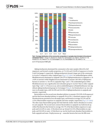
The proportion of Gammaproteobacteria was higher in copepods than in the water samples
(57±23% vs. 10±3.2% of sequences respectively) (Fig 2). In Acartia F, the proportion of Gam-
maproteobacteria was close to equal to that of Alphaproteobacteria, but in all other copepod
types, Gammaproteobacteria formed the largest part of the community. Proportion of Gam-
maproteobacteria was generally greater in starved Acartia and Centropages (63±9.7 and 70
±8.4, respectively) than in their full gut counterparts (40±24% in Acartia F and 34±21% in Cen-
tropages F) (Fig 2). In Calanus F, the proportion of Gammaproteobacteria was similar to or
higher (64–87% of sequences) than that in Acartia S and Centropages S.
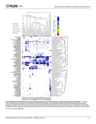
Several Gammaproteobacteria were found at higher proportions in copepods than in seawa-
ter. The genus Pseudoalteromonas formed 7.4±9.5% of Gammaproteobacteria in water and 32
±25% in copepods, being the most abundant and consistently dominant Gammaproteobacter-
ium overall in copepods (Fig 3). Glaciecola sp. consistently formed a large proportion in cope-
pods (17±21% of Gammaproteobacteria), and <0.5% of Gammaproteobacteria in all water
samples. A genus in Colwelliaceae within Alteromonadales had high abundances in both full
gut and starved copepods, and appeared to co-vary with abundances of Pseudoalteromonas
spp. (Fig 3, S3 Table). The most abundant genus within Vibrionaceae formed 3.7±8.3% of all
Stable and Transient Members in the Marine Copepod Microbiome
PLOS ONE | DOI:10.1371/journal.pone.0138967 September 22, 2015 4 / 17](https://image.slidesharecdn.com/c60e6df5-b2ed-4b19-8b27-a4a1d5040d4f-160113150935/85/journal-pone-0138967-4-320.jpg)
![gammaproteobacterial sequences in copepods. Photobacterium, however, was a significant
group in Centropages S (Figs 3 and 4). There were other groups within Gammaproteobacteria
that were absent or formed a very small proportion of sequences in seawater, but were present
in copepods at times at high proportions. These groups included Halomonadaceae, Moraxella-
ceae, and Marinobacter spp. (Fig 3, S3 Table) that were all found across many replicates in Cen-
tropages S, but were also found in other copepod types. Within Gammaproteobacteria,
Pseudoalteromonas sp. and Marinomonas sp. were significantly more abundant groups in Cen-
tropages S, unidentified genera of Colwelliaceae and Piscirickettsiaceae were significantly ele-
vated in Calanus F, and an Acitenobacter sp. was significantly elevated in Centropages F (LefSe,
p<0.05, Fig 4).
A few ‘seawater-associated’ groups were also identified within Gammaproteobacteria, hav-
ing consistently high abundances in seawater samples, and generally a low proportion in cope-
pods, with the exception of a few, mostly full gut copepods (Fig 3). A phylotype identified as
Candidatus Portiera formed a large component of the Gammaproteobacteria in seawater (27–
62%), and in general formed a low proportion (<2%) of the community in copepods, with the
exception of a few full gut copepods of all types and one starved Centropages where they
formed up to 18% of Gammaproteobacteria (S3 Table). The SAR92 clade Gammaproteobacter-
ium, represented by the strain HTCC2207 isolated from the Oregon coast [29], formed 3.3–
20.3% of Gammaproteobacteria in water samples and a smaller proportion (0–10%) in cope-
pods, being most abundant in a few full gut copepods of all copepod types.
Fig 1. Rarefaction curves with pooled samples for the different sample types. OTU, Operational Taxonomic Unit (97% similarity threshold). S, starved;
F, full gut; 1, Acartia S; 2, Acartia F; 3, Centropages S; 4, Centropages F, 5, Calanus F, 6, Seawater.
doi:10.1371/journal.pone.0138967.g001
Stable and Transient Members in the Marine Copepod Microbiome
PLOS ONE | DOI:10.1371/journal.pone.0138967 September 22, 2015 5 / 17](https://image.slidesharecdn.com/c60e6df5-b2ed-4b19-8b27-a4a1d5040d4f-160113150935/85/journal-pone-0138967-5-320.jpg)
![Multidimensional Scaling (MDS) analysis was conducted to compare communities between
the sample types. Communities in seawater were significantly or borderline significantly differ-
ent from all copepod types (p<0.01 for Acartia S, Centropages S and F, and Calanus F, and
p = 0.058 for Acartia F; ANOSIM) (S1 Fig). Communities in Centropages S were significantly
different from Acartia F and Centropages F (p = 0.044 and p = 0.016, respectively), while com-
munities in Acartia S were not significantly different from the communities in any other cope-
pod type. Communities in Acartia F and Centropages F were not significantly different, and the
difference between Calanus F and Acartia F was borderline significant (p = 0.061, ANOSIM).
Discussion
Copepod specific associations
Presence of A. longiremis and C. hamatus in the study area in early summer is supported by
previous observations in the GoM. Both A. longiremis and C. hamatus tend to have high abun-
dances in the spring (May-June) in the Northwestern Atlantic coastal waters, while C. hamatus
abundances decrease in the summer [30, 31]. In a previous study, A. longiremis dominated in
the surface waters in May, and the species dominance switched to A. hudsonica in late June
[32]. Although on the annual basis, C. typicus dominated over C. hamatus [33], the latter domi-
nated during spring and early summer in our study area [32]. The low abundances of C.
Fig 6. Redundancy analysis for the operational taxonomic unit level microbial community data.
doi:10.1371/journal.pone.0138967.g006
Stable and Transient Members in the Marine Copepod Microbiome
PLOS ONE | DOI:10.1371/journal.pone.0138967 September 22, 2015 11 / 17](https://image.slidesharecdn.com/c60e6df5-b2ed-4b19-8b27-a4a1d5040d4f-160113150935/85/journal-pone-0138967-11-320.jpg)
![finmarchicus compared to Acartia and Centropages in our samples were expected based on
past reports in surface waters in the area where C. finmarchicus has a tendency to remain at
deeper depths during summer [32].
The Gammaproteobacterium Pseudoalteromonas spp. (Pseudoalteromonadaceae, Vibrio-
nales) was generally the most abundant and consistent group in the starved copepods, suggest-
ing a stable association, and was present in all copepod taxa. Copepod environment may be a
key niche for this group in GoM. Sequences within Vibrionaceae (Vibrionales), including Vib-
rio spp., were detected but were unexpectedly low in comparison [34, 35]. Although detected
even at low temperatures, abundances of some Vibrio spp. have a significant positive correla-
tion with water temperature [36, 37]. Our results suggest that the majority of the bacteria form-
ing stable associations with GoM copepods are Pseudoaltermonas spp., however it is possible
that Vibrio spp. become more abundant later in the summer as water temperatures increase.
While Vibrio spp. are often thought to form the core copepod microbiome, as recently seen in
the subtropical Sargasso Sea waters [14], our results suggest they are replaced by other Gamma-
proteobacteria during the cold season in temperate waters. Some Pseudoalteromonas spp. have
chitinases [38], which could provide a carbon source from the copepod exoskeleton, as is the
case for many Vibrio spp., however our methods cannot distinguish whether the detected bac-
teria were present as endo- or ectobionts on the copepods. Alteromonadales are also consid-
ered R-strategist copiotrophs, that would benefit from a high nutrient environment
surrounding the copepod relative to seawater. They also contribute to antifouling on various
marine animals [39], and could play such roles also on copepods, which would make the Pseu-
doalteromonas-copepod association mutualistic. Such defensive roles of microbes in animal
and plant associations is considered one important form of symbiosis [40].
A few microbial groups had high specificity to one copepod type, indicating taxon-specific
niches. Marinomonas sp. was a prominent gammaproteobacterial group that was found at a
highest relative abundance in Centropages S, potentially forming a host-specific association. In
addition, many species within Photobacterium spp. (Vibrionaceae), observed at high relative
abundance in Centropages S, have bioluminescent properties, and may play multiple symbiotic,
commensal, or pathogenic roles on marine organisms [41]. Genera within Colwelliaceae
(Alteromonadales) and Piscirickettsiaceae each were observed that were significantly elevated
in Calanus F when compared to other copepod sample types. Like many marine bacteria, some
Colwellia spp. have chitinases [38], that would be beneficial in providing access to carbon on
the copepod exoskeleton. Piscirickettsiae family includes several genera with a range of meta-
bolic and ecological characteristics, some being fish pathogens and others that play specific
roles in biogeochemical cycling of sulphur and carbon. Further work should investigate the
roles of these groups in C. finmarchicus in more detail.
Influence of seasonality and food on copepod microbiome
The sampling time coincided with the initiation of summer stratification of the GoM water
mass [32]. As temperature increases, the life stage duration in the GoM copepods decreases,
with more frequent molting [42]. It is generally assumed that the exterior of the copepod is col-
onized by bacteria each time after molting, thus some of the temporal changes in the communi-
ties detected could be related with the length of time the microbial community had time to
colonize since last molting. The dominant groups found on copepods were detected also in sea-
water, suggesting rapid re-colonization is occurring. An example of such groups was Flavobac-
teria (Bacteroidetes), frequently recovered from copepods in previous studies [43], but it
appeared to have represented a transient community member in copepods in this study, given
its high relative abundance in seawater. Several other groups were found at high relative
Stable and Transient Members in the Marine Copepod Microbiome
PLOS ONE | DOI:10.1371/journal.pone.0138967 September 22, 2015 12 / 17](https://image.slidesharecdn.com/c60e6df5-b2ed-4b19-8b27-a4a1d5040d4f-160113150935/85/journal-pone-0138967-12-320.jpg)
![abundances in both seawater and copepods, suggesting the association of several bacterial
groups with copepods may be food associated, not host promoted.
Temperature is a major driver of bacterioplankton growth in temperate marine waters, thus
the increase in local temperature is likely to have influenced the ambient microbial abundance
and growth rates [44]. The community composition in seawater did not show prominent tem-
poral shifts over the study period, however, suggesting any temporal changes on the copepods
were not based on major changes in the surrounding bacterial community.
Persistent and significant differences were observed between the communities in full-gut
and starved copepods, demonstrating that temporary associations related to food play an
important part in the variability of bacterial community found in association of copepods.
Observed differences in full gut copepods among copepod species also strongly suggest food
preferences influence their microbiomes. The adults, nauplii and copepodite life stages of C.
finmarchicus are important grazers of primary producers and microzooplankton in the North
Atlantic waters [45], while A. longiremis is primarily herbivorous and C. hamatus utilizes
omnivory [46, 47]. Copepod feeding on picoplankton would be an unexpected path in the
marine microbial food web but could occur as part of dead or live particles or via aggregate
feeding [48, 49]. Direct bacterivory by copepods is generally thought to form only a minor por-
tion of carbon flow in aquatic food webs [50, 51], but the data from this study suggest bacteria
associated with food particles form a substantial portion of the copepod microbiome and possi-
bly part of their nutrition. This conclusion is supported by our recent study from coastal North
Atlantic waters in which the communities of N2-fixing bacteria in full gut copepods (primarily
Acartia) had high similarity with communities in seawater while the diazotrophic communities
in starved Acartia spp. were distinct [52]. In this study the particle-associated bacteria could
have been associated with phytoplankton or microzooplankton targeted as food, or less likely,
recently digested by such microzooplankton. The bacteria could also be in dead particles such
as fecal pellets that the copepods consume. Based on this study, in herbivorous and omnivorous
copepods, the pathway of carbon from such particle-associated bacteria directly to copepods
could be significant.
Besides the direct influence of food associated bacteria, conditions internally in the copepod
that are distinct from the surrounding seawater environment in terms of availability of oxygen
[53], acidity, and nutrients, could promote a specific microbial community responding to vari-
able food in situ [54]. The type and abundance of food resulted in differences in the cultivable
bacterial load from Acartia [16], suggesting that depending on type and quantity of food, the
core microbiome may differentially utilize carbon and nutrients. Changes in food quality dur-
ing the study may thus have also indirectly contributed to the observed variability. In addition,
C. hamatus can feed on both moving and suspended prey, but male and female clearance and
ingestion rates and food selectivities differ [46], which may have induced variability in the data
in this study, since females and males were not separated. Further, C. hamatus, like many other
copepods, feed on fecal pellets [55], which could contribute to the exchange of microbial com-
munity members across copepod taxa, and could have influenced the fact that some groups
known to be free-living (e.g. Synechococcus and SAR11) were at times still detected in copepods
24 h after starvation.
Overall our results suggest that these copepods have stable, potentially mutualistic associa-
tions with specific groups of Gammaproteobacteria, especially Pseudoalteromonas spp., during
the early summer in the Gulf of Maine. In addition, there are transient interactions with high
abundances of food-associated bacteria, including Rhodobacteraceae in Acartia that may con-
tribute to copepod nutrition. Further work should investigate the quantitative importance of
such pathway of bacterial carbon to these higher consumers not traditionally viewed as bacteri-
vores. Additionally, there are many less abundant bacterial groups that play potential taxon-
Stable and Transient Members in the Marine Copepod Microbiome
PLOS ONE | DOI:10.1371/journal.pone.0138967 September 22, 2015 13 / 17](https://image.slidesharecdn.com/c60e6df5-b2ed-4b19-8b27-a4a1d5040d4f-160113150935/85/journal-pone-0138967-13-320.jpg)