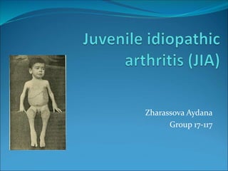
Juvenile idiopathic arthritis (JIA) is the most common type of arthritis in kids and teens. It typically causes joint pain and inflammation in the hands, knees, ankles, elbows and/or wrists. But, it may affect other body parts too . JIA used to be called juvenile rheumatoid arthritis (JRA), but the name changed because it is not a kid version of the adult disease.
2. a systemic chronic disease that develops in children under the age of 16,
characterized by a predominant destructive lesion of the joints, as well as
pathology of other organs and tissues with the formation of multiple organ
failure of varying severity.
Incidence 2-16:100,000 children under 16 years of age.
The prevalence is 0.05-0.6% (Baranov A. A., Alekseeva E. I., 2004).
The prevalence in the Russian Federation in children under 18 is
62.3:100,000 of the population, the primary incidence is 16.2:100,000.
Adolescents: prevalence 116.4 per 100,000.
primary incidence 28.3 per 100,000
Children 0-14 years old: prevalence 45.8 per 100,000.
primary incidence 12.6 per 100,000
3. Etiological factors
Hereditary factors: family susceptibility, markers of
predisposition, depending on sex and age.
Environmental factors: viral or mixed viral-bacterial
infection, joint trauma, insolation or hypothermia,
preventive vaccinations, especially against the
background of SARS or immediately after it.
4. Pathogenesis
Violation of microcirculation and damage to the cells lining the
synovial membrane.
Formation of altered IgGs (self-antigens)
Production by plasma cells of the synovial membrane of AT - antiIgG
(RF).
Autoantigen + antiIgG = CEC damaging effect on the vascular
endothelium and environment. Fabrics arthritis.
IL1, TNFα - inflammation, cartilage destruction, IL6 - hyperproduction
of CRP and fibrinogen.
Angiogenesis - increased destruction of cartilage - the formation of a
pannus (cloak), covering the surface of the cartilage - increased
destruction.
5. Classification of juvenile arthritis
Clinical and anatomical
characteristics of the disease
Clinical and immunological
characteristics of the disease
The degree of activity of the
process
Oligoarticular JIA (persistent or
progressive)
Polyarticular JIA (negative or
positive for rheumatoid
factor [RF])
Enthesitis-associated arthritis
Psoriatic JIA
Undifferentiated JIA
Systemic JIA
RF test is positive
RF test negative
rapid progression
slow progression
No noticeable progression
6. Classification of juvenile chronic arthritis
X-ray stage of arthritis The functional ability of the patient
I - periarticular osteoporosis; signs of effusion
into the joint cavity, compaction of
periarticular tissues, accelerated
growth of the epiphyses of the affected
joint
II - the same changes and narrowing of the
joint space
III - widespread osteoporosis, pronounced
osteochondral destruction,
dislocations, subluxations, systemic
bone growth disorder.
IV - changes inherent in I-III degree, and
ankylosis
1. Saved.
2. Violated by the state of the
musculoskeletal system:
A) the ability to self-service is preserved
B) the ability to self-service is partially lost
C) the ability to self-service is completely lost
1. 3. Violated by the condition of the eyes
and internal organs.
7.
8. Clinical picture
RA, predominantly articular form
Acute onset: fever, pain, swelling in one or more joints, often
symmetrical. Large joints (knees, ankles, wrists). The defeat of the
SHOP. Acute pain, swelling, 38-39°C, polymorphic allergic rash,
lymphadenopathy, hepatolienal syndrome. Anemia, ESR acceleration
up to 40-60 mm/h, shift to the left, increased IgG. More common in
preschoolers and younger students.
Subacute onset: arthritis of one joint (knee or ankle). Edema, dysfunction
without severe pain. Change in gait, morning stiffness up to 1 hour or
more. Uveitis. Several joints - oligoarticular form. The temperature is
normal, polyadenitis is moderate.
9.
10. RA, articular-visceral form:
acute onset, fever, polyarthritis with damage to small joints,
lymphadenopathy, hepatolienal syndrome, polyserositis, myocarditis,
anemia, a sharp acceleration of ESR. The rash is spotty, linear, rarely
maculopapular, hemorrhagic. hepatolienal syndrome.
Articular syndrome: arthralgia and exudative arthritis. Persistent changes
in the joints in 40% after 6 months, in 80-90% - after a year.
Deformities and contractures.
Macrophage activation syndrome (EBS, NSAIDs): activation and
proliferation of T cells, macrophages, decreased antiviral activity and
constant cellular activation - a systemic fatal inflammatory response
(γ-interferon, IL6, IL1, TNF-alpha).
The peak incidence is from 1 g to 5 years. The prognosis in 10-15 years is
unfavorable.
11. Complications
JIA with systemic onset:
Cardiopulmonary failure
Macrophage activation syndrome (hectic fever,
thrombocytopenia, leukopenia, decreased ESR, increased
fibrinogen content, bone marrow punctate contains a large
number of macrophages that phagocytize hematopoietic
cells).
Amyloidosis.
growth retardation.
infectious complications.
Flexion contractures (seropositive RA)
Disability
12. Damage of eyes during JIA
Uveitis 15-20%.
Localization: anterior
uveitis, peripheral uveitis,
posterior uveitis
panuveitis.
Downstream: acute,
subacute, chronic.
Depending on the number
of affected eyes:
unilateral, bilateral.
Complications: cataract,
corneal dystrophy,
vitreous fibrosis,
secondary glaucoma,
blindness.
13. Diagnostics
Clinical signs:
1. Arthritis lasting 3 months or more.
2. Arthritis of the second joint, which arose after 3 months and later.
3. Symmetrical damage to small joints.
4. Joint contractures.
5. Tenosynovitis or bursitis.
6. Muscular atrophy (often regional).
7. Morning stiffness.
8. Rheumatoid eye disease.
9. Rheumatoid nodules.
10. Effusion in the joint cavity.
Radiological signs:
1. Osteoporosis, small cystic restructuring of the bone structure of the epiphysis.
2. Narrowing of the joint space, bone erosion, ankylosis of the joints.
3. Violation of bone growth.
4. Damage to the cervical spine.
Laboratory signs:
1. Positive RF.
2. Positive synovial biopsy data.
14. Diagnostics
Depending on the number of identified positive signs, the degree of
probability of the presence of the disease is determined (with the
mandatory presence of arthritis):
3 signs - probable JRA;
· 4 features - certain JRA;
8 signs - classic JRA.
Differential Diagnosis:
rheumatoid arthritis
Reactive arthritis
Bechterew's disease
Reiter's disease
Traumatic arthritis (hemophilia)
15. Diagnosis
GBA, GUA, biochemical study.
Antinuclear factor, RF, immunogram.
ECG
Ultrasound of the abdominal organs, heart, joints.
X-ray examination of the chest.
X-ray examination of the joints
CT scan of the chest, joints.
Endoscopy.
17. Treatment
NSAIDs: diclofenac, acetylsalicylic acid, indomethacin, ibuprofen.
GC: oral prednisolone 0.2-0.5 mg / kg / day, methylprednisolone / in 10-15 mg /
kg, methylprednisolone and betamethasone - intra-articular (not more than 1
time in 1-3 months).
Basic preparations: quinoline (delagil, plaquenil), methotrexate 10-12
mg/m2/week, sulfasalazine 30-40 mg/kg/day, cyclosporine 4.4-4.5 mg/kg/day.
Immunotherapy (pentaglobin, intraglobin).
local therapy.
Antibiotics (aminoglycosides, cephalosporins 3.4, beta-lactams) 10-14 days.
Anticoagulants (heparin 100-150 U / kg), antiplatelet agents (pentoxifylline IV
20 mg / kg 2 times a day), fibrinolysis activators (nicotinic acid IV 5-10 mg 2
times a day).
Biological therapy: infliximab (monoclonal antibodies TNFα), rituximab (anti-
CD20 monoclonal antibodies), etanercept (genetically engineered protein that
binds TNFαß
18. Dispensary observation
Observation of a cardiorheumatologist
Physical examination once a month.
In the treatment of immunosuppressants: KLA,
biochemical blood test 1 time in 2 weeks, immunological
parameters 1 time in 3 months. (immunoglobulins, CRP,
RF, ANF), ECG 1 time in 3 months; Ultrasound, X-ray
examination 1 time in 6 months.
In the treatment of NSAIDs: EGDS 1 time in 6 months.
Ophthalmologist, slit lamp examination once every 3
months.
Form of disability.
Education at home (systemic beginning).
exercise therapy.
Vaccinations, the introduction of immunoglobulins are
contraindicated.
19. Markers of poor prognosis
The onset of the disease before 5 years;
Systemic variants of the debut of the disease;
Debut as oligoarthritis types 1 and 2;
Debut as a seropositive JRA variant;
Rapid (6 months) formation of a symmetrical generalized or
polyarticular articular syndrome;
Continuously relapsing course of the disease;
Significantly and persistent increase in ESR, the concentration of CRP,
IgG and RF in the blood serum;
Increasing functional insufficiency of the affected joints with limited
self-care during the first 6-12 months. illness.