1. Abstract
Conventional Organ transplantation often results in chronic organ rejection,
even when donor and recipient are matched at the tissue level. Genetic variation
between individuals gives rise to variable peptide antigens, which may be
recognized as “non-self” by T cells in the context of the MHC. Thus, novel
strategies are needed to overcome antigen induced organ rejection. One
potential strategy is xeno-transplantation, which is the use of non-human tissues
in transplantation. Preliminary studies have discovered that important antigens
are conserved in human and in murine cells. However, one antigen, H47, is not
detected on human cells although human cells possess and express the same
“H47 antigen” as seen in mouse cells. Although the human gene which encodes
H47 (VIMP) is transcriptionally expressed, one potential caveat is that the
precursor H47 protein is not processed to the H47 peptide antigen in human cell
systems. Current research is aimed at generating stable Transfectant cell lines,
which over-express HuVIMP and murine H-47 in human cell systems. These
cell lines will be subjected to a CML (T cell) killing assays at the Jackson Lab,
Bar Harbor, ME. A better understanding of antigen processing variations
between species can lead to a stronger understanding of cross species
transplantation barriers, which may be valuable in augmenting development of
novel xeno-transplantation strategies.
T-Cell Xeno-Antigen Recognition
Joseph Rovinsky
B.S. Biotechnology
Class of 2007
Principal Investigator: Dr. Peter Eden
Science Department
Introduction
Minor Histocompatibility Antigens
Proteins are constantly processed in the proteosome into peptide fragments
known as Minor Histocompatibility antigens. These antigens vary based
upon genomic differences between individuals within the same and across
species. Antigenic variation may elucidate a T Cell mediated attack when
presented by the MHC in normal immunosurveillance
MHC I
The Major Histocompatibility Complex I is a protein structure which
presents antigenic peptides for surveillance by Cytotoxic (CD8) T-Cells.
Cytotoxic T-Cells are specialized in there function to destroy cells which
present antigens which are recognized as foreign or “non-self
T-Cell /MHC Interaction(Figure 1)
Each T-Cell is specific for one MHC isoform. The MHC is encoded by
highly polymorphic genes, thus, the antigen peptides must be presented for
review in the context of the specific MHC molecule.
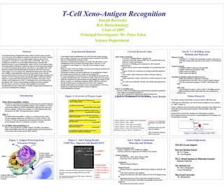
Figure 1. Antigen Processing from
Precursor Protein
NSF-RUI Grant Support
Harvard Medical School
Dr. Ye Yihong
Dr. Tom Rapopor
M.I.T. Broad Institute of Molecular Genetics
Shannon Chilewski
The Jackson Lab
Dr. Derry Roopenian
Greg Christianson
Marywood University
Dr. Michael Kiel
- Conventional organ transplantation can result in chronic organ rejection
due to antigen variations; even when donor/recipient matched at the MHC,
thus new transplantation strategies needed
- Alternative transplantation strategies are needed. One alternative is xeno-
transplantation, the use of tissues from other species. However, T-cell
recognition of “xeno-antigens” as “non-self” occurs, leading to organ
rejection
- Rules governing T-cell xeno-antigen expression, processing, presentation and
recognition are not well understood
- This project examines cross-species differences in transplantation antigen
processing and presentation of a unique barrier antigen H47
- It was previously discovered that mouse T-Cells specific for H4 and H7 will
recognize and kill human cells. H4 and H7 are thus conserved across species
- When the experiment was performed for the H47 antigen, the murine T cells
did not recognize several human lymphoid cell lines, and these cells lived
- The goal of this project is to understand why H47 is not recognized
- Results of this project may elucidate a better understanding of T-Cell xeno-
antigen processing and presentation, in order to augment
xenotransplantation strategies
Experimental Rationale
Figure 2. Overview of Project Goals
1 2 3 4 5 6
Figure 3. Sub-Cloning Results
VIMP Myc- Digested with BamH1/KPN
AIM I. Stable Transfection
-Cell Lines: CEMDB/CIRDB Lymphoid Cells
-Constructs: HuVIMP, Murine VIMP, H47A and H47B Mini-Genes
-Effectene Transfectant Reagent
-Previous T-Cell Killing assay conducted on cells transiently
transfected and
loaded with the various constructs/peptides. It is possible that only a
small
number of cells were transfected, preventing statistically significant
T-Cell
assay results. Stable transfectant cell lines will insure uniform
expression.
-Stable transfection “selects” based off of a selective pressure for cells
that
have only been transfected. All other cells will die when subjected to
an
antibiotic.
AIM II. T-Cell Killing Assay
-Previous CML (T-Cell Killing Assay) performed on transiently transfected
cells, results were inconclusive (Figure 4)
-Assay will be conducted in collaboration with the Jackson Lab,
Bar Harbor, ME which specializes in this technique
Current Research Aims
1. Effector: 21M/B10-A7 Target :CEMDB Transfected With HuVIMP
2. Effector: 21M/B10-A7 Target: CEMDB
3. Effector: 21M/B10-A7 Target: T2DB Loaded with mH47a
4. Effector: 21M/B10-A7 Target: T2DB Loaded with mH47b
5. Effector: 21M/B10-A7 Target: T2DB Loaded with Viral Peptides
Dark Blue=50:1 Red= 25:1 Yellow= 5:1 Light Blue= 0.5:1
Transfected Cells-Expected to
yield higher Percentage killed
(3).
Cells loaded with surface H47
effectively killed(4).
Stable Transfection will insure
uniform expression!
Figure 4. Preliminary T-Cell Killing Assay Results
Cell Lines (Suspension Cells)
-Human Lymphoid Cells (CEMDB and CIRDB)
-Cell Lines cultured in supplemented VcDMEM, 10^6 cells/mL
Genetic Constructs
-HuVIMP Myc-, H47a, H47b, Murine VIMP
-Expression Vector: pCDNA3.1 TOPO
Transfection
-Effectene Reagent (Qiagen)
Selective Pressure
-250 mg Neomycin, suspended in DMSO will be added to cells in 60 x 60
mm
-Cells will be split 48 hrs after addition
-Sample of transfectant cells will be subjected to RT-PCR (Qiagen) using
the
appropriate primers in order to determine transfection success
-After a two week culture, cells will be cryogenically preserved and
shipped to
the Jackson Lab, for further research
Aim I. Stable Transfection
Materials and Methods
Effectors (T-Cells)
-21M/B10-A7- T-Cell line that specifically recognizes and interacts
with MHC isoform DB, expressed on the surface of CEM/CIR cell
lines
Targets (Cell Lines)
-CEMDB/CEMDB Transfected with HuVIMP
-C1RDB Transfected with Murine VIMP
-T2DB Loaded with MH47A
-T2DB Loaded with MH47B
Chromium-51 Peptide Loading Procedural Overview
-T2DB cells incubated in radioactive chromium.
-Chromium binds to the MHC and can capture peptide antigens
-Cell’s then incubated in peptides “Loaded”, which bind to the
MHC
CML Assay
-Cells incubated for approx. 4 hours with T-Cells killing detected
-Different Effector/Target ratios used; this assay performed at
Jackson Lab
Aim II. T-Cell Killing Assay
Methods and Materials
Future Research
Acknowledgements
HuVIMP
1.)Lambda HindIII
2.)VIMP Clone Digest 1
3.)VIMP Clone Digest 10
4.)VIMP Clone Digest 9
5.)Blank
6.)Blank
VIMP Myc- successfully sub-cloned, removing Myc Tag Sequence
Restriction enzyme digestion (BamH1/KPN) liberated VIMP
fragment
DNA Sequencing results confirmed VIMP sequence without Myc
tag (Conducted at UCONN)
pcDNA3.1
Figure 5. Cellular Location of HuVIMP
The cellular location of HuVIMP was characterized in 2005 (Harvard)
VIMP interacts with Derlin-1, p97 and E3 protein complexes in the synthesis
of MHC I (Figure 5)
Is there a difference between humans and other organisms at the cellular level
which affects processing and presentation? Will potentially explore this
possibility!!!
Results of this research may elucidate novel information on antigen
processing/presentation across species, a useful application in developing xeno-
transplantation strategies.
T-Cells recognize
“foreign” antigens and
mediate an immune
attack
Antigenic differences
across species can lead to
rejection!