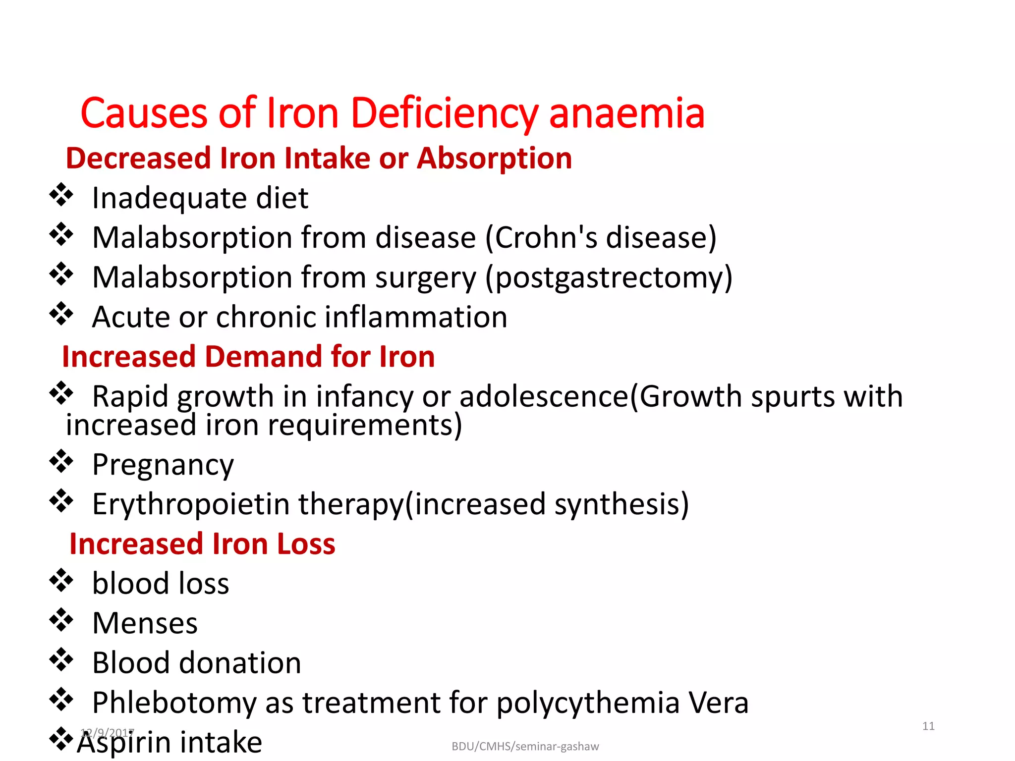
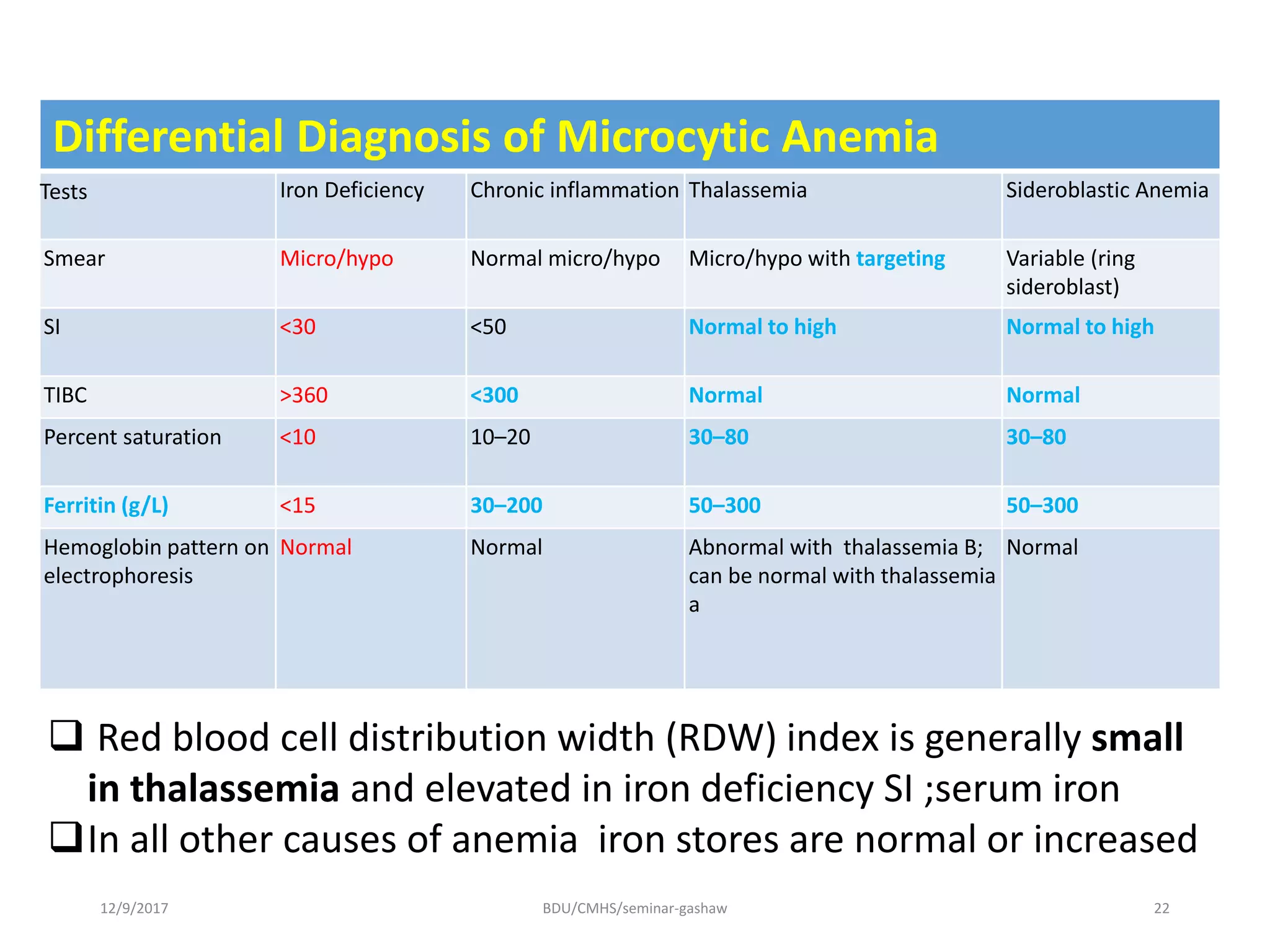
By: Gashaw-C1
12/9/2017 BDU/CMHS/seminar-gashaw 1
Outline
Definition
Epidemiology
Iron metabolism
Causes of iron deficiency anemia
Stages of IDA
Clinical presentation of IDA
Diagnosis of IDA
DDx
12/9/2017 BDU/CMHS/seminar-gashaw 2
Definition
A hypoproliferative anemia which
results from deficiency of iron to bone
marrow for normal erythtopoiesis.
Deficiency of iron is the most common
nutritional disorder in the world.
12/9/2017 BDU/CMHS/seminar-gashaw 3
Epidemiology
 Globally, 50% of anemia is attributable to iron
deficiency.
 Africa and parts of Asia bear 71% of the
global mortality burden
North America represents only 1.4% of the
total morbidity and mortality due to IDA
The WHO data on anemia 1997
2 billion people affected
Children & pregnant women most affected
12/9/2017 BDU/CMHS/seminar-gashaw 4
Iron metabolism
Iron is a critical element in the function of all cells
The major role of iron in mammals is to carry O2 as
part of hemoglobin
It is a critical element in iron-containing enzymes.
Free iron, is highly toxic in that it participates in
chemical reactions that generate free radicals such
as singlet O2 or OH–.
12/9/2017 BDU/CMHS/seminar-gashaw 5
12/9/2017 BDU/CMHS/seminar-gashaw 6
12/9/2017 BDU/CMHS/seminar-gashaw 7
Intestinal Absorption
 Iron containing heme = animal source such red meat , liver,
foods egg
non heme = grains, legumes & vegetables
It is absorbed specifically in the duodenum.
Enhancers: gastric HCL,citrate,mucin
Inhibitors; tea,coffee,phosphates,antacids, antibiotics (eg,
quinolones, tetracycline)
Milk is a poor source of iron, hence breast-fed babies need
iron supplements
Iron metabolism
12/9/2017 BDU/CMHS/seminar-gashaw 8
Proteins in Fe metabolism
 Transferrin(Tf):Transports Fe3+ through plasma
 Ferritin :Huge molecule for cellular storage of Fe
 Divalent Metal Transporter( DMT1)
 Ferroportin
 Hepcidin & Hephaestin
12/9/2017 BDU/CMHS/seminar-gashaw 9
Iron balance
Normal body content = 3-4gm
Daily iron loss = 1mg
 Adult male/women-absorb at least 1 mg /1.4 mg/d resp.
12/9/2017 BDU/CMHS/seminar-gashaw 10
Causes of Iron Deficiency anaemia
Decreased Iron Intake or Absorption
 Inadequate diet
 Malabsorption from disease (Crohn's disease)
 Malabsorption from surgery (postgastrectomy)
 Acute or chronic inflammation
Increased Demand for Iron
 Rapid growth in infancy or adolescence(Growth spurts with
increased iron requirements)
 Pregnancy
 Erythropoietin therapy(increased synthesis)
Increased Iron Loss
 blood loss
 Menses
 Blood donation
 Phlebotomy as treatment for polycythemia Vera
Aspirin intake12/9/2017
BDU/CMHS/seminar-gashaw
11
•Others causes IDA
•Pulmonary hemosiderosis
•Intravascular hemolysis
•Hemoglobinuria & hemosiderinuria
12/9/2017 BDU/CMHS/seminar-gashaw 12
Stages of Iron Deficiency
The progression to iron deficiency can be divided into three stages
1) Negative iron balance: -Depleted iron store without anemia
 The demands for (or losses of) iron exceed the body's ability to absorb iron
from the diet
 As long as iron stores are present and can be mobilized, the serum iron, total
iron-binding capacity (TIBC), and red cell protoporphyrin levels remain within
normal limits
 At this stage, red cell morphology and indices are normal.
12/9/2017 BDU/CMHS/seminar-gashaw 13
12/9/2017 BDU/CMHS/seminar-gashaw 14
2) Iron deficient erythropoiesis
 By definition, marrow iron stores are absent when the serum ferritin level is
<15 microg/L.
Gradually, the TIBC increases, as do red cell protoporphyrin levels. transferrin
saturation is 15-20%
The first appearance of microcytic cells, and hypochromic reticulocytes in
circulation
3) Iron deficiency anaemia
Gradually, the hemoglobin and hematocrit begin to fall, The transferrin
saturation at this point is 10–15%.
12/9/2017 BDU/CMHS/seminar-gashaw 15
When moderate anemia is present (hemoglobin 10–13
g/dL), the bone marrow remains hypoproliferative.
With more severe anemia (hemoglobin 7–8 g/dL),
hypochromia and microcytosis become more prominent,
 The erythroid marrow becomes increasingly ineffective.
Laboratory studies in the evolution of iron deficiency
12/9/2017 BDU/CMHS/seminar-gashaw 16
Clinical Presentation of Iron Deficiency
Signs related to iron deficiency depend on
the severity and chronicity of the anemia.
Increased likelihood- Pregnancy, adolescence, periods of rapid
growth, and an intermittent history of blood loss of any kind
• Iron deficiency in an adult male means GI blood loss until proven
otherwise
•Usual signs of anemia—fatigue, pallor, and reduced exercise capacity
17BDU/CMHS/seminar-gashaw12/9/2017
•In advanced state,
Cheilosis (fissures at the corners
of the mouth), or angular
stomatitis)
koilonychia (spooning of the
fingernails)
 Atrophic glossitis (red, glazed,
smooth tongue).
• Pica and pagophagia — perverted
appetite to clay or dirt (geophagia),
paper products, or starch
(amylophagia), ice BDU/CMHS/seminar-gashaw 1812/9/2017
PATERSON-KELLY/PLUMMER-VINSON
SYNDROME
•Oesophageal web
•Dysphagia
•Atrophic glossitis
12/9/2017 BDU/CMHS/seminar-gashaw 19
Diagnosis of iron deficiency anemia
History
• Gynecologic(including fertility)
• History of surgery(bleeding,gastrectomy)
• Dietary habit
• Bleeding(trauma,family history)
• Drugs(aspirin)
• Blood donation,phlebotomy
P/E
Investigations:-
 CB
 peripheral smear ,
stool exam iron study (occult blood ,ova of parasites)
Endoscopy ,colonoscopy
BM study,therapeutic trial
12/9/2017 BDU/CMHS/seminar-gashaw 20
Lab Features of IDA
RBC morphology:Microcytic-hypochromic
SI <30
 High TIBC >360
saturation levels below 10 to15%
 serum ferritin falls to <15 g/L.
Absent iron stores
12/9/2017 BDU/CMHS/seminar-gashaw 21
Differential Diagnosis of Microcytic Anemia
Tests Iron Deficiency Chronic inflammation Thalassemia Sideroblastic Anemia
Smear Micro/hypo Normal micro/hypo Micro/hypo with targeting Variable (ring
sideroblast)
SI <30 <50 Normal to high Normal to high
TIBC >360 <300 Normal Normal
Percent saturation <10 10–20 30–80 30–80
Ferritin (g/L) <15 30–200 50–300 50–300
Hemoglobin pattern on
electrophoresis
Normal Normal Abnormal with thalassemia B;
can be normal with thalassemia
a
Normal
12/9/2017 BDU/CMHS/seminar-gashaw 22
 Red blood cell distribution width (RDW) index is generally small
in thalassemia and elevated in iron deficiency SI ;serum iron
In all other causes of anemia iron stores are normal or increased
12/9/2017 BDU/CMHS/seminar-gashaw 23
TREATMENT
Treatment approach
• Treatment of the underlying cause
• Red Cell Transfusion
• Administration of iron
• Amount of iron needed is calculated from:
• Body weight (kg) x 2.3 x (15–pt's HGB, g/dL) + 500 or 1000 mg (for stores).
12/9/2017 BDU/CMHS/seminar-gashaw 24
References
Harrison 18th edition.
Robins and Cotran 8th edition.
The web.
12/9/2017 BDU/CMHS/seminar-gashaw 25
12/9/2017 BDU/CMHS/seminar-gashaw 26

Iron defficiency anemia

  • 1.
  • 2.
    Outline Definition Epidemiology Iron metabolism Causes ofiron deficiency anemia Stages of IDA Clinical presentation of IDA Diagnosis of IDA DDx 12/9/2017 BDU/CMHS/seminar-gashaw 2
  • 3.
    Definition A hypoproliferative anemiawhich results from deficiency of iron to bone marrow for normal erythtopoiesis. Deficiency of iron is the most common nutritional disorder in the world. 12/9/2017 BDU/CMHS/seminar-gashaw 3
  • 4.
    Epidemiology  Globally, 50%of anemia is attributable to iron deficiency.  Africa and parts of Asia bear 71% of the global mortality burden North America represents only 1.4% of the total morbidity and mortality due to IDA The WHO data on anemia 1997 2 billion people affected Children & pregnant women most affected 12/9/2017 BDU/CMHS/seminar-gashaw 4
  • 5.
    Iron metabolism Iron isa critical element in the function of all cells The major role of iron in mammals is to carry O2 as part of hemoglobin It is a critical element in iron-containing enzymes. Free iron, is highly toxic in that it participates in chemical reactions that generate free radicals such as singlet O2 or OH–. 12/9/2017 BDU/CMHS/seminar-gashaw 5
  • 6.
  • 7.
    12/9/2017 BDU/CMHS/seminar-gashaw 7 IntestinalAbsorption  Iron containing heme = animal source such red meat , liver, foods egg non heme = grains, legumes & vegetables It is absorbed specifically in the duodenum. Enhancers: gastric HCL,citrate,mucin Inhibitors; tea,coffee,phosphates,antacids, antibiotics (eg, quinolones, tetracycline) Milk is a poor source of iron, hence breast-fed babies need iron supplements
  • 8.
  • 9.
    Proteins in Femetabolism  Transferrin(Tf):Transports Fe3+ through plasma  Ferritin :Huge molecule for cellular storage of Fe  Divalent Metal Transporter( DMT1)  Ferroportin  Hepcidin & Hephaestin 12/9/2017 BDU/CMHS/seminar-gashaw 9
  • 10.
    Iron balance Normal bodycontent = 3-4gm Daily iron loss = 1mg  Adult male/women-absorb at least 1 mg /1.4 mg/d resp. 12/9/2017 BDU/CMHS/seminar-gashaw 10
  • 11.
    Causes of IronDeficiency anaemia Decreased Iron Intake or Absorption  Inadequate diet  Malabsorption from disease (Crohn's disease)  Malabsorption from surgery (postgastrectomy)  Acute or chronic inflammation Increased Demand for Iron  Rapid growth in infancy or adolescence(Growth spurts with increased iron requirements)  Pregnancy  Erythropoietin therapy(increased synthesis) Increased Iron Loss  blood loss  Menses  Blood donation  Phlebotomy as treatment for polycythemia Vera Aspirin intake12/9/2017 BDU/CMHS/seminar-gashaw 11
  • 12.
    •Others causes IDA •Pulmonaryhemosiderosis •Intravascular hemolysis •Hemoglobinuria & hemosiderinuria 12/9/2017 BDU/CMHS/seminar-gashaw 12
  • 13.
    Stages of IronDeficiency The progression to iron deficiency can be divided into three stages 1) Negative iron balance: -Depleted iron store without anemia  The demands for (or losses of) iron exceed the body's ability to absorb iron from the diet  As long as iron stores are present and can be mobilized, the serum iron, total iron-binding capacity (TIBC), and red cell protoporphyrin levels remain within normal limits  At this stage, red cell morphology and indices are normal. 12/9/2017 BDU/CMHS/seminar-gashaw 13
  • 14.
    12/9/2017 BDU/CMHS/seminar-gashaw 14 2)Iron deficient erythropoiesis  By definition, marrow iron stores are absent when the serum ferritin level is <15 microg/L. Gradually, the TIBC increases, as do red cell protoporphyrin levels. transferrin saturation is 15-20% The first appearance of microcytic cells, and hypochromic reticulocytes in circulation 3) Iron deficiency anaemia Gradually, the hemoglobin and hematocrit begin to fall, The transferrin saturation at this point is 10–15%.
  • 15.
    12/9/2017 BDU/CMHS/seminar-gashaw 15 Whenmoderate anemia is present (hemoglobin 10–13 g/dL), the bone marrow remains hypoproliferative. With more severe anemia (hemoglobin 7–8 g/dL), hypochromia and microcytosis become more prominent,  The erythroid marrow becomes increasingly ineffective.
  • 16.
    Laboratory studies inthe evolution of iron deficiency 12/9/2017 BDU/CMHS/seminar-gashaw 16
  • 17.
    Clinical Presentation ofIron Deficiency Signs related to iron deficiency depend on the severity and chronicity of the anemia. Increased likelihood- Pregnancy, adolescence, periods of rapid growth, and an intermittent history of blood loss of any kind • Iron deficiency in an adult male means GI blood loss until proven otherwise •Usual signs of anemia—fatigue, pallor, and reduced exercise capacity 17BDU/CMHS/seminar-gashaw12/9/2017
  • 18.
    •In advanced state, Cheilosis(fissures at the corners of the mouth), or angular stomatitis) koilonychia (spooning of the fingernails)  Atrophic glossitis (red, glazed, smooth tongue). • Pica and pagophagia — perverted appetite to clay or dirt (geophagia), paper products, or starch (amylophagia), ice BDU/CMHS/seminar-gashaw 1812/9/2017
  • 19.
  • 20.
    Diagnosis of irondeficiency anemia History • Gynecologic(including fertility) • History of surgery(bleeding,gastrectomy) • Dietary habit • Bleeding(trauma,family history) • Drugs(aspirin) • Blood donation,phlebotomy P/E Investigations:-  CB  peripheral smear , stool exam iron study (occult blood ,ova of parasites) Endoscopy ,colonoscopy BM study,therapeutic trial 12/9/2017 BDU/CMHS/seminar-gashaw 20
  • 21.
    Lab Features ofIDA RBC morphology:Microcytic-hypochromic SI <30  High TIBC >360 saturation levels below 10 to15%  serum ferritin falls to <15 g/L. Absent iron stores 12/9/2017 BDU/CMHS/seminar-gashaw 21
  • 22.
    Differential Diagnosis ofMicrocytic Anemia Tests Iron Deficiency Chronic inflammation Thalassemia Sideroblastic Anemia Smear Micro/hypo Normal micro/hypo Micro/hypo with targeting Variable (ring sideroblast) SI <30 <50 Normal to high Normal to high TIBC >360 <300 Normal Normal Percent saturation <10 10–20 30–80 30–80 Ferritin (g/L) <15 30–200 50–300 50–300 Hemoglobin pattern on electrophoresis Normal Normal Abnormal with thalassemia B; can be normal with thalassemia a Normal 12/9/2017 BDU/CMHS/seminar-gashaw 22  Red blood cell distribution width (RDW) index is generally small in thalassemia and elevated in iron deficiency SI ;serum iron In all other causes of anemia iron stores are normal or increased
  • 23.
  • 24.
    TREATMENT Treatment approach • Treatmentof the underlying cause • Red Cell Transfusion • Administration of iron • Amount of iron needed is calculated from: • Body weight (kg) x 2.3 x (15–pt's HGB, g/dL) + 500 or 1000 mg (for stores). 12/9/2017 BDU/CMHS/seminar-gashaw 24
  • 25.
    References Harrison 18th edition. Robinsand Cotran 8th edition. The web. 12/9/2017 BDU/CMHS/seminar-gashaw 25
  • 26.

Editor's Notes

  • #4 hypoproliferative anemias. This category includes early iron deficiency (before hypochromic microcytic red cells develop), acute and chronic inflammation (including many malignancies), renal disease, hypometabolic states such as protein malnutrition and endocrine deficiencies, and anemias from marrow damage. The anemia of inflammation, similar to iron deficiency, is related in part to abnormal iron metabolism.
  • #6 Although the amount of iron required by individual tissues varies during development. At the same time, the body must protect itself from free iron, which is highly toxic in that it participates in chemical reactions that generate free radicals such as singlet O2 or OH–. Iron is also included the cytochrome system in mitochondria. Iron overload • Disturbances resulting from raised iron concentrations are less frequent. Known as hemochromatoses, these conditions can have genetic causes, or may be due to repeated administration of blood transfusions. As the body has practically no means of excreting iron, more and more stored iron is deposited in the organs over time in patients with untreated hemochromatosis, ultimately leading to severe disturbances of organ function.
  • #7 • Most Iron in the food that we ingested is in the ferric form ( Fe+3 ) but Iron can only be resorbed by the bowel in bivalent form (i. e., as Fe2+). • For this reason, reducing agents in food such as ascorbate (vitamin C ) promote iron uptake. • In this case, when we ingest heme iron it will be absorbed quickly because of appearance of Hemoglobin Carrier Protein ( HCP ) in the apical part of enterocyte • Hephaestin is membrane bound- ferri oxidase enzyme, which contain copper, converts Fe2+ to Fe3+ at the basal surface prior to binding apotransferrin. • Hepicidin controls iron body status by affecting ferroportin-1 concentration. • Ceruloplasmin- circulated ferri oxidase (copper containing protein), convert Fe+2 into Fe+3 in circulation for binding apotransferrin.
  • #8  Foods from animal source such as liver, and eggs are good sources of iron in a form that the body can readily use. But too often, people cannot afford these foods, or the foods are culturally unacceptable. Iron is also found in grains, legumes, and vegetables, but in a form less easily absorbed unless taken at the same time with meat or food rich in vitamin C, or processed in a way to enhance the absorption of iron. Thus, plant-based diets in developing countries are often deficient in absorbable iron.   To maintain a normal iron balance, about 1 mg of iron must be absorbed from the diet every day. Infants, children, and adolescents may be unable to maintain normal iron balance because of the demands of body growth and lower dietary intake of iron. During the last two trimesters of pregnancy, daily iron requirements increase to 5–6 mg. That is the reason why iron supplements are strongly recommended for pregnant women in developed countries Iron salts should not be given with food because phosphates, phytates, and tannates in food bind the iron and impair its absorption A number of factors can inhibit the absorption of iron salts, including the use of antacids, certain antibiotics (eg, quinolones, tetracycline), and the ingestion of iron along with cereals, dietary fiber, tea, coffee, eggs, or milk.Iron should be given two hours before, or four hours after, ingestion of antacids.Iron is best absorbed as the ferrous (Fe2+) salt in a mildly acidic medium. As a result, we usually add a 250 mg ascorbic acid tablet at the time of iron administration to enhance the degree of iron absorpt Enhancers :Gastric HCL, vit C, citrates ,muci Inhibitors :Tannates(tea,coffee),TTC .phosphates,antacids ,oxalates phytates(plant foods) • Most Iron in the food that we ingested is in the ferric form ( Fe+3 ) but Iron can only be resorbed by the bowel in bivalent form (i. e., as Fe2+). • For this reason, reducing agents in food such as ascorbate (vitamin C ) promote iron uptake. • In this case, when we ingest heme iron it will be absorbed quickly because of appearance of Hemoglobin Carrier Protein ( HCP ) in the apical part of enterocyte • In case of ingest non- heme iron , firstly it should be transfer from Fe+3 to Fe+2 by reductase agent ( ascorbic acid or ferric reductase Dcytb1 ) , after that it will be transport to inside the enterocyte by divalent material transport ( Dmt1 ). • Iron enter the enterocyte 1- some will be storage in the enterocyte as ferritin. 2- some will transport through the basolateral surface of enterocyte by ferroprotein.
  • #9 Iron absorption is regulated by hepcidin, a small circulating peptide that is synthesized and released from the liver in response to increases in intrahepatic iron levels. Alterations in hepcidin have a central role in diseases involving disturbances of iron metabolism. As will be described subsequently, the anemia of chronic disease is caused in part by inflammatory mediators that increase hepatic hepcidin production. ] Luminal nonheme iron is mostly in the Fe[3]+ (ferric) state and must first be reduced to Fe[2]+ (ferrous) iron by ferrireductases, such as b cytochromes and STEAP3. Fe[2]+ iron is then transported across the apical membrane by divalent metal transporter 1 (DMT1). The absorption of nonheme iron is variable and often inefficient, being inhibited by substances in the diet that bind and stabilize Fe[3]+ iron and enhanced by substances that stabilize Fe[2] Hepcidin inhibits iron transfer from the enterocyte to plasma by binding to ferriportin and causing it to be endocytosed and degraded. As a result, as hepcidin levels rise, iron becomes trapped within duodenal cells in the form of mucosal ferritin and is lost as these cells are sloughed. Thus, when the body is replete with iron, high hepcidin levels inhibit its absorption into the blood. Conversely, with low body stores of iron, hepcidin synthesis falls and this in turn facilitates iron absorption. By inhibiting ferriportin, hepcidin not only reduces iron uptake from enetrocytes but also suppresses iron release from macrophages, which are an important source of the iron that is used by erythroid precursors to make hemoglobin. This, as we shall see, is important in the pathogenesis of anemia of chronic diseases. iron destined for the circulation, is transported from the cytoplasm across the basolateral enterocyte membrane by ferriportin. This process is coupled to the oxidation of Fe[2]+ iron to Fe[3]+ iron, which is carried out by the iron oxidases hephaestin and ceruloplasmin. Newly absorbed Fe[3]+ iron binds rapidly to the plasma protein transferrin, which delivers iron to red cell progenitors in the marrow (see Fig. 14-21 ). Both DMT1 and ferriportin are widely distributed in the body and are involved in iron transport in other tissues as well. For example, DMT1 also mediates the uptake of “functional” iron (derived from endocytosed transferrin) across lysosomal membranes into the cytosol of red cell precursors in the bone marrow, and ferriportin plays an important role in the release of storage iron from macrophages. • Most Iron in the food that we ingested is in the ferric form ( Fe+3 ) but Iron can only be resorbed by the bowel in bivalent form (i. e., as Fe2+). • For this reason, reducing agents in food such as ascorbate (vitamin C ) promote iron uptake. • In this case, when we ingest heme iron it will be absorbed quickly because of appearance of Hemoglobin Carrier Protein ( HCP ) in the apical part of enterocyte • Hephaestin is membrane bound- ferri oxidase enzyme, which contain copper, converts Fe2+ to Fe3+ at the basal surface prior to binding apotransferrin. • Hepicidin controls iron body status by affecting ferroportin-1 concentration. • Ceruloplasmin- circulated ferri oxidase (copper containing protein), convert Fe+2 into Fe+3 in circulation for binding apotransferrin.
  • #10 Transferin;Synthesized in the liver Increased production in deficiency states Measured as Tf or TIBC 1/3rd saturated normally Fe/Tf = 33% Excess is changed to Hemosiderin Hemosiderin is not easy accessible for metabolism Measured as apoferritin in the plasma Ferritin ;Plasma level reflects body iron store 1ng of ferritin= 10mg of total iron store • Most Iron in the food that we ingested is in the ferric form ( Fe+3 ) but Iron can only be resorbed by the bowel in bivalent form (i. e., as Fe2+). • For this reason, reducing agents in food such as ascorbate (vitamin C ) promote iron uptake. • In this case, when we ingest heme iron it will be absorbed quickly because of appearance of Hemoglobin Carrier Protein ( HCP ) in the apical part of enterocyte • Hephaestin is membrane bound- ferri oxidase enzyme, which contain copper, converts Fe2+ to Fe3+ at the basal surface prior to binding apotransferrin. • Hepicidin controls iron body status by affecting ferroportin-1 concentration. • Ceruloplasmin- circulated ferri oxidase (copper containing protein), convert Fe+2 into Fe+3 in circulation for binding apotransferrin.
  • #11 About 80% of the functional iron is found in; hemoglobin; myoglobin and iron-containing enzymes such as catalase and the cytochromes contain the rest. The storage pool represented by hemosiderin and ferritin contains about 15% to 20% of total body iron. Iron in the body is recycled extensively between the functional and storage pools. Normal body iron content: -Hemoglobin in circulating red cells — approximately 2 grams -Iron containing proteins (eg, myoglobin, cytochromes, catalase) — 400 mg - Plasma iron bound to transferrin — 3 to 7 mg -The remainder is storage iron in the form of ferritin or hemosiderin
  • #12  .Infants, who are at high risk due to the very small amounts of iron in milk. Human breast milk provides only about 0.3 mg/L of iron. Cow's milk contains about twice as much iron, but its bioavailability is poor. .Gastrectomy impairs iron absorption by decreasing hydrochloric acid and transit time through the duodenum. Specific items in the diet, as is evident from the preceding discussion, can also affect absorption. .Chronic blood loss is the most common cause of iron deficiency in the Western world. External hemorrhage or bleeding into the gastrointestinal, urinary, or genital tracts depletes iron reserves. Iron deficiency in adult men and postmenopausal women in the Western world must be attributed to gastrointestinal blood loss until proven otherwise. -Others causes IDA Pulmonary hemosiderosis Intravascular hemolysis Hemoglobinuria & hemosiderinuria
  • #14 Stage 1 blood loss, pregnancy (in which the demands for red cell production by the fetus outstrip the mother's ability to provide iron), rapid growth spurts in the adolescent, or inadequate dietary iron intake. Blood loss in excess of 10–20 mL of red cells per day is greater than the amount of iron that the gut can absorb from a normal diet. Under these circumstances the iron deficit must be made up by mobilization of iron from RE storage sites. During this period, iron stores—reflected by the serum ferritin level or the appearance of stainable iron on bone marrow aspirations—decrease
  • #15 As long as the serum iron remains within the normal range, hemoglobin synthesis is unaffected despite the dwindling iron stores. Once the transferrin saturation falls to 15–20%, hemoglobin synthesis becomes impaired.
  • #16 Consequently, with severe prolonged iron-deficiency anemia, erythroid hyperplasia of the marrow develops, rather than hypoproliferation. Anemia appears only when iron stores are completely depleted and is accompanied by low serum iron, ferritin, and transferrin saturation levels.
  • #17 Table 103-3 Iron Store Measurements Iron Stores Marrow Iron Stain, 0-4+ Serum Ferritin, g/L 0 1–300 mg 300–800 mg 800–1000 mg 1–2 g Iron overload 0 Trace to 1+ 2+ 3+ 4+ — <15 15–30 30–60 60–150 >150 >500–1000 Protoporphyrin is an intermediate in the pathway to heme synthesis. Under conditions in which heme synthesis is impaired, protoporphyrin accumulates within the red cell. This reflects an inadequate iron supply to erythroid precursors to support hemoglobin synthesis. Normal values are <30 g/dL of red cells. In iron deficiency, values in excess of 100 g/dL are seen. The most common causes of increased red cell protoporphyrin levels are absolute or relative iron deficiency and lead poisoning.
  • #18 The dominating signs and symptoms frequently relate to the underlying cause of the anemia certain clinical conditions carry an increased likelihood of iron deficiency. Pregnancy, adolescence, periods of rapid growth, and an intermittent history of blood loss of any kind should alert the clinician to possible iron deficiency. A cardinal rule is that the appearance of iron deficiency in an adult male means gastrointestinal blood loss until proven otherwise. Signs related to iron deficiency depend on the severity and chronicity of the anemia in addition to the usual signs of anemia—fatigue, pallor, and reduced exercise capacity
  • #21 SEARCH FOR THE SOURCE History is vital Gynecologic History of surgery Dietary habit Stool exam Occult blood Ova of parasites
  • #22  Lack of stainable iron in erythroid precursors and marrow macrophages is considered to be the "gold standard" for the diagnosis of iron deficiency the normal range for TIBC is 300–360 g/dL the normal range for the serum iron is 50–150 g/dL;. Transferrin saturation, which is normally 25–50%, is obtained by the following formula: serum iron x 100 ÷ TIBC transferrin saturation to below 15%.
  • #23 Thalassemias; These are differentiated from iron deficiency most readily by serum iron values; Anemia of chronic inflammation ; Usually the anemia of chronic inflammation is normocytic and normochromic. Normal or high serum feritin level Myelodysplastic syndromes ; myelodysplasia have impaired hemoglobin synthesis with mitochondrial dysfunction, resulting in impaired iron incorporation into heme caused by:alcochol ;mitoconrial poison so decrease protoporphyin production lead poisoning ;it denature enzymes for protoporphyin production ;ALAD ,ferokalitase vit b 12 deficincy ( isoniazid )need as cofactor for ALAS enzyme