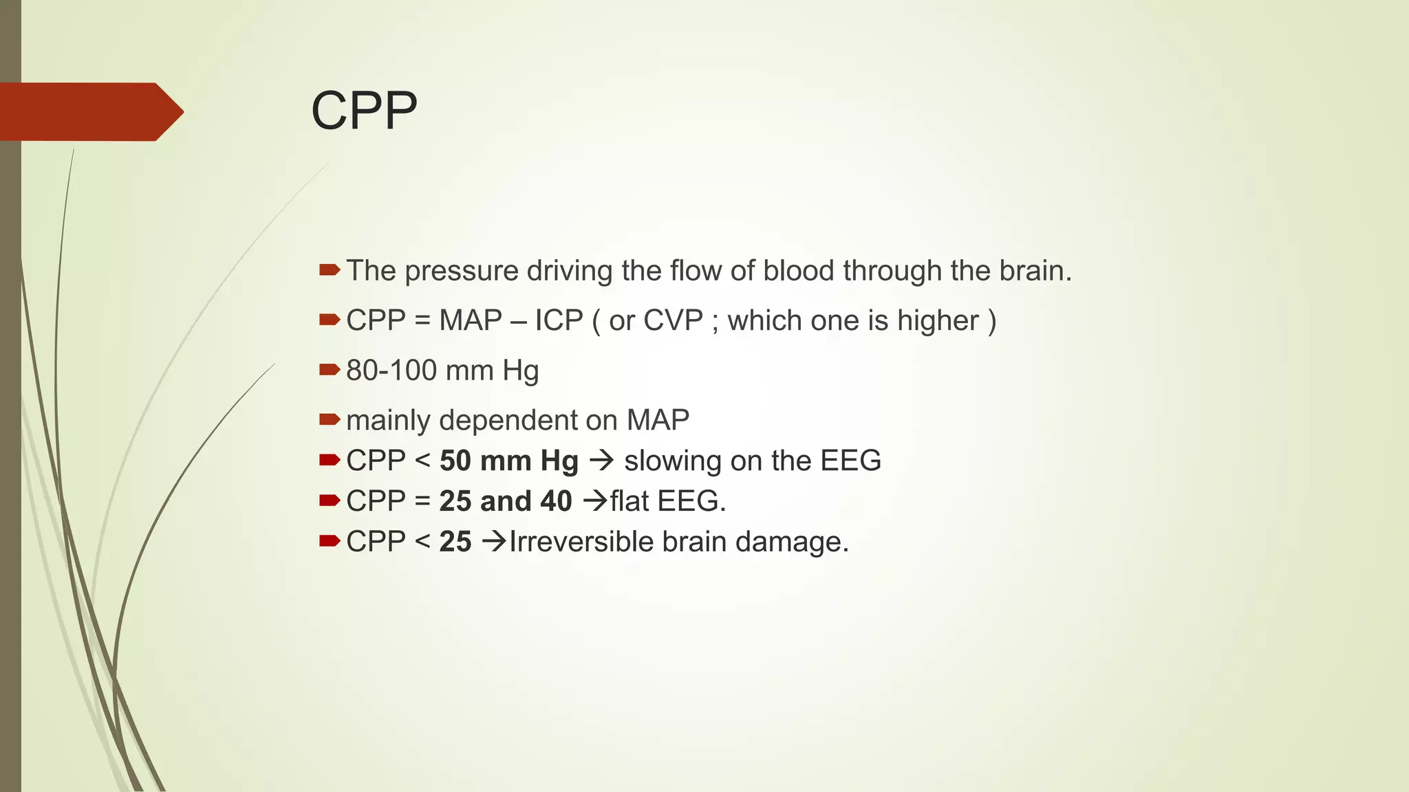
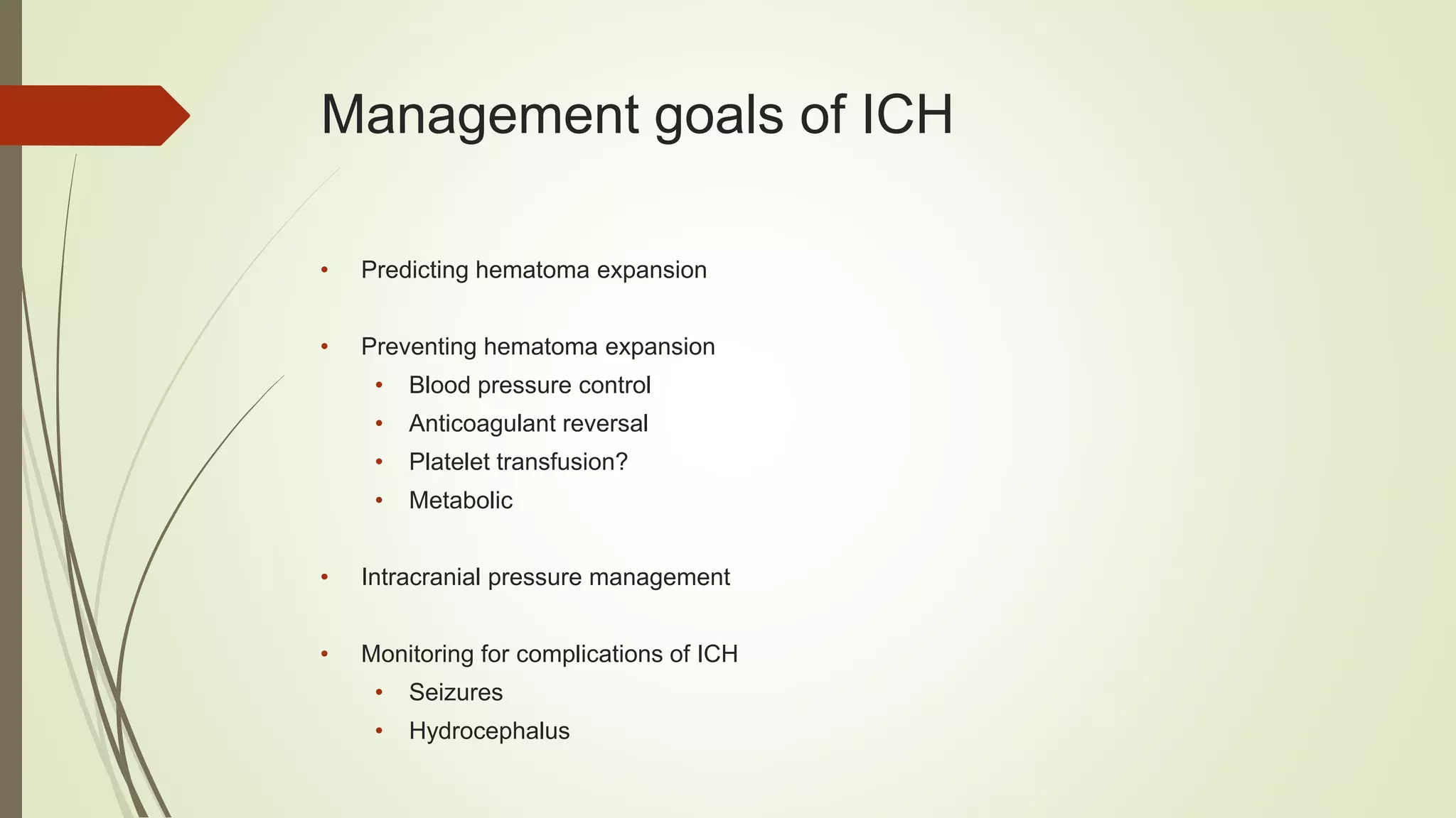
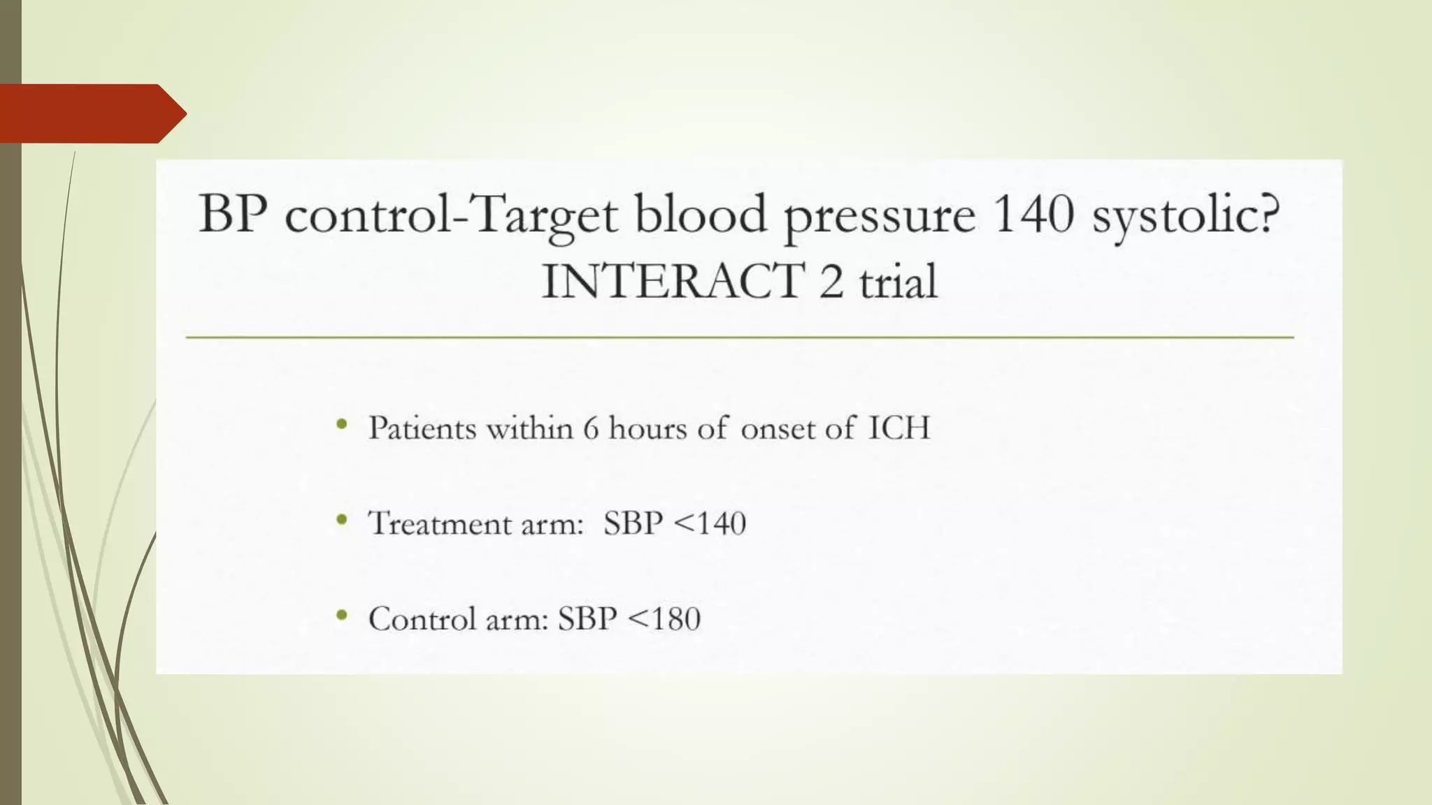
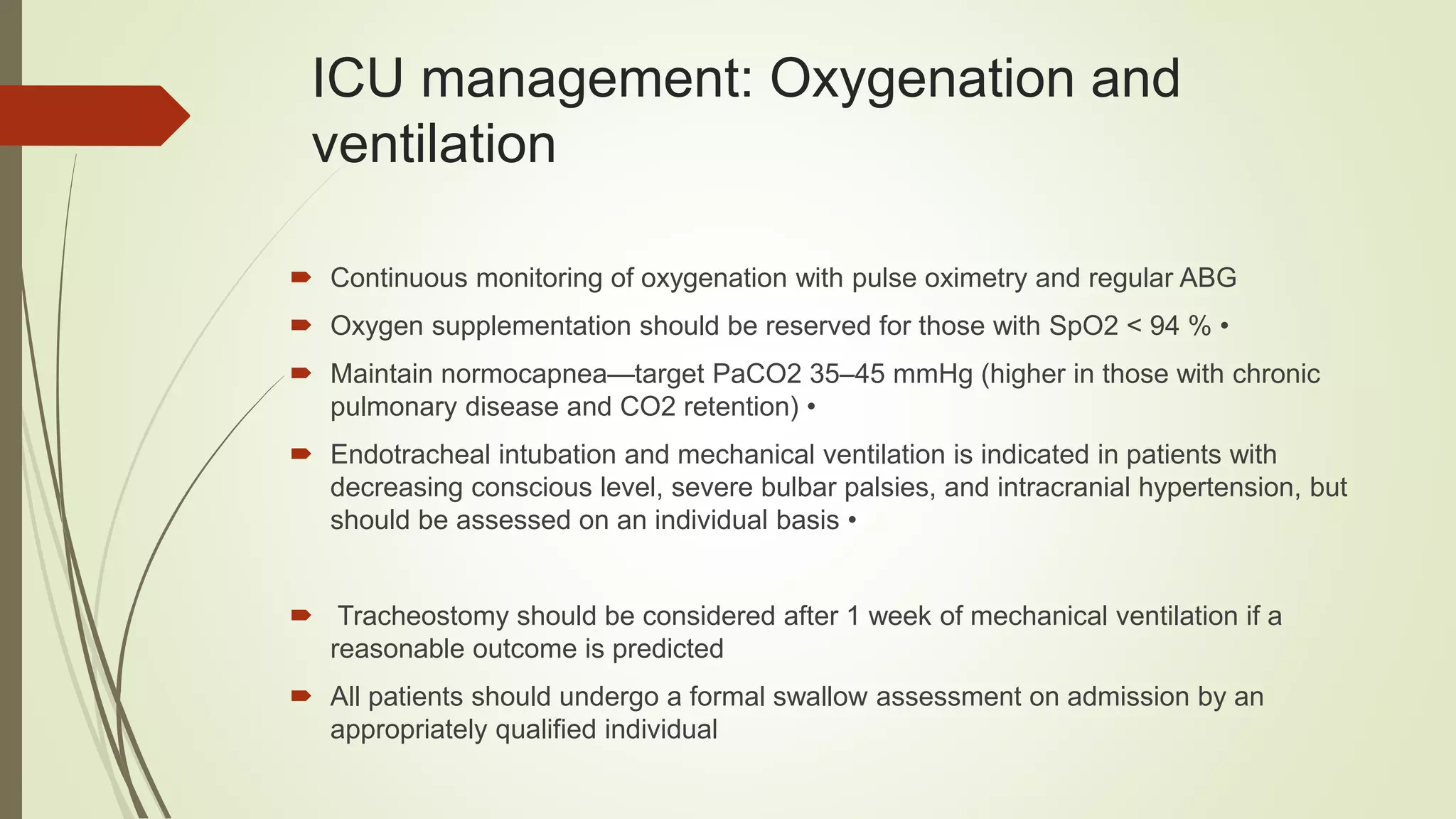
Intracerebral hemorrhage, SAH,
ischemic stroke, decompressive
craniectomy, seizures.
Presenter: Malek alai
Moderator: Dr. Sohel, Dr. Adel
Intracranial hemorrhage
 Terminology
• Intracranial hemorrhage: Any hemorrhage within
cranial vault, Epidural, subdural, SAH, intraventricular, parenchymal
• Intracerebral hemorrhage: Bleeding in the brain parenchyma
ICP
 Cranial vault: rigid structure with a fixed total volume
( Brain 80%, Blood 12% and CSF 8%)
 ICP: supratentorial CSF pressure measured in the lateral ventricles or over the
cerebral cortex.
 normally 10 mm Hg (5 – 15)
ICP
> usually a small rise in one of
the volume components is well
tolerated.
> A point is reached at which
further increases produce rises
in ICP.
CBF
• varies from 10-300 ml/100g/minute of brain tissue.
• Avg CBF = 50 ml/100g/min
• CBF in gray matter = 80
• CBF in white matter = 20
• Total CBF in adults averages 750mL/min ( 15-20% of Cardiac Output).
• < 20-25 - cerebral impairment ( Slowing on EEG).
• < 15-20 - isoelectric line on EEG.
• < 10 - irreversible brain damage.
CPP
The pressure driving the flow of blood through the brain.
CPP = MAP – ICP ( or CVP ; which one is higher )
80-100 mm Hg
mainly dependent on MAP
CPP < 50 mm Hg  slowing on the EEG
CPP = 25 and 40 flat EEG.
CPP < 25 Irreversible brain damage.
Management goals of ICH
• Predicting hematoma expansion
• Preventing hematoma expansion
• Blood pressure control
• Anticoagulant reversal
• Platelet transfusion?
• Metabolic
• Intracranial pressure management
• Monitoring for complications of ICH
• Seizures
• Hydrocephalus
Is it expanding?
hours from onset
0-3 3-6 6-12
Godoy, D. A. et al. Stroke 2006
Preventing hematoma expansion
 Blood pressure control
 Anticoagulant reversal
 ? Platelet transfusion
 Metabolic
BP Control:
 AHA Statement:
> For ICH patients presenting with SBP between 150-220 mm Hg and with out
contraindication to acute BP treatment, lowering of SBP to 140 mm Hg is safe
(class1; Level of Evidence A) and can be effective for improving functional out
come (class 2a; Level of Evidence B)
> For ICH patients presenting with SBP>220 mm Hg, it may be reasonable to
consider aggressive reduction of BP with a continuous intravenous infusion and
frequent BP monitoring (class 2a; Level of Evidence C)
Reversal of anticoagulants:
Warfarin:
 the 2018 guideline from the American Society of Hematology (ASH) and the 2012
guideline from the American College of Chest Physicians (ACCP), both of which
recommend the following:
 Hold warfarin.
 Administer a 4-factor PCC; if a PCC is not available, a plasma product such as
fresh frozen plasma (FFP)
 Administer intravenous vitamin K
Why FFP is not the first choice:
 Higher volume
 Longer time for INR normalization 30 hr vs 30 min
 Risk of transfusion reaction
 Has advantage of being readily available
PCC dose:
Vit K:
 Vitamin K should be given because the half-life of PCC is very short (hours).
 Vitamin K 10 mg is given by slow intravenous infusion, no faster than 1 mg/min to
minimize anaphylactic risk
 The effect of vitamin K on the INR takes approximately 12 to 24 hours; thus, all
patients should also receive PCC.
 Vitamin K administration can be repeated every 12 hours for persistent INR
elevation, and daily INR should be obtained to assess for this need
AHA Statement:
• Patients with ICH whose INR is elevated because of VKA should have
their VKA withheld, receive therapy to replace vitamin K-dependent factors
and correct the INR and receive intravenous vitamin K (class 1; Level of
Evidence C).
• PCCs may have fewer complications and correct the INR more rapidly
than FFP and might be considered over FFP (class 2b; Level of Evidence
B)
• rF7a does not replace all clotting factors and so is not recommended
NOAC:
> For patients with ICH who are taking dabigatran, treatment with FEIBA( activated
PCC), other PCCs, or rF7a might be considered on an individual basis. Activated charcoal
might be used if most recent dose was taken <2 hours earlier, hemodialysis can be used
as well.
> Idarucizumab is monoclonal ab designed for the reversal of dabigatran 5g IV (2 doses
of 2.5gms, no more than 15 mins apart)
> Intracranial bleeding associated with a direct factor Xa inhibitor, can be treated
with andexanet alfa (a reversal agent for factor Xa inhibitors) or 4-factor PCC
Platelet transfusion for Aspirin use?
282 ICH cases imaged at onset and at 72 hours, including 70 (25%) taking
antiplatelet medication
No difference in baseline hematoma volume
No difference in hematoma growth at 72 hours
No difference in need for surgical evacuation
No difference in Rankin score at 90 days
No difference in mortality
Platelet Transfusion:
AHA Statement:
 The usefulness of platelet transfusions in ICH patients with a history of antiplatelet
use is uncertain (class 2b: Level of Evidence C)
 Patients with a severe coagulation factor deficiency or severe thrombocytopenia
should receive appropriate factor replacement therapy or platelets, respectively
(class 1; Level of Evidence C)
Factor 7:
AHA Statement: ( FAST trial)
 Although rfactor7a can limit the
extent of hematoma expansion in
noncoagulopathic ICH patients,
there is an increase in
thromboembolic risk with rF7a and
no clear clinical benefit in
unselected patients. This rF7a is
not recommended (class 3; Level of
Evidence A)
Intracranial pressure management
Basic measures:
 Elevation of the head of the bed to 30 degrees
 Mild sedation, as needed for comfort
 Avoidance of endotracheal tube holder and securement device ties, constrictive
central line dressings, or twisting of the head that might constrict cervical veins
 Use of normal saline initially for maintenance and replacement fluids; hypotonic
fluids are contraindicated
 Glucocorticoids should not be used to lower the ICP in most patients with ICH
Intracranial pressure management
Osmotic therapy
 hypertonic saline or mannitol, There is no compelling evidence to support the
superiority of either agent, although some but not all traumatic brain injury studies
suggest that hypertonic saline is more effective
 Manitol is given as initial bolus of 0.5 to 1 g/kg, followed by repeated infusions of
0.25 to 0.5 g/kg as needed, generally every four to twelve hours with monitoring of
serum osmolality.
 Hypertonic saline ( 3%-23.4%), 3 percent NaCl may be titrated to an initial sodium
goal of approximately 145 to 155 mEq/L, 23% used as supplemental boluses.
Intracranial pressure management
 CSF drainage ( EVD)
 Decompressive cranioectomy
 Salvage therapy: medication coma, neuromuscular blockade and hyperventilation
 hyperventilation:
- (PaCO2) goal of 30 to 35 mmHg is suggested
 More aggressive hyperventilation (ie, a PaCO2 of 26 to 30 mmHg) may result in
brain ischemia and worse outcomes
 the effect of hyperventilation on ICP only lasts for a few hours.
Surgery:
• Patients with cerebellar hemorrhage who are deteriorating neurologically
or have brain stem compression or hydrocephalus should under go
surgical removal as soon as possible (class 1; Level C)
• Initial treatment of these patients with ventricular drainage rather than
surgical evacuation is not recommended (class 3; Level C)
• For most patients with supratentorial ICH, usefulness of surgery is not well
established (class2b; Level A)
Medical care:
 Glucose should be monitored. Both hyperglycemia and hypoglycemia should be
avoided (class 1; Level C)
 Treatment of fever after ICH may be reasonable (class 2b; Level C)
 Systemic screening for MI with ECG and cardiac enzyme testing after ICH is
reasonable (class 2a; Level C)
 A formal dysphagia screen should be performed in all patients before initiating oral
intake to reduce pneumonia risk (class 1; Level B)
Medical care:
 Intermittent pneumatic compression must be used immediately (class 1; Level A)
 Graduated compression stockings are not beneficial to reduce DVT or improve out
comes (class 3; Level A)
 After 1-4 days from onset LMW heparin or unfractionated heparin.
Post stroke Seizure
 Seizures affect 9% of stroke patients
 Seizures occurred within 24 hours of the stroke in 43 percent of patients
 Intermittent EEG monitoring is sufficient generally
 When to treat — Given the relatively low frequency of recurrent seizures after stroke,
and an absence of absolute predictors of poststroke epilepsy, the decision of when to
treat patients for a poststroke seizure is difficult.
 The efficacy of specific antiseizure drugs for poststroke seizures has not been rigorously
assessed in controlled trials, so there is no evidence to support one specific
antiseizure drug over another
Ischemic stroke
Etiology and classification of acute
ischemic stroke
According to one study:
 29 % of strokes relate to cardioembolic disorders, particularly atrial fibrillation (AF),
 16 % to large vessel cervical or intracranial atherosclerosis with stenosis,
 16 % to lacunar disease,
 and 3 % to other causes such as migraine, malignancy, and hypercoagulable
states .
 Although the etiology is unclear in one-third of patients, many of these likely have
undiagnosed paroxysmal AF.
Reperfusion therapy
 There is class I evidence supporting the use of intravenous rt-PA as soon
as possible but within 4.5 h of stroke onset
 exclusion of a hemorrhagic stroke by noncontrast (CT) scan should be done first
 A pooled analysis of four trials confirms that the odds ratio of a favorable
3-month outcome decreases as time to treatment increases.
 Endovascular therapy (including intraarterial thrombolysis, mechanical
embolectomy, and angioplasty/stenting) represents an alternative therapy to
intravenous rt-PA, but US guidelines restrict its use to those who are not candidates
for rt-PA, or who fail to improve after full rt-PA therapy
Reperfusion therapy
 It is generally accepted that intraarterial thrombolysis should be performed within 6 h
and thrombectomy within 8 h of symptom onset.
 Several trials have shown endovascular therapy (either combined with intravenous
thrombolysis or alone) to be no better than intravenous thrombolysis in terms of
outcomes.
 Patients should have cranial imaging in the first 24 h following reperfusion
therapy to detect hemorrhage or other complications
Intensive care management of acute
ischemic stroke
 The ICU management of AIS focuses on monitoring and optimization of
systemic physiological homeostasis, and monitoring and management of
intracranial complications
General indications for intensive care unit admission
following acute ischemic stroke
 Need for intubation and/or mechanical ventilation due to Decreased conscious level
(GCS B8) or evidence of brainstem dysfunction or any other cause of a threatened airway
 Adjuvant therapy for intracranial hypertension or significant cerebral edema
 Acute respiratory failure, for example, due to pulmonary edema (neurogenic or
cardiogenic)
 Generalized tonic–clonic seizures or status epilepticus
 Severe stroke (National Institutes of Health Stroke Score)
 Reperfusion therapy (intravenous or intraarterial).
 if multiorgan failure present
 Large middle cerebral artery infarct volume that predicts a malignant course
 Persistent extremes of blood pressure
 Postoperatively following decompressive craniectomy
ICU management: Oxygenation and
ventilation
 Continuous monitoring of oxygenation with pulse oximetry and regular ABG
 Oxygen supplementation should be reserved for those with SpO2 < 94 % •
 Maintain normocapnea—target PaCO2 35–45 mmHg (higher in those with chronic
pulmonary disease and CO2 retention) •
 Endotracheal intubation and mechanical ventilation is indicated in patients with
decreasing conscious level, severe bulbar palsies, and intracranial hypertension, but
should be assessed on an individual basis •
 Tracheostomy should be considered after 1 week of mechanical ventilation if a
reasonable outcome is predicted
 All patients should undergo a formal swallow assessment on admission by an
appropriately qualified individual
Hemodynamic and fluid management
 Approximately 80 % of AIS patients are hypertensive [ SBP>140 mmHg] at
presentation.
 Severe hypertension likely contributes to cardiorespiratory complications and
promotes cytotoxic edema and hemorrhagic transformation
 There is a U-shaped relationship between BP and outcome after AIS, with both
high and low BP having adverse effects on outcome
 Although high BP is independently associated with poor outcome after AIS, the
effect of acute blood pressure lowering is not clear. Some studies suggest improved,
some unchanged , and others worsened long-term outcomes.
Hemodynamic and fluid management
 severe hypotension will compromise cerebral perfusion and potentially
increase infarct volume. Some patients may benefit from BP augmentation, for
example, those with severe carotid stenosis.
 European, US, and UK guidelines vary in their approaches to BP management
after AIS, reflecting the controversy surrounding optimal BP targets
 BP lowering is not indicated in those not undergoing thrombolysis unless [220/120
mmHg or in the presence of significant comorbidities, particularly severe cardiac
failure, aortic dissection, or hypertensive encephalopathy.
Hemodynamic and fluid management
 BP lowering should be cautious, i.e.<15 % in the first 24 h in those not receiving
thrombolysis. BP should be lowered <185/110 mmHg before and for at least 24
h after thrombolysis
 Intravenous labetalol and nicardipine are reasonable first-line agents to lower
BP, avoid medications difficult to titrate.
 SBP<90 should be treated with fluid resuscitation in the first instance, and with
vasoactive agents such as norepinephrine if unresponsive to volume replacement.
Hemodynamic and fluid management
 Fluid balance should be carefully monitored and managed to maintain euvolemia.
 a meta analysis of 18 trials suggested that intentional hemodilution does not
improve outcome
 European and US guidelines recommend the use of 0.9 %, dextrose-containing
fluids should always be avoided except in the presence of hypoglycemia.
 large RCT found no benefit OF 25% ALBUMIN on outcomes at 90 days compared
with saline.
 Fluid replacement should be monitored closely and special caution exercised in
those with cardiovascular disease and cerebral edema.
Myocardial complications
 commonly coexist, either as a trigger or as a result of the stroke itself.
 Dysrhythmias are present in 57 % of patients after AIS , elevated cardiac troponin
levels in up to 17.5 % , and at least 12 % have abnormal left ventricular function on
echocardiography
 A history of cardiovascular disease may predispose to sudden death.
 All AIS patients on ICU should undergo continuous electrocardiography (EKG)
and have echocardiography at least once during the course.
Glycemic control
 Hyperglycemia occurs in more than 40 % of AIS patients and, as a marker of
illness severity, is associated with increased cortical toxicity, larger infarct
volumes, and susceptibility to infection.
 Poststroke hyperglycemia is independently associated with increased
mortality and morbidity at 90 days, and postthrombolysis ICH
 ASA guidance and thresholds for initiating glycemic treatment, aiming to maintain
serum glucose between 140 and 180 mg/dl (8.0–10.0 mmol/l), with
intravenous insulin infusion whilst avoiding large swings in glucose levels.
Fever
 Pyrexia affects up to 50 %of patients after AIS, and is independently associated
with poor outcome (several studies)
 REASON of fever should be investigated and treated
 Larger trials are needed to determine if pharmacologic temperature reduction
improves outcome from acute stroke.
 Induced hypothermia is not currently recommended for patients with ischemic
stroke, outside of clinical trials
Anticoagulation, antiplatelet therapy,
and thromboprophylaxis
 Administer oral high-dose aspirin (160–325 mg loading dose) within 48 h of AIS but
delay for 24 h in those receiving thrombolysis •
 Early mobilization reduces the risk of thromboembolic complications of AIS
 All immobilized AIS patients on ICU should be treated with prophylactic-dose
subcutaneous LMWH to prevent DVT and mechanical intermittent calf
compression. Treatment should be started early, but LMWH should not be started
until 24 h following thrombolysis.
anemia
 Was identified in 97.2 % of severe AIS patients managed on the ICU •
 A hemoglobin value of 10 g/dl or below might be the most appropriate transfusion
trigger in AIS •
 Although anemia should be avoided, aggressive transfusion approaches are not
currently recommended.
Hemorrhagic transformation
 Hemorrhagic transformation should be managed in the first instance by stopping
any remaining intravenous rt-PA (if applicable), repeating cranial imaging, and
monitoring coagulation status. •
 The role of surgery is unclear, but decompressive surgery/hematoma evacuation
may be indicated in large superficial hematomas and/or those causing significant
mass effect •
 The use of cryoprecipitate, fresh frozen plasma, and recombinant factor VII is not
supported by robust evidence
Cerebral Edema and Hemorrhage
 Frequent monitoring of clinical status is superior to ICP monitoring in detecting
those requiring medical or surgical management for intracranial worsening •
 Patients at high risk of clinical deterioration, particularly malignant MCA infarction
and edematous cerebellar AIS, should be managed in a center with immediate
access to neurosurgical facilities •
 Suspect the need for urgent surgical intervention in those with malignant MCA
infarction or cerebellar stroke if pupillary abnormalities develop, there is a drop in
the Glasgow Coma Score by more than 1 point, and/or there is progression of
edema on cranial imaging
Cerebral Edema and Hemorrhage
 Decompressive craniectomy is indicated in patients aged 18–60 years with dominant and
nondominant hemisphere malignant MCA infarction and should be performed as soon as
possible (but within 48 h) of clinical or radiological deterioration •
 Outcomes are worse with increasing age, but decisions should be made on an individual
basis, and intervention in those [60 years should be considered •
 Ventriculostomy should be performed as soon as possible for obstructive hydrocephalus
associated with cerebellar AIS, and accompanied by suboccipital decompressive
craniectomy. •
 Hypertonic saline appears to be superior to mannitol at lowering ICP after AIS, but both
agents should only be used as a temporizing measure prior to surgical decompression.
 Prophylactic use of osmotic diuretics before the detection of cerebral edema is not
recommended
Decompressive craniectomy:
Indication , technique, present
status and controversies
Learning Objectives
The learner will be able to:
 –explain the difference between craniotomy and craniectomy
 –describe the implications for a craniectomy
 –summarize the risks and complications related to craniectomy
definitions
 •Craniotomy defines a procedure where the cranial cavity is accessed through
removal of bone to perform a variety of brain surgeries. Once the surgery is
completed, the bone flap is returned to its previous position.
 •Craniectomydiffers from craniotomy in that the bone is not replaced to its
previous position; instead it is stored for future insertion or may be discarded
(depending on pathology –e.g. infection). This results in a cranial defect.
 –If the bone flap needs to be discarded, it is replaced with a custom-made implant.
Indication of DC
 Traumatic brain injury
 Malignant cerebral infarction
 Others – Cerebral venous sinus thrombosis, intracerebral
 hematoma, metabolic encepahlopathies .
TBI in Adults
 Various studies demonstrate DC improves ICP, cerebral
oxygenation, cerebral compliance
 No evidence to support the routine use of secondary DC to reduce
unfavourable outcome in adults with severe TBI and refractory high ICP.
J. Neurosurg. / Volume 108 / May 2008, Effect of decompressive craniectomy on intracranial
pressure and cerebrospinal compensation following traumatic brain injury J.
Neurosurg. / Volume 108 / May 2008, Cerebral oxygenation, vascular reactivity, and
neurochemistry following decompressive craniectomy for severe traumatic brain injury
 DECRA trail - In adults with severe diffuse traumatic brain injury and
refractory intracranial hypertension, early bifrontotemporoparietal
decompressive craniectomy decreased intracranial pressure and the length
of stay in the ICU but was associated with more unfavourable outcomes .
Decompressive Craniectomy in Diffuse Traumatic Brain Injury . N Engl J Med. 2011
Apr21;364(16):1493-502.
Malignant cerebral infarction
 Large territorial parenchymal infarction with post ischemic edema and associated with
uncal or axial herniation.
 Occlusion of proximal MCA, more than 50% of supplied territory involved.
 Treatment – medical management or surgery (decompressive craniectomy)
 Surgery – clearly reduce mortality however survivors suffered high morbidity .
Rishi Gupta, E. Sander Connolly, Stephan Mayer and Mitchell S.V Elkind. Hemicraniectomy for
MassiveMiddle Cerebral Artery Territory Infarction : ASystematic Review Stroke 2004, 35:539-543:
3 RCTs conducted and results published.
 DECIMAL (DEcompressive Craniectomy In MALignant middle cerebral
artery infarction)
 DESTINY (DEcompressive Surgery for Treatment of INfarction of
malignant middle cerebral arterY)
 HAMLET ( Hemicraniectomy After Middle cerebral artery infarction with
Life –threatening Edema Trial )
Results
 Effect on mortality – all 3 trial shows significant reduction in mortality.
 Absolute reduction in mortality is 49.9%
 absolute reduction in risk of bad functional outcome was 41.9% .
 Conclusion - surgical decompression with in 48 hrs of onset of stroke reduced
risk of significant morbidity.
Timing of surgery – optimal time window ?
 HeADDFIRST (Hemicraniectomy And Durotomy on Deterioration From
Infarction Related Swelling Trial) window period – 96 hrs
 HeMMI trial (Hemicraniectomy for Malignant Middle cerebral artery
Infarcts) time period of 72 hrs.
Conclusion (present status)
 Pooled analysis of all trials provides Class I evidence for the performance of
early decompressive craniectomy in the setting of large unilateral infarcts
(volume > 145 cc) within 48 hours of the ischemic event.
 Further studies are needed to better define quality of life issues at long-term
follow-up as well as age limit issues.
Neurosurg Focus 30 (6):E18, 2011 Decompressive hemicraniectomy after
malignant middle cerebral artery infarction: rationale and controversies Omar M. Arnaout, M.D., Salah G. Aoun,
M.D., H. Hunt Batjer, M.D., and Bernard R. Bendo k, M.D. Department of Neurological Surgery, FeinbergSchool
of Medicine and McGaw Medical Centre, North-western University, Chicago,Illinois
Technique of decompressive craniectomy
 Fronto-temporo-parietal approach
 Bifrontal decompressive approach
 Temporal approach
Complications
The major risks of craniectomy include the following:
 •bleeding
 •infection
 •seizures
 •abnormalities in cerebrospinal fluid (CSF) absorption
 •further damage to the brain
 •stroke
 •death
questions

Intracerebral hemorrhage, SAH, ischemic stroke,[412].pptx

  • 1.
    Intracerebral hemorrhage, SAH, ischemicstroke, decompressive craniectomy, seizures. Presenter: Malek alai Moderator: Dr. Sohel, Dr. Adel
  • 2.
    Intracranial hemorrhage  Terminology •Intracranial hemorrhage: Any hemorrhage within cranial vault, Epidural, subdural, SAH, intraventricular, parenchymal • Intracerebral hemorrhage: Bleeding in the brain parenchyma
  • 7.
    ICP  Cranial vault:rigid structure with a fixed total volume ( Brain 80%, Blood 12% and CSF 8%)  ICP: supratentorial CSF pressure measured in the lateral ventricles or over the cerebral cortex.  normally 10 mm Hg (5 – 15)
  • 9.
    ICP > usually asmall rise in one of the volume components is well tolerated. > A point is reached at which further increases produce rises in ICP.
  • 10.
    CBF • varies from10-300 ml/100g/minute of brain tissue. • Avg CBF = 50 ml/100g/min • CBF in gray matter = 80 • CBF in white matter = 20 • Total CBF in adults averages 750mL/min ( 15-20% of Cardiac Output). • < 20-25 - cerebral impairment ( Slowing on EEG). • < 15-20 - isoelectric line on EEG. • < 10 - irreversible brain damage.
  • 12.
    CPP The pressure drivingthe flow of blood through the brain. CPP = MAP – ICP ( or CVP ; which one is higher ) 80-100 mm Hg mainly dependent on MAP CPP < 50 mm Hg  slowing on the EEG CPP = 25 and 40 flat EEG. CPP < 25 Irreversible brain damage.
  • 13.
    Management goals ofICH • Predicting hematoma expansion • Preventing hematoma expansion • Blood pressure control • Anticoagulant reversal • Platelet transfusion? • Metabolic • Intracranial pressure management • Monitoring for complications of ICH • Seizures • Hydrocephalus
  • 14.
    Is it expanding? hoursfrom onset 0-3 3-6 6-12
  • 16.
    Godoy, D. A.et al. Stroke 2006
  • 17.
    Preventing hematoma expansion Blood pressure control  Anticoagulant reversal  ? Platelet transfusion  Metabolic
  • 21.
    BP Control:  AHAStatement: > For ICH patients presenting with SBP between 150-220 mm Hg and with out contraindication to acute BP treatment, lowering of SBP to 140 mm Hg is safe (class1; Level of Evidence A) and can be effective for improving functional out come (class 2a; Level of Evidence B) > For ICH patients presenting with SBP>220 mm Hg, it may be reasonable to consider aggressive reduction of BP with a continuous intravenous infusion and frequent BP monitoring (class 2a; Level of Evidence C)
  • 22.
    Reversal of anticoagulants: Warfarin: the 2018 guideline from the American Society of Hematology (ASH) and the 2012 guideline from the American College of Chest Physicians (ACCP), both of which recommend the following:  Hold warfarin.  Administer a 4-factor PCC; if a PCC is not available, a plasma product such as fresh frozen plasma (FFP)  Administer intravenous vitamin K
  • 24.
    Why FFP isnot the first choice:  Higher volume  Longer time for INR normalization 30 hr vs 30 min  Risk of transfusion reaction  Has advantage of being readily available
  • 25.
  • 26.
    Vit K:  VitaminK should be given because the half-life of PCC is very short (hours).  Vitamin K 10 mg is given by slow intravenous infusion, no faster than 1 mg/min to minimize anaphylactic risk  The effect of vitamin K on the INR takes approximately 12 to 24 hours; thus, all patients should also receive PCC.  Vitamin K administration can be repeated every 12 hours for persistent INR elevation, and daily INR should be obtained to assess for this need
  • 27.
    AHA Statement: • Patientswith ICH whose INR is elevated because of VKA should have their VKA withheld, receive therapy to replace vitamin K-dependent factors and correct the INR and receive intravenous vitamin K (class 1; Level of Evidence C). • PCCs may have fewer complications and correct the INR more rapidly than FFP and might be considered over FFP (class 2b; Level of Evidence B) • rF7a does not replace all clotting factors and so is not recommended
  • 28.
    NOAC: > For patientswith ICH who are taking dabigatran, treatment with FEIBA( activated PCC), other PCCs, or rF7a might be considered on an individual basis. Activated charcoal might be used if most recent dose was taken <2 hours earlier, hemodialysis can be used as well. > Idarucizumab is monoclonal ab designed for the reversal of dabigatran 5g IV (2 doses of 2.5gms, no more than 15 mins apart) > Intracranial bleeding associated with a direct factor Xa inhibitor, can be treated with andexanet alfa (a reversal agent for factor Xa inhibitors) or 4-factor PCC
  • 29.
    Platelet transfusion forAspirin use? 282 ICH cases imaged at onset and at 72 hours, including 70 (25%) taking antiplatelet medication No difference in baseline hematoma volume No difference in hematoma growth at 72 hours No difference in need for surgical evacuation No difference in Rankin score at 90 days No difference in mortality
  • 30.
    Platelet Transfusion: AHA Statement: The usefulness of platelet transfusions in ICH patients with a history of antiplatelet use is uncertain (class 2b: Level of Evidence C)  Patients with a severe coagulation factor deficiency or severe thrombocytopenia should receive appropriate factor replacement therapy or platelets, respectively (class 1; Level of Evidence C)
  • 31.
    Factor 7: AHA Statement:( FAST trial)  Although rfactor7a can limit the extent of hematoma expansion in noncoagulopathic ICH patients, there is an increase in thromboembolic risk with rF7a and no clear clinical benefit in unselected patients. This rF7a is not recommended (class 3; Level of Evidence A)
  • 32.
    Intracranial pressure management Basicmeasures:  Elevation of the head of the bed to 30 degrees  Mild sedation, as needed for comfort  Avoidance of endotracheal tube holder and securement device ties, constrictive central line dressings, or twisting of the head that might constrict cervical veins  Use of normal saline initially for maintenance and replacement fluids; hypotonic fluids are contraindicated  Glucocorticoids should not be used to lower the ICP in most patients with ICH
  • 33.
    Intracranial pressure management Osmotictherapy  hypertonic saline or mannitol, There is no compelling evidence to support the superiority of either agent, although some but not all traumatic brain injury studies suggest that hypertonic saline is more effective  Manitol is given as initial bolus of 0.5 to 1 g/kg, followed by repeated infusions of 0.25 to 0.5 g/kg as needed, generally every four to twelve hours with monitoring of serum osmolality.  Hypertonic saline ( 3%-23.4%), 3 percent NaCl may be titrated to an initial sodium goal of approximately 145 to 155 mEq/L, 23% used as supplemental boluses.
  • 34.
    Intracranial pressure management CSF drainage ( EVD)  Decompressive cranioectomy  Salvage therapy: medication coma, neuromuscular blockade and hyperventilation  hyperventilation: - (PaCO2) goal of 30 to 35 mmHg is suggested  More aggressive hyperventilation (ie, a PaCO2 of 26 to 30 mmHg) may result in brain ischemia and worse outcomes  the effect of hyperventilation on ICP only lasts for a few hours.
  • 35.
    Surgery: • Patients withcerebellar hemorrhage who are deteriorating neurologically or have brain stem compression or hydrocephalus should under go surgical removal as soon as possible (class 1; Level C) • Initial treatment of these patients with ventricular drainage rather than surgical evacuation is not recommended (class 3; Level C) • For most patients with supratentorial ICH, usefulness of surgery is not well established (class2b; Level A)
  • 36.
    Medical care:  Glucoseshould be monitored. Both hyperglycemia and hypoglycemia should be avoided (class 1; Level C)  Treatment of fever after ICH may be reasonable (class 2b; Level C)  Systemic screening for MI with ECG and cardiac enzyme testing after ICH is reasonable (class 2a; Level C)  A formal dysphagia screen should be performed in all patients before initiating oral intake to reduce pneumonia risk (class 1; Level B)
  • 37.
    Medical care:  Intermittentpneumatic compression must be used immediately (class 1; Level A)  Graduated compression stockings are not beneficial to reduce DVT or improve out comes (class 3; Level A)  After 1-4 days from onset LMW heparin or unfractionated heparin.
  • 38.
    Post stroke Seizure Seizures affect 9% of stroke patients  Seizures occurred within 24 hours of the stroke in 43 percent of patients  Intermittent EEG monitoring is sufficient generally  When to treat — Given the relatively low frequency of recurrent seizures after stroke, and an absence of absolute predictors of poststroke epilepsy, the decision of when to treat patients for a poststroke seizure is difficult.  The efficacy of specific antiseizure drugs for poststroke seizures has not been rigorously assessed in controlled trials, so there is no evidence to support one specific antiseizure drug over another
  • 39.
  • 40.
    Etiology and classificationof acute ischemic stroke According to one study:  29 % of strokes relate to cardioembolic disorders, particularly atrial fibrillation (AF),  16 % to large vessel cervical or intracranial atherosclerosis with stenosis,  16 % to lacunar disease,  and 3 % to other causes such as migraine, malignancy, and hypercoagulable states .  Although the etiology is unclear in one-third of patients, many of these likely have undiagnosed paroxysmal AF.
  • 41.
    Reperfusion therapy  Thereis class I evidence supporting the use of intravenous rt-PA as soon as possible but within 4.5 h of stroke onset  exclusion of a hemorrhagic stroke by noncontrast (CT) scan should be done first  A pooled analysis of four trials confirms that the odds ratio of a favorable 3-month outcome decreases as time to treatment increases.  Endovascular therapy (including intraarterial thrombolysis, mechanical embolectomy, and angioplasty/stenting) represents an alternative therapy to intravenous rt-PA, but US guidelines restrict its use to those who are not candidates for rt-PA, or who fail to improve after full rt-PA therapy
  • 42.
    Reperfusion therapy  Itis generally accepted that intraarterial thrombolysis should be performed within 6 h and thrombectomy within 8 h of symptom onset.  Several trials have shown endovascular therapy (either combined with intravenous thrombolysis or alone) to be no better than intravenous thrombolysis in terms of outcomes.  Patients should have cranial imaging in the first 24 h following reperfusion therapy to detect hemorrhage or other complications
  • 43.
    Intensive care managementof acute ischemic stroke  The ICU management of AIS focuses on monitoring and optimization of systemic physiological homeostasis, and monitoring and management of intracranial complications
  • 44.
    General indications forintensive care unit admission following acute ischemic stroke  Need for intubation and/or mechanical ventilation due to Decreased conscious level (GCS B8) or evidence of brainstem dysfunction or any other cause of a threatened airway  Adjuvant therapy for intracranial hypertension or significant cerebral edema  Acute respiratory failure, for example, due to pulmonary edema (neurogenic or cardiogenic)  Generalized tonic–clonic seizures or status epilepticus  Severe stroke (National Institutes of Health Stroke Score)  Reperfusion therapy (intravenous or intraarterial).  if multiorgan failure present  Large middle cerebral artery infarct volume that predicts a malignant course  Persistent extremes of blood pressure  Postoperatively following decompressive craniectomy
  • 45.
    ICU management: Oxygenationand ventilation  Continuous monitoring of oxygenation with pulse oximetry and regular ABG  Oxygen supplementation should be reserved for those with SpO2 < 94 % •  Maintain normocapnea—target PaCO2 35–45 mmHg (higher in those with chronic pulmonary disease and CO2 retention) •  Endotracheal intubation and mechanical ventilation is indicated in patients with decreasing conscious level, severe bulbar palsies, and intracranial hypertension, but should be assessed on an individual basis •  Tracheostomy should be considered after 1 week of mechanical ventilation if a reasonable outcome is predicted  All patients should undergo a formal swallow assessment on admission by an appropriately qualified individual
  • 46.
    Hemodynamic and fluidmanagement  Approximately 80 % of AIS patients are hypertensive [ SBP>140 mmHg] at presentation.  Severe hypertension likely contributes to cardiorespiratory complications and promotes cytotoxic edema and hemorrhagic transformation  There is a U-shaped relationship between BP and outcome after AIS, with both high and low BP having adverse effects on outcome  Although high BP is independently associated with poor outcome after AIS, the effect of acute blood pressure lowering is not clear. Some studies suggest improved, some unchanged , and others worsened long-term outcomes.
  • 47.
    Hemodynamic and fluidmanagement  severe hypotension will compromise cerebral perfusion and potentially increase infarct volume. Some patients may benefit from BP augmentation, for example, those with severe carotid stenosis.  European, US, and UK guidelines vary in their approaches to BP management after AIS, reflecting the controversy surrounding optimal BP targets  BP lowering is not indicated in those not undergoing thrombolysis unless [220/120 mmHg or in the presence of significant comorbidities, particularly severe cardiac failure, aortic dissection, or hypertensive encephalopathy.
  • 48.
    Hemodynamic and fluidmanagement  BP lowering should be cautious, i.e.<15 % in the first 24 h in those not receiving thrombolysis. BP should be lowered <185/110 mmHg before and for at least 24 h after thrombolysis  Intravenous labetalol and nicardipine are reasonable first-line agents to lower BP, avoid medications difficult to titrate.  SBP<90 should be treated with fluid resuscitation in the first instance, and with vasoactive agents such as norepinephrine if unresponsive to volume replacement.
  • 49.
    Hemodynamic and fluidmanagement  Fluid balance should be carefully monitored and managed to maintain euvolemia.  a meta analysis of 18 trials suggested that intentional hemodilution does not improve outcome  European and US guidelines recommend the use of 0.9 %, dextrose-containing fluids should always be avoided except in the presence of hypoglycemia.  large RCT found no benefit OF 25% ALBUMIN on outcomes at 90 days compared with saline.  Fluid replacement should be monitored closely and special caution exercised in those with cardiovascular disease and cerebral edema.
  • 50.
    Myocardial complications  commonlycoexist, either as a trigger or as a result of the stroke itself.  Dysrhythmias are present in 57 % of patients after AIS , elevated cardiac troponin levels in up to 17.5 % , and at least 12 % have abnormal left ventricular function on echocardiography  A history of cardiovascular disease may predispose to sudden death.  All AIS patients on ICU should undergo continuous electrocardiography (EKG) and have echocardiography at least once during the course.
  • 51.
    Glycemic control  Hyperglycemiaoccurs in more than 40 % of AIS patients and, as a marker of illness severity, is associated with increased cortical toxicity, larger infarct volumes, and susceptibility to infection.  Poststroke hyperglycemia is independently associated with increased mortality and morbidity at 90 days, and postthrombolysis ICH  ASA guidance and thresholds for initiating glycemic treatment, aiming to maintain serum glucose between 140 and 180 mg/dl (8.0–10.0 mmol/l), with intravenous insulin infusion whilst avoiding large swings in glucose levels.
  • 52.
    Fever  Pyrexia affectsup to 50 %of patients after AIS, and is independently associated with poor outcome (several studies)  REASON of fever should be investigated and treated  Larger trials are needed to determine if pharmacologic temperature reduction improves outcome from acute stroke.  Induced hypothermia is not currently recommended for patients with ischemic stroke, outside of clinical trials
  • 53.
    Anticoagulation, antiplatelet therapy, andthromboprophylaxis  Administer oral high-dose aspirin (160–325 mg loading dose) within 48 h of AIS but delay for 24 h in those receiving thrombolysis •  Early mobilization reduces the risk of thromboembolic complications of AIS  All immobilized AIS patients on ICU should be treated with prophylactic-dose subcutaneous LMWH to prevent DVT and mechanical intermittent calf compression. Treatment should be started early, but LMWH should not be started until 24 h following thrombolysis.
  • 54.
    anemia  Was identifiedin 97.2 % of severe AIS patients managed on the ICU •  A hemoglobin value of 10 g/dl or below might be the most appropriate transfusion trigger in AIS •  Although anemia should be avoided, aggressive transfusion approaches are not currently recommended.
  • 55.
    Hemorrhagic transformation  Hemorrhagictransformation should be managed in the first instance by stopping any remaining intravenous rt-PA (if applicable), repeating cranial imaging, and monitoring coagulation status. •  The role of surgery is unclear, but decompressive surgery/hematoma evacuation may be indicated in large superficial hematomas and/or those causing significant mass effect •  The use of cryoprecipitate, fresh frozen plasma, and recombinant factor VII is not supported by robust evidence
  • 56.
    Cerebral Edema andHemorrhage  Frequent monitoring of clinical status is superior to ICP monitoring in detecting those requiring medical or surgical management for intracranial worsening •  Patients at high risk of clinical deterioration, particularly malignant MCA infarction and edematous cerebellar AIS, should be managed in a center with immediate access to neurosurgical facilities •  Suspect the need for urgent surgical intervention in those with malignant MCA infarction or cerebellar stroke if pupillary abnormalities develop, there is a drop in the Glasgow Coma Score by more than 1 point, and/or there is progression of edema on cranial imaging
  • 57.
    Cerebral Edema andHemorrhage  Decompressive craniectomy is indicated in patients aged 18–60 years with dominant and nondominant hemisphere malignant MCA infarction and should be performed as soon as possible (but within 48 h) of clinical or radiological deterioration •  Outcomes are worse with increasing age, but decisions should be made on an individual basis, and intervention in those [60 years should be considered •  Ventriculostomy should be performed as soon as possible for obstructive hydrocephalus associated with cerebellar AIS, and accompanied by suboccipital decompressive craniectomy. •  Hypertonic saline appears to be superior to mannitol at lowering ICP after AIS, but both agents should only be used as a temporizing measure prior to surgical decompression.  Prophylactic use of osmotic diuretics before the detection of cerebral edema is not recommended
  • 58.
    Decompressive craniectomy: Indication ,technique, present status and controversies
  • 59.
    Learning Objectives The learnerwill be able to:  –explain the difference between craniotomy and craniectomy  –describe the implications for a craniectomy  –summarize the risks and complications related to craniectomy
  • 60.
    definitions  •Craniotomy definesa procedure where the cranial cavity is accessed through removal of bone to perform a variety of brain surgeries. Once the surgery is completed, the bone flap is returned to its previous position.  •Craniectomydiffers from craniotomy in that the bone is not replaced to its previous position; instead it is stored for future insertion or may be discarded (depending on pathology –e.g. infection). This results in a cranial defect.  –If the bone flap needs to be discarded, it is replaced with a custom-made implant.
  • 62.
    Indication of DC Traumatic brain injury  Malignant cerebral infarction  Others – Cerebral venous sinus thrombosis, intracerebral  hematoma, metabolic encepahlopathies .
  • 65.
    TBI in Adults Various studies demonstrate DC improves ICP, cerebral oxygenation, cerebral compliance  No evidence to support the routine use of secondary DC to reduce unfavourable outcome in adults with severe TBI and refractory high ICP. J. Neurosurg. / Volume 108 / May 2008, Effect of decompressive craniectomy on intracranial pressure and cerebrospinal compensation following traumatic brain injury J. Neurosurg. / Volume 108 / May 2008, Cerebral oxygenation, vascular reactivity, and neurochemistry following decompressive craniectomy for severe traumatic brain injury
  • 66.
     DECRA trail- In adults with severe diffuse traumatic brain injury and refractory intracranial hypertension, early bifrontotemporoparietal decompressive craniectomy decreased intracranial pressure and the length of stay in the ICU but was associated with more unfavourable outcomes . Decompressive Craniectomy in Diffuse Traumatic Brain Injury . N Engl J Med. 2011 Apr21;364(16):1493-502.
  • 67.
    Malignant cerebral infarction Large territorial parenchymal infarction with post ischemic edema and associated with uncal or axial herniation.  Occlusion of proximal MCA, more than 50% of supplied territory involved.  Treatment – medical management or surgery (decompressive craniectomy)  Surgery – clearly reduce mortality however survivors suffered high morbidity . Rishi Gupta, E. Sander Connolly, Stephan Mayer and Mitchell S.V Elkind. Hemicraniectomy for MassiveMiddle Cerebral Artery Territory Infarction : ASystematic Review Stroke 2004, 35:539-543:
  • 68.
    3 RCTs conductedand results published.  DECIMAL (DEcompressive Craniectomy In MALignant middle cerebral artery infarction)  DESTINY (DEcompressive Surgery for Treatment of INfarction of malignant middle cerebral arterY)  HAMLET ( Hemicraniectomy After Middle cerebral artery infarction with Life –threatening Edema Trial )
  • 69.
    Results  Effect onmortality – all 3 trial shows significant reduction in mortality.  Absolute reduction in mortality is 49.9%  absolute reduction in risk of bad functional outcome was 41.9% .  Conclusion - surgical decompression with in 48 hrs of onset of stroke reduced risk of significant morbidity.
  • 70.
    Timing of surgery– optimal time window ?  HeADDFIRST (Hemicraniectomy And Durotomy on Deterioration From Infarction Related Swelling Trial) window period – 96 hrs  HeMMI trial (Hemicraniectomy for Malignant Middle cerebral artery Infarcts) time period of 72 hrs.
  • 71.
    Conclusion (present status) Pooled analysis of all trials provides Class I evidence for the performance of early decompressive craniectomy in the setting of large unilateral infarcts (volume > 145 cc) within 48 hours of the ischemic event.  Further studies are needed to better define quality of life issues at long-term follow-up as well as age limit issues. Neurosurg Focus 30 (6):E18, 2011 Decompressive hemicraniectomy after malignant middle cerebral artery infarction: rationale and controversies Omar M. Arnaout, M.D., Salah G. Aoun, M.D., H. Hunt Batjer, M.D., and Bernard R. Bendo k, M.D. Department of Neurological Surgery, FeinbergSchool of Medicine and McGaw Medical Centre, North-western University, Chicago,Illinois
  • 72.
    Technique of decompressivecraniectomy  Fronto-temporo-parietal approach  Bifrontal decompressive approach  Temporal approach
  • 73.
    Complications The major risksof craniectomy include the following:  •bleeding  •infection  •seizures  •abnormalities in cerebrospinal fluid (CSF) absorption  •further damage to the brain  •stroke  •death
  • 77.