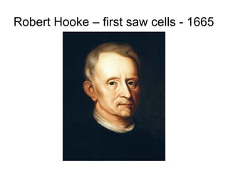
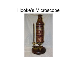
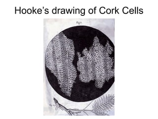
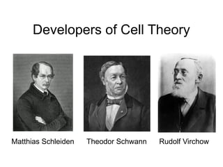
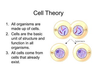
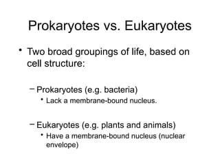
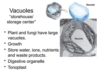
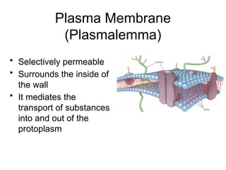
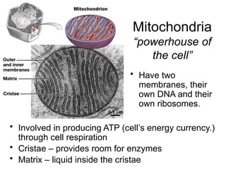
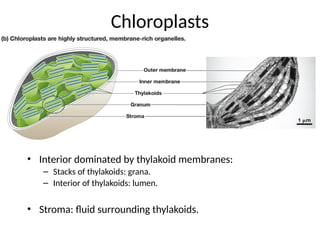
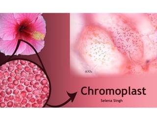
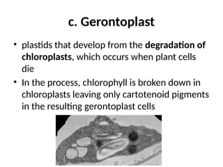
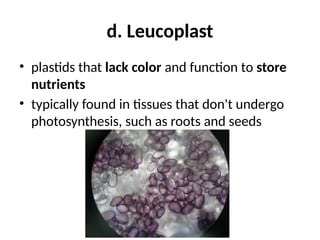
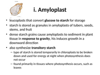
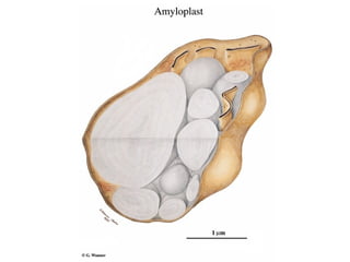
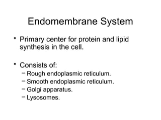
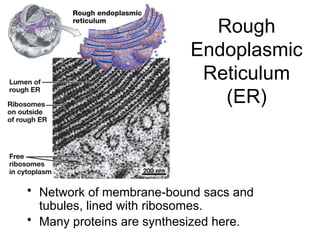
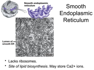
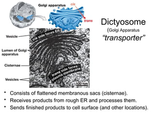
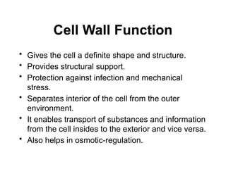
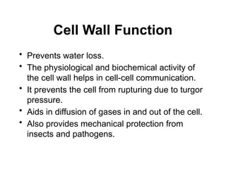
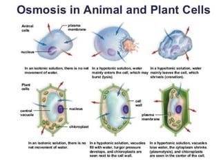
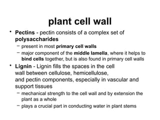
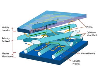
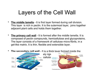
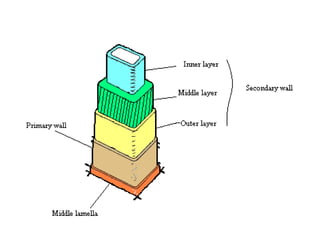
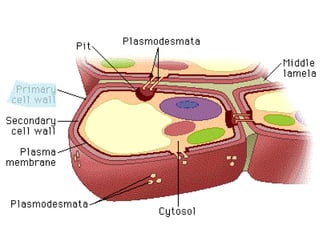
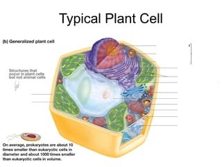
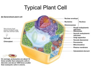
Inside the Cell
Robert Hooke – first saw cells - 1665
Hooke’s Microscope
Hooke’s drawing of Cork Cells
Development of Cell Theory of Life
• 1838 – Matthias Schleiden – stated all
plants composed of cells
• 1839 – Theodor Schwann – stated all
animals also composed of cells – thus
claimed all living things composed of
cells
• 1858 – Rudolf Virchow – all cells come
from preexisting cells
Developers of Cell Theory
Matthias Schleiden Theodor Schwann Rudolf Virchow
Cell Theory
1. All organisms are
made up of cells.
2. Cells are the basic
unit of structure and
function in all
organisms.
3. All cells come from
cells that already
exist.
Prokaryotes vs. Eukaryotes
• Two broad groupings of life, based on
cell structure:
– Prokaryotes (e.g. bacteria)
• Lack a membrane-bound nucleus.
– Eukaryotes (e.g. plants and animals)
• Have a membrane-bound nucleus (nuclear
envelope)
Protoplasm
• A substance all cells are made of
• Mass of
– Protein
– Lipids
– Nucleic acid
– And water within the cell
– Except for the wall, everything in the it, cell
composed the organelles
Nucleus
“control unit”
• Contains a cell’s genetic information:
– Stored in the DNA sequences of chromosomes.
Nucleus
• Bounded by double membrane: nuclear envelope.
• Contains a nucleolus: where ribosomal RNA is synthesized.
Nucleus
• Nucleoplasm
– DNA
– Enzymes for repair and
reading DNA
– Histone proteins –
supports the DNA
– RNA of several types
– Water and substances
necessary for nuclear
metabolism
Vacuoles
“storehouse/
storage center”
• Plant and fungi have large
vacuoles.
• Growth
• Store water, ions, nutrients
and waste products.
• Digestive organelle
• Tonoplast
Plasma Membrane
(Plasmalemma)
• Selectively permeable
• Surrounds the inside of
the wall
• It mediates the
transport of substances
into and out of the
protoplasm
Cytoplasm
• Protoplasm less nucleus and vacuole
• Contains the following structures:
Mitochondria
“powerhouse of
the cell”
• Involved in producing ATP (cell’s energy currency.)
through cell respiration
• Cristae – provides room for enzymes
• Matrix – liquid inside the cristae
• Have two
membranes, their
own DNA and their
own ribosomes.
Types of Plastids
• Plastids are organelles that function primarily in nutrient
synthesis and storage of biological molecules
• located in the cell cytoplasm and are surrounded by a
double lipid membrane
• also have their own DNA
• Some plastids contain pigments and are colorful, while
others lack pigments and are colorless
• develop from immature, undifferentiated cells called
proplastids
• Proplastids mature into four types of specialized plastids:
a. chloroplasts, b. chromoplasts, c. gerontoplasts, and d.
leucoplasts.
a. Chloroplast
• green plastids responsible for photosynthesis and
energy production through glucose synthesis
• contains chlorophyll, a green pigment that
absorbs light energy
• commonly found in specialized cells called guard
cells located in plant leaves and stems
• Guard cells open and close tiny pores called
stomata to allow for gas exchange required for
photosynthesis
Chloroplasts
“food factory”
• Found in plant and algal cells.
• Have two membranes and their own DNA.
Chloroplasts
• Interior dominated by thylakoid membranes:
– Stacks of thylakoids: grana.
– Interior of thylakoids: lumen.
• Stroma: fluid surrounding thylakoids.
b. Chromoplast
• colorful plastids responsible for cartenoid
pigment production and storage
• Carotenoids produce red, yellow, and orange
pigments
• primarily located in ripened fruit, flowers,
roots, and leaves of angiosperms
b. Chromoplast
• responsible for tissue coloration in plants,
which serves to attract pollinators
• Some chloroplasts found in unripened fruit
convert to chromoplasts as the fruit matures
• Amyloplasts can also be converted to
chromoplasts by first transitioning to
amylochromoplasts (plastids containing starch
and carotenoids) and then to chromoplasts.
c. Gerontoplast
• plastids that develop from the degradation of
chloroplasts, which occurs when plant cells
die
• In the process, chlorophyll is broken down in
chloroplasts leaving only cartotenoid pigments
in the resulting gerontoplast cells
d. Leucoplast
• plastids that lack color and function to store
nutrients
• typically found in tissues that don't undergo
photosynthesis, such as roots and seeds
Types of leucoplasts include:
i. Amyloplast
• leucoplasts that convert glucose to starch for storage
• starch is stored as granules in amyloplasts of tubers, seeds,
stems, and fruit
• dense starch grains cause amyloplasts to sediment in plant
tissue in response to gravity, his induces growth in a
downward direction
• also synthesize transitory starch
– type of starch is stored temporarily in chloroplasts to be broken
down and used for energy at night when photosynthesis does
not occur
– found primarily in tissues where photosynthesis occurs, such as
leaves
ii. Elaioplasts
• leucoplasts that synthesize fatty acids and
store oils in lipid-filled microcompartments
called plastoglobuli
• important to the proper development of
pollen grains
iii. Etioplast
• light-deprived chloroplasts that do not contain
chlorophyll, but have the precursor pigment
for chlorophyll production
• Once exposed to light, chlorophyll production
occurs and etioplasts are converted to
chloroplasts
iv. Proteinoplasts
• also called aleuroplasts
• these leucoplasts store protein and are often
found in seeds
• Composed of proteins and RNA.
• Immersed in the protoplasm
• Involved in protein synthesis.
Ribosomes
“protein factory"
Endomembrane System
• Primary center for protein and lipid
synthesis in the cell.
• Consists of:
– Rough endoplasmic reticulum.
– Smooth endoplasmic reticulum.
– Golgi apparatus.
– Lysosomes.
Rough
Endoplasmic
Reticulum
(ER)
• Network of membrane-bound sacs and
tubules, lined with ribosomes.
• Many proteins are synthesized here.
Smooth
Endoplasmic
Reticulum
• Lacks ribosomes.
• Site of lipid biosynthesis. May store Ca2+ ions.
Dictyosome
(Golgi Apparatus
“transporter”
• Consists of flattened membranous sacs (cisternae).
• Receives products from rough ER and processes them.
• Sends finished products to cell surface (and other locations).
• Have a relatively acidic lumen (interior: pH = 5).
• Contain many digestive enzymes (acid hydrolases):
– Involved in digesting various molecules into monomers.
Lysosomes
“cell suicide bag”
Delivering Material
to Lysosomes
Receptor-
mediated
Endocytosis
Peroxisomes
the “liver” organelle
• Peroxisomal enzymes:
– Help metabolize fatty
acids into smaller
molecules.
– Help detoxify many
toxins, including alcohol.
– Include catalase, which
breaks down H2O2.
Microtubules
• Cytoskeleton - fibers which gives the
cell shape and structural stability
• Catch and guides vesicle to where they
are needed
• Means of motility of organelles and cells
Cell Wall
• Cell Wall:
– Protects the cell.
– Found in fungi, algae, and plants.
Cell Wall Definition
• a rigid layer that surrounds some types of
cells
• located outside the cell membrane whose
main function is to provide:
– rigidity
– strength
– protection against mechanical stress and
– infection
Cell Wall Definition
• also provides the cell with limited plasticity
that prevents the cell from rupturing due to
the turgor pressure
• Cutinized, preventing water loss and also
aid in cell-cell communication
• characteristic feature to cells of plants,
bacteria, fungi, many algae and some
archaea
• protozoans and animals do not have a cell
wall
Cell Wall Function
• Gives the cell a definite shape and structure.
• Provides structural support.
• Protection against infection and mechanical
stress.
• Separates interior of the cell from the outer
environment.
• It enables transport of substances and information
from the cell insides to the exterior and vice versa.
• Also helps in osmotic-regulation.
Cell Wall Function
• Prevents water loss.
• The physiological and biochemical activity of
the cell wall helps in cell-cell communication.
• It prevents the cell from rupturing due to turgor
pressure.
• Aids in diffusion of gases in and out of the cell.
• Also provides mechanical protection from
insects and pathogens.
Cell Wall Structure
• The composition of the cell wall differs from
one species to the other:
• bacteria – peptidoglycans
• archean - glycoproteins and polysaccharides
• Fungi - glucosamine and chitin
• Algae - it is composed of glycoproteins and
polysaccharides
• plant cell wall - cellulose, hemicellulose,
glycoproteins, pectins and lignin
Plant Cell Wall
• Presence of Cell wall is the major
difference between plant cell and animal
cell.
• What is its function?
• What are its components?
plant cell wall
• Cellulose - composed of linear chains of
covalently linked glucose residues
– very stable chemically and extremely
insoluble
– primary cell wall consists one glucose
polymer of roughly 6000 glucose units
– secondary wall their number increased to 13 -
16000 units
– Cellulose chains form crystalline structures
called microfibrils.
plant cell wall
• Hemicellulose
Any of several branched polysaccharides that
are composed of a variety of
different monosaccharides
– forms a
matrix with cellulose and lignin or pectin
in plant cell walls
– It is produced commercially from corn fiber.
• Glycoproteins - a type of protein molecule
that has had a carbohydrate attached to it
plant cell wall
• Pectins - pectin consists of a complex set of
polysaccharides
– present in most primary cell walls
– major component of the middle lamella, where it helps to
bind cells together, but is also found in primary cell walls
• Lignin - Lignin fills the spaces in the cell
wall between cellulose, hemicellulose,
and pectin components, especially in vascular and
support tissues
– mechanical strength to the cell wall and by extension the
plant as a whole
– plays a crucial part in conducting water in plant stems
Layers of the Cell Wall
• The middle lamella - It is first layer formed during cell division.
This layer is rich in pectin. It is the outermost layer, joins together
adjacent plant cells and holds them together.
• The primary cell wall - It is formed after the middle lamella. It is
composed of pectin compounds, hemicellulose and glycoproteins.
The layer consists of a framework of cellulose micro-fibrils, in a
gel-like matrix. It is thin, flexible and extensible layer.
• The secondary cell wall - It is a thick layer formed inside the
primary cell wall. It is extremely rigid and provides strength. It is
composed of cellulose, hemicellulose and lignin.
Typical Plant Cell
Typical Plant Cell

Inside the Cell show.ppsxvkfofjfjfkfofdndnfjken

  • 1.
  • 2.
    Robert Hooke –first saw cells - 1665
  • 3.
  • 4.
  • 5.
    Development of CellTheory of Life • 1838 – Matthias Schleiden – stated all plants composed of cells • 1839 – Theodor Schwann – stated all animals also composed of cells – thus claimed all living things composed of cells • 1858 – Rudolf Virchow – all cells come from preexisting cells
  • 6.
    Developers of CellTheory Matthias Schleiden Theodor Schwann Rudolf Virchow
  • 7.
    Cell Theory 1. Allorganisms are made up of cells. 2. Cells are the basic unit of structure and function in all organisms. 3. All cells come from cells that already exist.
  • 8.
    Prokaryotes vs. Eukaryotes •Two broad groupings of life, based on cell structure: – Prokaryotes (e.g. bacteria) • Lack a membrane-bound nucleus. – Eukaryotes (e.g. plants and animals) • Have a membrane-bound nucleus (nuclear envelope)
  • 9.
    Protoplasm • A substanceall cells are made of • Mass of – Protein – Lipids – Nucleic acid – And water within the cell – Except for the wall, everything in the it, cell composed the organelles
  • 10.
    Nucleus “control unit” • Containsa cell’s genetic information: – Stored in the DNA sequences of chromosomes.
  • 11.
    Nucleus • Bounded bydouble membrane: nuclear envelope. • Contains a nucleolus: where ribosomal RNA is synthesized.
  • 12.
    Nucleus • Nucleoplasm – DNA –Enzymes for repair and reading DNA – Histone proteins – supports the DNA – RNA of several types – Water and substances necessary for nuclear metabolism
  • 13.
    Vacuoles “storehouse/ storage center” • Plantand fungi have large vacuoles. • Growth • Store water, ions, nutrients and waste products. • Digestive organelle • Tonoplast
  • 14.
    Plasma Membrane (Plasmalemma) • Selectivelypermeable • Surrounds the inside of the wall • It mediates the transport of substances into and out of the protoplasm
  • 15.
    Cytoplasm • Protoplasm lessnucleus and vacuole • Contains the following structures:
  • 16.
    Mitochondria “powerhouse of the cell” •Involved in producing ATP (cell’s energy currency.) through cell respiration • Cristae – provides room for enzymes • Matrix – liquid inside the cristae • Have two membranes, their own DNA and their own ribosomes.
  • 17.
    Types of Plastids •Plastids are organelles that function primarily in nutrient synthesis and storage of biological molecules • located in the cell cytoplasm and are surrounded by a double lipid membrane • also have their own DNA • Some plastids contain pigments and are colorful, while others lack pigments and are colorless • develop from immature, undifferentiated cells called proplastids • Proplastids mature into four types of specialized plastids: a. chloroplasts, b. chromoplasts, c. gerontoplasts, and d. leucoplasts.
  • 19.
    a. Chloroplast • greenplastids responsible for photosynthesis and energy production through glucose synthesis • contains chlorophyll, a green pigment that absorbs light energy • commonly found in specialized cells called guard cells located in plant leaves and stems • Guard cells open and close tiny pores called stomata to allow for gas exchange required for photosynthesis
  • 20.
    Chloroplasts “food factory” • Foundin plant and algal cells. • Have two membranes and their own DNA.
  • 21.
    Chloroplasts • Interior dominatedby thylakoid membranes: – Stacks of thylakoids: grana. – Interior of thylakoids: lumen. • Stroma: fluid surrounding thylakoids.
  • 22.
    b. Chromoplast • colorfulplastids responsible for cartenoid pigment production and storage • Carotenoids produce red, yellow, and orange pigments • primarily located in ripened fruit, flowers, roots, and leaves of angiosperms
  • 23.
    b. Chromoplast • responsiblefor tissue coloration in plants, which serves to attract pollinators • Some chloroplasts found in unripened fruit convert to chromoplasts as the fruit matures • Amyloplasts can also be converted to chromoplasts by first transitioning to amylochromoplasts (plastids containing starch and carotenoids) and then to chromoplasts.
  • 25.
    c. Gerontoplast • plastidsthat develop from the degradation of chloroplasts, which occurs when plant cells die • In the process, chlorophyll is broken down in chloroplasts leaving only cartotenoid pigments in the resulting gerontoplast cells
  • 26.
    d. Leucoplast • plastidsthat lack color and function to store nutrients • typically found in tissues that don't undergo photosynthesis, such as roots and seeds
  • 27.
  • 28.
    i. Amyloplast • leucoplaststhat convert glucose to starch for storage • starch is stored as granules in amyloplasts of tubers, seeds, stems, and fruit • dense starch grains cause amyloplasts to sediment in plant tissue in response to gravity, his induces growth in a downward direction • also synthesize transitory starch – type of starch is stored temporarily in chloroplasts to be broken down and used for energy at night when photosynthesis does not occur – found primarily in tissues where photosynthesis occurs, such as leaves
  • 30.
    ii. Elaioplasts • leucoplaststhat synthesize fatty acids and store oils in lipid-filled microcompartments called plastoglobuli • important to the proper development of pollen grains
  • 31.
    iii. Etioplast • light-deprivedchloroplasts that do not contain chlorophyll, but have the precursor pigment for chlorophyll production • Once exposed to light, chlorophyll production occurs and etioplasts are converted to chloroplasts
  • 32.
    iv. Proteinoplasts • alsocalled aleuroplasts • these leucoplasts store protein and are often found in seeds
  • 33.
    • Composed ofproteins and RNA. • Immersed in the protoplasm • Involved in protein synthesis. Ribosomes “protein factory"
  • 34.
    Endomembrane System • Primarycenter for protein and lipid synthesis in the cell. • Consists of: – Rough endoplasmic reticulum. – Smooth endoplasmic reticulum. – Golgi apparatus. – Lysosomes.
  • 35.
    Rough Endoplasmic Reticulum (ER) • Network ofmembrane-bound sacs and tubules, lined with ribosomes. • Many proteins are synthesized here.
  • 36.
    Smooth Endoplasmic Reticulum • Lacks ribosomes. •Site of lipid biosynthesis. May store Ca2+ ions.
  • 37.
    Dictyosome (Golgi Apparatus “transporter” • Consistsof flattened membranous sacs (cisternae). • Receives products from rough ER and processes them. • Sends finished products to cell surface (and other locations).
  • 38.
    • Have arelatively acidic lumen (interior: pH = 5). • Contain many digestive enzymes (acid hydrolases): – Involved in digesting various molecules into monomers. Lysosomes “cell suicide bag”
  • 39.
  • 40.
  • 41.
    Peroxisomes the “liver” organelle •Peroxisomal enzymes: – Help metabolize fatty acids into smaller molecules. – Help detoxify many toxins, including alcohol. – Include catalase, which breaks down H2O2.
  • 42.
    Microtubules • Cytoskeleton -fibers which gives the cell shape and structural stability • Catch and guides vesicle to where they are needed • Means of motility of organelles and cells
  • 43.
    Cell Wall • CellWall: – Protects the cell. – Found in fungi, algae, and plants.
  • 44.
    Cell Wall Definition •a rigid layer that surrounds some types of cells • located outside the cell membrane whose main function is to provide: – rigidity – strength – protection against mechanical stress and – infection
  • 45.
    Cell Wall Definition •also provides the cell with limited plasticity that prevents the cell from rupturing due to the turgor pressure • Cutinized, preventing water loss and also aid in cell-cell communication • characteristic feature to cells of plants, bacteria, fungi, many algae and some archaea • protozoans and animals do not have a cell wall
  • 47.
    Cell Wall Function •Gives the cell a definite shape and structure. • Provides structural support. • Protection against infection and mechanical stress. • Separates interior of the cell from the outer environment. • It enables transport of substances and information from the cell insides to the exterior and vice versa. • Also helps in osmotic-regulation.
  • 48.
    Cell Wall Function •Prevents water loss. • The physiological and biochemical activity of the cell wall helps in cell-cell communication. • It prevents the cell from rupturing due to turgor pressure. • Aids in diffusion of gases in and out of the cell. • Also provides mechanical protection from insects and pathogens.
  • 50.
    Cell Wall Structure •The composition of the cell wall differs from one species to the other: • bacteria – peptidoglycans • archean - glycoproteins and polysaccharides • Fungi - glucosamine and chitin • Algae - it is composed of glycoproteins and polysaccharides • plant cell wall - cellulose, hemicellulose, glycoproteins, pectins and lignin
  • 51.
    Plant Cell Wall •Presence of Cell wall is the major difference between plant cell and animal cell. • What is its function? • What are its components?
  • 52.
    plant cell wall •Cellulose - composed of linear chains of covalently linked glucose residues – very stable chemically and extremely insoluble – primary cell wall consists one glucose polymer of roughly 6000 glucose units – secondary wall their number increased to 13 - 16000 units – Cellulose chains form crystalline structures called microfibrils.
  • 54.
    plant cell wall •Hemicellulose Any of several branched polysaccharides that are composed of a variety of different monosaccharides – forms a matrix with cellulose and lignin or pectin in plant cell walls – It is produced commercially from corn fiber. • Glycoproteins - a type of protein molecule that has had a carbohydrate attached to it
  • 56.
    plant cell wall •Pectins - pectin consists of a complex set of polysaccharides – present in most primary cell walls – major component of the middle lamella, where it helps to bind cells together, but is also found in primary cell walls • Lignin - Lignin fills the spaces in the cell wall between cellulose, hemicellulose, and pectin components, especially in vascular and support tissues – mechanical strength to the cell wall and by extension the plant as a whole – plays a crucial part in conducting water in plant stems
  • 58.
    Layers of theCell Wall • The middle lamella - It is first layer formed during cell division. This layer is rich in pectin. It is the outermost layer, joins together adjacent plant cells and holds them together. • The primary cell wall - It is formed after the middle lamella. It is composed of pectin compounds, hemicellulose and glycoproteins. The layer consists of a framework of cellulose micro-fibrils, in a gel-like matrix. It is thin, flexible and extensible layer. • The secondary cell wall - It is a thick layer formed inside the primary cell wall. It is extremely rigid and provides strength. It is composed of cellulose, hemicellulose and lignin.
  • 62.
  • 63.

Editor's Notes

  • #11 Nucleolus makes rRNA, and is where the r proteins are made in cytoplasm and are imported into the nucleolus to combine with the rRNA and make ribosomomal subunits->ribosomes.
  • #12 Nucleolus makes rRNA, and is where the r proteins are made in cytoplasm and are imported into the nucleolus to combine with the rRNA and make ribosomomal subunits->ribosomes.
  • #16 Has two membranes, inner and outer. The fluid in the inner membrane is the matrix. The space between the inner and outer is the intermembrane space. The foldings of the inner membrane are the cristae.
  • #21 The lumen is the blue
  • #27 https://www.thoughtco.com/amyloplast-definition-4142136
  • #33 Free ribosomes-not associated with any specific membrane and bound ribosomes-associated with the ER. In euks the cytoplasm is everything in the cell other than the nucleus, the fluid of the cytoplasm is the cytosol.
  • #34 Share vesicles or membranes with each other
  • #35 When proteins are made by bound ribosomes, they tend to be exported to the cell surface to exit cells. Things that are secreted outside the cell are made by bound. Lysosome proteins are made by bound. Any other free proteins are made by free ribosomes.
  • #37 Proteins don’t get synthesized here, but get modified or cleaved. They’re then packaged in new vesicles, and leave the golgi to their final destination.
  • #39 Theyre involved in autophagy, eat/merge damaged or no longer needed organelle. It recycles the materials. Involved in phagocytosis, where it reaches out, grabs the particle/bacterial cell, engulfs it. Then the lysosomes fuse with it and digest it. Pseudopodium, the parts of the membrane that surrounds and grabs the particle.
  • #40 Mostly know #1,2 Specific materials are being brought in
  • #41 Both animal and plant cells have there The “liver” organelle Breaks H peroxide into H and O, or water
  • #50 Peptidoglycans - provides rigidity to the cell wall Chitin - This is a long carbohydrate polymer that also occurs in the exoskeletons of insects, spiders, and other arthropods. The chitin adds rigidity and structural support to the thin cells of the fungus, and makes fresh mushrooms crisp.
  • #54 https://www.thoughtco.com/glycoprotein-definition-and-function-4134331
  • #62 Plants have cell walls, cholorplasts, vacuole, and euks don’t.